Aggregation and ROS Mediated Platelet Apoptosis
Kodagahalli S. Rakesh1., Swamy Jagadish1., Ajjampura C. Vinayaka1., Mahadevappa Hemshekhar2
, Manoj Paul2, Ram M. Thushara2, Mahalingam S. Sundaram2, Toreshettahally R. Swaroop1,
Chakrabhavi D. Mohan1, Basappa3, Marilinganadoddi P. Sadashiva1, Kempaiah Kemparaju2,
Kesturu S. Girish2,4*, Kanchugarakoppal S. Rangappa1*
1DOS in Chemistry, University of Mysore, Manasagangotri, Mysore, India,2DOS in Biochemistry, University of Mysore, Manasagangotri, Mysore, India,3Laboratory of Chemical Biology, Department of Chemistry, Bangalore University, Bangalore, India,4Department of Studies and Research in Biochemistry, Tumkur University, Tumkur, India
Abstract
Thrombocytopenia is a serious issue connected with the pathogenesis of several human diseases including chronic inflammation, arthritis, Alzheimer’s disease, cardiovascular diseases (CVDs) and other oxidative stress-associated pathologies. The indiscriminate use of antibiotics and other biological drugs are reported to result in thrombocytopenia, which is often neglected during the treatment regime. In addition, augmented oxidative stress induced by drugs and pathological conditions has also been shown to induce thrombocytopenia, which seems to be the most obvious consequence of elevated rate of platelet apoptosis. Thus, blocking oxidative stress-induced platelet apoptosis would be of prime importance in order to negotiate thrombocytopenia and associated human pathologies. The current study presents the synthesis and platelet protective nature of novel ibuprofen derivatives. The potent anti-oxidant ibuprofen derivative 4f was selected for the study and the platelet protective efficacy and platelet aggregation inhibitory property has been demonstrated. The compound 4f dose dependently mitigates the oxidative stress-induced platelet apoptosis in both platelet rich plasma and washed platelets. The platelet protective nature of compound 4f was determined by assessing various apoptotic markers such as ROS generation, cytosolic Ca2+levels, PS externalization, cytochrome C translocation, Caspase activation, mitochondrial membrane depolarization, cytotoxicity, LDH leakage and tyrosine phosphorylation of cytosolic proteins. Furthermore, compound 4f dose dependently ameliorated agonist induced platelet aggregation. Therefore, compound 4f can be estimated as a potential candidate in the treatment regime of pathological disorders associated with platelet activation and apoptosis. In addition, compound 4f can be used as an auxiliary therapeutic agent in pathologies associated with thrombocytopenia.
Citation:Rakesh KS, Jagadish S, Vinayaka AC, Hemshekhar M, Paul M, et al. (2014) A New Ibuprofen Derivative Inhibits Platelet Aggregation and ROS Mediated Platelet Apoptosis. PLoS ONE 9(9): e107182. doi:10.1371/journal.pone.0107182
Editor:Dermot Cox, Royal College of Surgeons, Ireland
ReceivedMay 7, 2014;AcceptedAugust 6, 2014;PublishedSeptember 19, 2014
Copyright:ß2014 Rakesh et al. This is an open-access article distributed under the terms of the Creative Commons Attribution License, which permits unrestricted use, distribution, and reproduction in any medium, provided the original author and source are credited.
Data Availability:The authors confirm that all data underlying the findings are fully available without restriction. Data are available from the Mysore University Institutional Data Access/Ethics Committee for researchers who meet the criteria for access to confidential data.
Funding:KS Rangappa thanks DST-JSPS (DST/INT/JAP/P-79/09, Dated: 20/05/2009), KS Rakesh thanks BRNS (No. 2009/37/40/BRNS/2266, Dated: 23/11/2009), S Jagadish thanks UGC-MRP (F. No. 39-106/2010 SR, Dated: 24/12/2010), AC Vinayaka thanks CSIR (SRF-Ref: 9/119 (0819) 2KR-EMR-I) for financial assistance. The funders had no role in study design, data collection and analysis, decision to publish, or preparation of the manuscript.
Competing Interests:The authors have declared that no competing interests exist.
* Email: [email protected] (K. S. Rangappa); [email protected] (KSG)
.These authors have contributed equally to this work.
Introduction
Several human diseases including chronic inflammation, diabetes, arthritis, cardiovascular diseases (CVDs) and other oxidative stress-associated pathologies are also being linked to mean platelet volume and reduced platelet count, in other words thrombocytopenia [1–4]. The augmented oxidative stress as evidenced by elevated reactive oxygen species (ROS) is shown to induce thrombocytopenia, which seems to be the most obvious consequence of elevated rate of platelet apoptosis [5]. Platelets are simple anuclear cells, yet execute a plethora of physiological actions such as hemostasis, thrombosis and wound healing [6]. It is no more surprising that the anuclear platelets end their life through apoptosis like any other nucleated cell. Several studies
have demonstrated that platelets possess the necessary cellular machinery to undergo apoptosis [7,8]. Besides aging, several other factors like chemical and physical agonists, oxidative stress-induced pathological conditions are also shown to trigger apoptosis in platelets [5,9,10]. They undergo apoptosis via intrinsic apoptotic pathway; however, very few studies have reported the extrinsic pathway too [5,11]. The altered platelet number and functions would certainly lead to bleeding disorders and throm-botic diseases. The distorted platelet functions are also responsible for the multifactorial diseases including coronary heart disease (CHD) and other cardiovascular diseases (CVDs), inflammatory and immune reactions [12–14].
particularly, hydrogen peroxide (H2O2). The principal target of
oxidative stress is mitochondrial damage resulting in the down-regulation of electron transport chain, which in turn increases ROS generation and formation of mitochondrial permeability transition pore (MPTP). This would significantly induce depolar-ization of mitochondrial membrane potential (DYm) causing leakage of cytochrome c (cyt c) into the cytoplasm. Furthermore, the leaked cyt c in association with pro-apoptotic factors like Apoptotic Protease Activating Factor-1 (Apaf-1) mediate the activation of caspase-9 (elicitor caspase), which in turn activates caspase-3 (executioner caspase). Finally, phosphatidylserine (PS) externalization occurs, which is a signal for phagocytosis [7,8,15]. Besides, the process of platelet apoptosis also releases PS-positive membrane fractions called microparticles (MPs), which signifi-cantly contribute to the pathogenesis of atherosclerosis, central nervous system damage and neoplasia [16].
Thus, blocking oxidative stress-induced platelet apoptosis would be of prime importance in order to combat thrombocytopenia and associated human pathologies. Hence, there is a need for potent molecules that can protect platelets from the premature death. Till date there are only two reports describing the inhibition of platelet apoptosis by phytochemicals namely, cinnamtannin-B1 and crocin [17,18]. Both the molecules ameliorate the oxidative stress-induced platelet apoptosis by modulating ROS mediated mito-chondrial damage. Ibuprofen is a non-steroidal anti-inflammatory drug (NSAID) used to treat the pathological conditions related to inflammation. It exerts its anti-inflammatory action by blocking cyclooxygenase-1 non-specifically and thereby thromboxane production [19]. In the recent past, studies reported that Ibuprofen augments the anticancer activity of cisplatin in lung adenocarcinoma cells by blocking heat shock protein 70 [20]. However, laboratory testing of drug-induced immune thrombo-cytopenia (DIIT) reported development of immune thrombocyto-penia by ibuprofen [21]. Based on these facts, a series of novel ibuprofen derivatives 4(a-f) are synthesized by modifying the functional groups and evaluated for improved anti-oxidant activity. Among the derivatives, the potent anti-oxidant compound 4f was investigated for the protective efficacy against platelet apoptosis as a clinical implication to thrombocytopenia.
Materials and Methods
Chemicals/Reagents
Calcium ionophore (A23187), benzamidine hydrochloride, N-acetyl-Leu-Glu-His-Asp trifluoro methylcoumarin LEHD-FMC), acetyl-Asp-Glu-Val-Asp-7-amido-4-methylcoumarin (AC-DEVD-AMC), sodium orthovanadate, dimethyl sulfoxide (DMSO), fluorescein isothiocyanate (FITC)-labeled annexin V, 5-(and-6)-chloromethyl-29,79-dichlorodihydrofluorescein diacetate acetylester (CM-H2DCFDA), CHAPS, rhodamine 123, leupeptin hydrochoride, N-(2-Hydroxyethyl) piperazine-N9-ethanesulfonic acid (HEPES), fura-2/AM, monoclonal anti-phosphotyrosine antibody, acridine orange 10-nonyl bromide (NAO) and dithio-threitol (DTT) were from Sigma Chemicals, St. Louis (USA). Monoclonal anti-cytochrome c antibody and anti-b-actin were from Epitomics Burlingame, CA (USA). Anti-Caspase-3 antibody was from Santa Cruz Biotechnology, Inc. Texas (USA). Collagen type-I was from Chrono-log Corporation, Pennsylvania (USA). 3-(4,5-dimethylthiazol-2-yl)-2,5-diphenyltetrazolium bromide (MTT) and 1, 1-diphenyl-2-picrylhdrazyl (DPPH) were from HiMedia Laboratories, Mumbai (India). Lactate dehydrogenase (LDH) kit was from AGAPPE diagnostics Ltd., Kerala (India).c -glutamyl p-nitroanilide and glycylglycine were from Sisco
Research laboratories Pvt Ltd., Mumbai (India). All other reagents were of analytical grade.
Synthesis and characterization of Ibuprofen derivatives
The starting materials were commercially available and used as received without further purification. Reactions were monitored by TLC using precoated sheets of silica gel (G/UV-254) of 0.25 mm thickness (Merck 60F254) using UV light for visualiza-tion. The melting points were determined on Selaco melting point apparatus and are uncorrected.1H and13C NMR spectra were recorded on an NMR spectrometer operating at 400 and 100 MHz, respectively, using the residual solvent peaks as reference relative to SiMe4. Mass spectra were recorded using
electrospray ionization (ESI) mass spectrometry. The C, H, and N analysis were performed using CE-400 CHN analyzer. Infrared spectra were recorded on Shimadzu FT-IR model 8300 spectro-photometer.
Ethyl 2-[4-(2-methylpropyl)phenyl]propanoate (2)
A solution of Ibuprofen 1 (2 g, 9.6 mmol) and 0.5 mL concentrated sulfuric acid in ethanol (20 mL) was refluxed for 6 h. The solvent was removed under reduced pressure and the residue was taken up in CHCl3 (30 mL). The solution was
extracted with saturated aqueous NaHCO3 solution (2615 mL)
and water. The organic layer was dried over anhydrous Na2SO4,
filtered and solvent was evaporated under reduced pressure, affording 2 (2.1 g, 95%) as a clear oil.
2-[4-(2-methylpropyl)phenyl]propanehydrazide (3)
The mixture of ethyl ester of Ibuprofen 2 (2 g, 8.5 mmol) and hydrazine hydrate (0.8 mL, 16 mmol) in absolute ethanol (30 mL) was refluxed for 10 h. The solvent was removed under reduced pressure and the residue was added to ice cold water. The solid separated was filtered, washed and dried. The solid was purified by recrystallization from ethanol to get pure product 3 (1.7 g, 96%) as a white solid.
2-[4-(2-methylpropyl)phenyl]-N ’-(phenylsulfonyl)propanehydrazide (4a-f)
To a stirred solution of 2-[4-(2-methylpropyl)phenyl]propane-hydrazide (0.15 g, 0.68 mmol), substituted benzene sulfonyl chloride (0.68 mmol) in dichlorormethane (5 mL), triethylamine (0.14 mL, 1.0 mmol) was added at 0uC. The reaction mixture was brought to room temperature and further stirred for 2 h. After the completion of reaction, the reaction mixture was extracted with ethyl acetate (2610 mL). The combined organic extracts were
washed with water (365 mL), brine (1610 mL) and dried over anhydrous Na2SO4, filtered and solvent was evaporated under
reduced pressure to get crude products 4 (a-f), which were purified by column chromatography over silica gel using hexanes-EtOAc as eluent.
2-[4-(2-methylpropyl)phenyl]-N ’-(phenylsulfonyl)propanehydrazide (4a)
Yield 89%, white solid; MP 112–114uC; IR (KBr) n,3320,
2951, 1675, 1585, 1124;1H NMR (400 MHz, CDCl3)d7.82 (d,
J = 6.4 Hz, 1H, NH), 7.72–7.57 (m, 5H, Ar-H), 7.21 (d, J = 6.4 Hz, 1H, NH), 7.07 (d, J = 7.6 Hz, 2H, Ar-H), 7.01 (d, J = 8.4 Hz, 2H, Ar-H), 3.42 (q, J = 6.1 Hz, 1H, CH), 2.44 (d, J = 7.6 Hz, 2H, ArCH2), 1.85 (m, 1H, CH), 1.26 (d, J = 7.2 Hz,
3H, Me), 0.88 (dd, J = 6.6 Hz, 1.0 Hz, 6H, Me2); 13
C NMR (100 MHz, CDCl3) d 177.8, 141.2, 136.4, 132.4, 130.8, 129.6,
for C19H24N2O3S: C 63.31, H 6.71, N 7.77. Found: C 63.32, H
6.72, N 7.78.
N’-[(4-fluorophenyl)sulfonyl]-2-[4-(2-methylpropyl) phenyl]propanehydrazide (4b)
Yield 91%, white solid, MP 121–123uC; IR (KBr) n,3312,
2960, 1679, 1592, 1139;1H NMR (400 MHz, CDCl3)d7.85 (d,
J = 6.2 Hz, 1H, NH), 7.79 (d, J = 7.8 Hz, 2H, Ar-H), 7.62 (d, J = 7.8 Hz, 2H, Ar-H), 7.26 (d, J = 6.2 Hz, 1H, NH), 7.07 (d, J = 7.6 Hz, 2H, Ar-H), 7.01 (d, J = 8.4 Hz, 2H, Ar-H), 3.42 (q, J = 6.1 Hz, 1H, CH), 2.44 (d, J = 7.6 Hz, 2H, ArCH2), 1.85 (m,
1H, CH), 1.26 (d, J = 7.2 Hz, 3H, Me), 0.88 (dd, J = 6.6 Hz, 1.0 Hz, 6H, Me2);13C NMR (100 MHz, CDCl3)d177.8, 168.8,
141.2, 136.4, 131.8, 129.6, 128.4, 127.2, 117.3, 44.9, 44.4, 30.2, 22.4, 22.3, 17.9; Anal. Calcd for C19H23FN2O3S: C 60.30, H
6.13, N 7.40. Found: C 60.31, H 6.15, N 7.43.
N ’-[(4-bromophenyl)sulfonyl]-2-[4-(2-methylpropyl)phenyl]propanehydrazide (4c)
Yield 81%, red solid, MP 116–118uC; IR (KBr)n,3340, 2949,
1668, 1598, 1162; 1H NMR (400 MHz, CDCl3) d 7.85 (d,
J = 6.2 Hz, 1H, NH), 7.72 (d, 2H, J = 7.2 Hz, Ar-H), 7.68 (d, 2H, J = 7.2 Hz, Ar-H), 7.25 (d, J = 6.2 Hz, 1H, NH), 7.07 (d, J = 7.6 Hz, 2H, Ar-H), 7.01 (d, J = 8.4 Hz, 2H, Ar-H), 3.42 (q, J = 6.1 Hz, 1H, CH), 2.44 (d, J = 7.6 Hz, 2H, ArCH2), 1.85 (m,
1H, CH), 1.26 (d, J = 7.2 Hz, 3H, Me), 0.88 (dd, J = 6.6 Hz, 1.0 Hz, 6H, Me2);
13
C NMR (100 MHz, CDCl3)d177.6, 141.1,
135.8, 131.1, 130.4, 129.6, 128.3, 127.1, 126.9, 44.9, 44.4, 30.2, 22.4, 22.3, 17.9; Anal. Calcd for C19H23BrN2O3S: C 51.94, H
5.28, N 6.38. Found: C 51.95, H 5.31, N 6.39.
N
’-[(4-nitrophenyl)sulfonyl]-2-[4-(2-methylpropyl)phenyl]propanehydrazide (4d)
Yield 78%, yellow solid, MP 142-144uC; IR (KBr) n,3319,
2971, 1678, 1582, 1145;1H NMR (400 MHz, CDCl3)d7.88 (d,
J = 6.2 Hz, 1H, NH), 7.71–7.63 (m, 4H, Ar-H), 7.29 (d, J = 6.2 Hz, 1H, NH), 7.07 (d, J = 7.6 Hz, 2H, Ar-H), 7.01 (d, J = 8.4 Hz, 2H, Ar-H), 3.40 (q, J = 6.4 Hz, 1H, CH), 2.44 (d, J = 7.6 Hz, 2H, ArCH2), 1.85 (m, 1H, CH), 1.28 (d, J = 7.2 Hz,
3H, Me), 0.88 (dd, J = 6.6 Hz, 1.0 Hz,6H, Me2); 13C NMR
(100 MHz, CDCl3) d177.8, 152.8, 144.1, 141.2, 136.4, 129.6,
128.6, 127.2, 125.1, 44.9, 44.4, 30.2, 22.4, 22.3, 17.9; Anal. Calcd for C19H23N3O5S: C 56.28, H 5.72, N 10.36. Found: C 56.29, H
5.74, N 10.38.
N ’-[(2,5-dichlorophenyl)sulfonyl]-2-[4-(2-methylpropyl)phenyl]propanehydrazide (4e)
Yield 85%, white solid, MP 102–104uC; IR (KBr) n,3364,
2983, 1658, 1518, 1084;1H NMR (400 MHz, CDCl3)d7.82 (d,
J = 6.4 Hz, 1H, NH), 7.90 (s, 1H, Ar-H), 7.71 (d, J = 8.0 Hz, 1H, Ar-H), 7.65(d, J = 8.0 Hz, 1H, Ar-H) 7.24 (d, J = 6.4 Hz, 1H, NH), 7.07 (d, J = 7.6 Hz, 2H, Ar-H), 7.01 (d, J = 8.4 Hz, 2H, Ar-H), 3.40 (q, J = 6.4 Hz, 1H, CH), 2.44 (d, J = 7.6 Hz, 2H, ArCH2),
1.85 (m, 1H, CH), 1.28 (d, J = 7.2 Hz, 3H, Me), 0.88 (dd, J = 6.6 Hz, 1.0 Hz, 6H, Me2);13C NMR (100 MHz, CDCl3) d
177.8, 143.4, 141.2, 136.4, 134.2, 132.8, 131.3, 130.8, 129.6, 128.5, 127.2, 44.9, 44.4, 30.2, 22.4, 22.3, 17.9; Anal. Calcd for C19H22Cl2N2O3S: C 53.15, H 5.16, N 6.52. Found: C 53.16, H
5.18, N 6.54.
N ’-[(4-methoxyphenyl)sulfonyl]-2-[4-(2-methylpropyl)phenyl]propanehydrazide (4f)
Yield 94%, White solid; MP 96–98uC; IR (KBr)n,3315, 2953,
1673, 1597, 1154; 1H NMR (400 MHz, CDCl3) d 7.82 (d,
J = 6.4 Hz, 1H, NH), 7.68 (dt, J = 8.4 Hz, 3.2 Hz, 2.0 Hz, 2H, Ar-H), 7.24 (d, J = 6.4 Hz, 1H, NAr-H), 7.07 (d, J = 7.6 Hz, 2H, Ar-Ar-H), 7.01 (d, J = 8.4 Hz, 2H, Ar-H), 6.84 (dt, J = 8.4 Hz, 3.2 Hz, 2.0 Hz, 2H, Ar-H), 3.84 (s, 3H, OMe), 3.40 (q, J = 6.4 Hz, 1H, CH), 2.44 (d, J = 7.6 Hz, 2H, ArCH2), 1.85 (m, 1H, CH), 1.28 (d,
J = 7.2 Hz, 3H, Me), 0.88 (dd, J = 6.6 Hz, 1.0 Hz, 6H, Me2);13C
NMR (100 MHz, CDCl3) d 177.8, 163.8, 141.2, 136.4, 130.8,
129.6, 127.2, 127.1, 114.1, 55.6, 44.9, 44.4, 30.2, 22.4, 22.3, 17.9; MS m/z 391 [M+H]; Anal. Calcd for C20H26N2O4S: C 61.52, H
6.71, N 7.17. Found: C 61.54, H 6.73, N 7.18.
Determination of reducing ability
Reducing ability of Ibuprofen and its derivatives (4a, 4b, 4c, 4d, 4e and 4f) along with quercetin as positive control were determined according to the method of Hsieh and Yan [22] with slight modifications. Briefly, samples (0–100mM) were mixed with 2.5 mL of 200 mM sodium phosphate buffer, pH 6.6 containing 1% potassium ferricyanide and the mixture was incubated at 50uC for 20 min. At the end of incubation, 2.5 mL of 5% TCA was added and centrifuged at 4506gfor 10 min. Further, 2.5 mL of
supernatant was taken and mixed with 0.5 mL of aqueous 0.1% ferric chloride. The absorbance was measured at 700 nm against blank using UV/Vis spectrophotometer (BioMate 3S, Thermo scientifics).
Antioxidant activity
The free radical scavenging activity of Ibuprofen and its derivatives (4a, 4b, 4c, 4d, 4e and 4f) along with quercetin as positive control were determined using 1, 1-diphenyl-2-picrylh-drazyl (DPPH) radical according to the method of Yamaguchi et al. [23] with slight modifications. Briefly, Samples (0–100mM)
were taken in test tubes with 1 mL of freshly prepared 0.1 mM DPPH solution and the final volume was made up to 2 mL using methanol. Samples were incubated for 20 min at room temper-ature in dark and the resulting absorbance was recorded at 517 nm against blank using UV/Vis spectrophotometer.
Preparation of platelet-rich plasma and washed platelets
Venous blood was drawn from healthy drug-free human volunteers (non-smokers) approved by the Institutional Human Ethical Committee (IHEC-UOM No. 95/Ph.D/2013-14) Univer-sity of Mysore, Mysore. Written consent were obtained from the healthy volunteers as per the guidelines of Institutional Human Ethical Committee (IHEC-UOM No. 95/Ph.D/2013-14). It was immediately mixed with acid citrate dextrose (ACD) anticoagulant (85 mM sodium citrate, 78 mM citric acid and 111 mM D-glucose) in the ratio 6:1 (blood: ACD v/v). The anti-coagulated whole blood was then centrifuged at 906g for 15 min and the supernatant thus obtained was the platelet-rich plasma (PRP). The PRP was centrifuged at 1,7006gfor 15 min at 37uC. The platelet
pellet thus obtained was suspended and incubated for 10 min in Tyrode’s albumin buffer [145 mM NaCl, 5 mM KCl, 10 mM HEPES, 0.5 mM Na2HPO4, 1 mM MgCl2, 6 mM glucose, and
final suspension using platelet poor plasma/Tyrode’s albumin buffer (pH 7.4) [24].
Determination of endogenously generated reactive oxygen species (ROS)
Endogenous ROS production in platelets was determined according to the method of Lopez et al. [25] with slight modifications using CMH2DCFDA, a ROS-sensitive fluorescent probe. PRP as well as washed platelet suspensions were independently treated with calcium ionophore (A23187, 10mM) as agonist. For inhibition studies, pre-loaded platelets with agonist were incubated with 4f in increasing doses (0–100mM) and the
final volume was made up to 200mL with HEPES-buffered saline [HBS, 145 mM NaCl, 10 mM HEPES, 10 mM D-glucose, 5 mM KCl, 1 mM MgSO4and supplemented with 0.1% bovine serum
albumin (BSA), pH 7.45] and incubated at 37uC for 1 h. The control (untreated) and treated platelets were then incubated with 10mM CMH2DCFDA for 30 min at 37uC, fluorescence was recorded using Varioskan multimode plate reader (Thermo Scientifics, USA) by exciting the samples at 488 nm and measuring the resulting fluorescence at 530 nm.
Estimation of intracellular calcium
Intracellular Ca2+ concentration was measured in PRP and washed platelets according to the method of Asai et al. [26] with slight modifications. Briefly, PRP and washed platelet suspensions were independently treated with A23187 (10mM) as agonist. For inhibition studies, pre-loaded platelets with agonist were incubated with 4f in increasing doses (0–100mM) and the final volume was made up to 200mL with modified Tyrode’s solution (150 mM
NaCl, 2.7 mM KCl, 1.2 mM KH2PO4, 1.2 mM MgSO4, 1.0 mM
CaCl2, 10 mM HEPES with 0.1% bovine serum albumin, pH 7.4)
and incubated for 1 h at 37uC to induce the release of Ca2+from the intracellular Ca2+ stores. Samples were then incubated for 45 min at room temperature with 2mM fura-2/AM, a
fluores-cence Ca2+indicator. The cells were subsequently washed twice with modified Tyrode’s solution to remove the unbound dye and finally the platelet pellet was suspended in modified Tyrode’s solution. The fura-2/AM absorption was determined by exciting the samples at 340 and 380 nm and the resulting fluorescence was measured at 500 nm. Data were presented as absorption ratios (340/380 nm).
Assesment of cardiolipin peroxidation
NAO, a fluorescent probe was used to detect peroxidation of cardiolipin. NAO loses its affinity for peroxidised cardiolipin resulting in decreased fluorescence [27]. Both PRP and washed platelets were processed as discussed above with either A23187 (10mM) as agonist or pre-loaded platelets with agonist were
incubated with 4f in increasing concentrations (0–100mM) for
inhibition studies. After incubation, samples were loaded with NAO (5mM) for 30 min at 37uC. After incubation fluorescence was recorded by exciting the samples at 499 nm and emission was recorded at 530 nm.
Determination of changes in mitochondrial membrane potential (DYm)
Changes in DYm were determined using cationic dye, rhodamine 123 in which the intensity of the fluorescence decreases proportionally with decrease inDYm[28]. Both PRP and washed platelets were processed as discussed above with either A23187 (10mM) as agonist or pre-loaded platelets with agonist were incubated with 4f in increasing concentrations (0–100mM) for
inhibition studies. After incubation, samples were then loaded with 0.2mM rhodamine 123 followed by 15 min incubation at 37uC
and the fluorescence was recorded by exciting the samples at 502 nm and the resulting emission was recorded at 527 nm.
Assay of Caspases activity
PRP and washed platelets were processed as discussed previously with A23187 (10mM) as agonist and 4f in increasing doses for inhibition studies along with A23187 as agonist. Platelet lysate was prepared by adding an equal volume of 2X Triton buffer (2% Triton X100, 2 mM EGTA, 100 mM TrisHCl -pH 7.2, 10mg/mL leupeptin, 2 mM PMSF, 10 mM benzami-dine, 2 mM Na3VO4) to the treated and control platelets and
allowed to undergo lysis for 30 min at 4uC. The lysate was centrifuged at 160006gfor 5 min. The pellet thus obtained is the cytoskeleton-rich (Triton-insoluble) fraction, which was subjected to caspase activity.
Caspase activity was determined by incubating cell lysate in a microtitre plate with substrate solution (20 mM HEPES, pH 7.4, 2 mM EDTA, 0.1% CHAPS, 5 mM DTT and 8.25mM caspase substrate (AC-DEVD-AMC for caspase-3 and AC-LEHD-AFC for caspase-9) for 2 h at 37uC. Substrate cleavage was measured with a multimode plate reader (excitation wavelength 360 nm and emission at 460 nm) [29].
Determination of PS externalization
Both PRP and washed platelets were processed as discussed above with A23187 (10mM) as agonist. For inhibition studies,
pre-loaded platelets with agonist were incubated with 4f in increasing concentrations (0–100mM) and incubated at 37uC for 1 h.
Further, samples of treated and control PRP/washed platelets were transferred to equal volume of ice-cold 1% (v/v) glutaral-dehyde in HBS for 10 min, and then incubated for 10 min with FITC labeled-annexin V (0.6mg/mL) in HBS. The cells were
collected by centrifugation for 60 s at 30006gand resuspended in
HBS. Cell staining was measured in a multimode plate reader by exciting the samples at 496 nm and emission was recorded at 516 nm [30].
Detection of cytochrome c release
Detection of protein tyrosine phosphorylation and caspase-3 by immunoblots
Washed platelets (56108/ml) were stimulated by adding collagen (1mg/ml) as agonist for protein tyrosine phosphorylation,
A23187 (10mM) as agonist for caspase-3 activation along with
different doses of 4f (0–100mM) for inhibition studies. After 3 min of incubation for protein tyrosine phosphorylation and 1 h for caspase-3 activation, platelet suspensions were lysed by adding 10mL of HEPES buffer containing 10% SDS, 20 mM
N-ethylmaleimide, 20 mM sodiumo-vanadate, 50 mM EDTA and 10 mM PMSF. Following centrifugation, the supernatants were separated on SDS-PAGE (10%) and electroblotted on to a PVDF membrane. After blocking with 5% skimmed milk powder in TBST, the blots were probed with monoclonal anti-phosphotyr-osine antibody (1:1,000) for protein tyranti-phosphotyr-osine phosphorylation and anti-caspase-3 antibody for caspase-3 in TBST for 3 h. Blots were then incubated with horseradish-peroxidase (HRP)-conjugated anti-IgG antibody (1:10,000) in TBST and blots were finally developed by ECL method [24] b-actin was used as loading control.
Determination of cytotoxicity by MTT assay
MTT [3-(4,5-dimethylthiazol-2-yl)-2,5-diphenyltetrazolium bromide] colorimetric assay was performed to assess cell viability. PRP was taken separately in polystyrene 96-well microtiter plates and treated with A23187 (10mM) as agonist and 4f (0–100mM) for inhibition studies along with agonist and the final volume was made up to 200mL with HBS. After 1 h of incubation 250mM of
MTT was added and incubated for additional 3 h. Thereafter, MTT was removed and remaining formazan crystals were completely dissolved in DMSO. Afterwards, the absorbance was recorded at 570 nm, using Varioskan multimode plate reader [31].
Measurement of LDH leakage
PRP was treated with different doses of 4f (0–100mM) along with A23187 (10mM) as agonist for 1 hr. Following, platelets were
pelleted by centrifugation at 17006g for 10 min. Supernatants
were used to detect LDH release by kit method, according to the manufacturer’s protocol. The assay was performed in a time course of decrease in NADH absorbance at 340 nm for 3 min.
Measurement ofc-glutamyltransferase (GGT) activity
To determine the GGT activity, washed platelets were suspended in tubes with 100mL HBS, each containing 26106platelets/mL. Further, platelets were treated with A23187 (10mM) as agonist and for inhibition studies 4f in increasing
concentrations (0–100mM) along with agonist and incubated for 1 h. After incubation, platelets were pelleted, suspended in distilled water and lysed by sonication. The resulting lysate was used to determine GGT activity. Platelet-GGT activity was done accord-ing to the method of Sener et al. [32], which included assay mixture containing 4 mMc-glutamylp-nitroanilide and 40 mM glycylglycine in 185 mM Tris-HCl buffer, pH 8.2. The results were calculated using molar extinction coefficient ofp-nitroanilide (9,900 M21cm21) at 405 nm and expressed as mMp-nitroanilide formed/min/mg protein.
Platelet aggregation
Platelet aggregation was determined by turbidimetric method with a dual channel Chrono-log model 700-2 aggregometer (Havertown, USA). Briefly, 250mL of PRP were taken in siliconized glass cuvette and pre-incubated for 3 min at 37uC
with different concentrations of compound 4f (0–100mM), and the
aggregation was initiated by the addition of collagen (2mg/mL)/ ADP (10mM)/epinephrine (10mM). The aggregation was then followed with constant stirring at 1200 rpm for 6 min [24].
Platelet adhesion assay
Platelet adhesion assay was carried out according to the method of Bellavite et al. [33]. Briefly, collagen was immobilized on to 96-well polystyrene microtiter plates by adding 20mg of collagen type I in 200mL phosphate buffered saline (PBS, 10 mM, pH 7.4) to each well and left overnight at 4uC. Following which, 200mL of 1% (w/v) BSA in PBS was added to block the wells and incubated at 37uC for 1 h. The wells were then washed three times with PBS. In the first set of experiment, 4f (0–100mM) was directly added to
the collagen-coated wells, pre-incubated for 10 min, washed three times with PBS and then PRP was added. In the second set of experiment, PRP pre-treated with 4f (0–100mM) for 10 min at 37uC was added to the collagen-coated wells. Total reaction volume was made up to 200mL with PBS. The reaction mixture
was incubated at 37uC for 90 min and then washed three times with PBS. The adherent platelets were then lysed with 150mL lysis
buffer (100 mM citrate buffer pH 5.4 containing 5 mM p -nitrophenyl phosphate and 0.1% Triton X-100) at 37uC for 90 min. The reaction was terminated by inactivating the platelet membrane acid phosphatase activity with the addition of 100mL stopping reagent (2 N NaOH). The color developed was measured at 405 nm. Platelet adhesion was expressed as percent adhesion, considering PBS-treated platelet suspension as 100%.
Protein estimation
Protein estimation was carried out according to the method of Lowry et al. [34] using BSA as standard.
Statistical analysis
Results were expressed as mean 6 SEM of five independent experiments. Statistical significance among groups was determined by one way analysis of variance (ANOVA) followed by Tukey’s test for comparison of mean [n = 5,p,0.05 (#),p,0.01 (**/##),p,
0.001 (***/###);*: significant compared to control platelets and
#: significant compared to A23187 treated platelets].
Results
Synthesis of new Ibuprofen derivatives
The novel derivatives of Ibuprofen 1 described in this study, were prepared by the reaction sequence as shown in Fig. 1. Esterification of parent compound (Ibuprofen) using ethanol in presence of sulfuric acid furnished ethyl 2-[4-(2-methylpropyl)-phenyl]propanoate. Upon refluxing the resulting ester 2 with alcoholic hydrazine hydrate gave 2-(4-isobutylphenyl)propanehy-drazide (3). Further, sulfonation of 3 with various sulfonyl chlorides generated N’-(2-(4-isobutylphenyl)propanoyl)-4-methox-ybenzenesulfonohydrazide 4(a-f). The synthesized compounds were chemically characterized (Table 1).
Free radical scavenging potential of Ibuprofen and its derivatives
reducing ability of Ibuprofen and its derivatives on Fe3+to Fe2+ was evaluated by ferric reducing power assay. It was observed that the compound 4f had significant and dose dependently increasing reducing ability (Fig. 2A). Further, DPPH radical scavenging potential of Ibuprofen and its derivatives were also evaluated. Compound 4f showed marked radical scavenging potential owing it to be a potent anti-oxidant molecule compared with other derivatives of Ibuprofen (Fig. 2B). Based on these results, compound 4f was proved to possess strong anti-oxidant property and appeared to be promising for the evaluation of its anti-apoptotic nature and platelet aggregation inhibitory properties. Quercetin is being used as a standard anti-oxidant compound to compare the efficacy of test molecules.
Compound 4f alleviates A23187 induced ROS and intracellular calcium levels
Platelets being anuclear generally undergo apoptosis via
intrinsic pathway mediated by mitochondria where mitochondrial generated ROS play imperative role in triggering apoptosis. Therefore, in order to investigate the anti-apoptotic properties of compound 4f, it is ideal to analyze the ROS scavenging properties
of 4f. Platelets treated with standard agonist A23187, evoked significant ROS levels with 138% in PRP and 119% in washed platelets respectively (Fig. 3A). Compound 4f was able to significantly reduce A23187 evoked ROS in a concentration dependent manner with marked reduction of ROS levels was observed at 50 and 100mM concentrations. Platelets undergoing
oxidative stress are associated with elevated levels of intracellular calcium which drives platelets towards intrinsic apoptotic pathway. Consequently, 4f was scrutinized for its effects on dwindling intracellular calcium levels in platelets where, A23187 was used as an agonist in increasing intracellular calcium concentration in platelets. Compound 4f was able to reduce A23187 induced increase in intracellular calcium in a dose dependent manner and significant reduction was observed at 100mM in both PRP and washed platelets (Fig. 3B).
Effect of compound 4f onDYmdepolarization and peroxidation of cardiolipin
As mitochondria are the primary sources of ROS production, making them highly susceptible to ROS attacks which includes dissipation of DYm and peroxidation of cardiolipin which is a
Figure 1. Reaction sequence for the synthesis of new Ibuprofen derivatives. doi:10.1371/journal.pone.0107182.g001
Table 1. Physical and chemical characteristics of new Ibuprofen derivatives.
Entry Ibuprofen derivatives Yield (%)
Melting point
(6C) Molecular formula Molecular weight
4a 2-[4-(2-methylpropyl)phenyl]-N’-(phenylsulfonyl)propanehydrazide 89 112–114 C19H24N2O3S 360
4b N’-[(4-fluorophenyl)sulfonyl]-2-[4-(2-methylpropyl) phenyl]propanehydrazide
91 121–123 C19H23FN2O3S 378
4c N’-[(4-bromophenyl)sulfonyl]-2-[4-(2-methylpropyl)phenyl]propanehydrazide
81 116–118 C19H23BrN2O3S 438
4d N’-[(4-nitrophenyl)sulfonyl]-2-[4-(2-methylpropyl)phenyl]propanehydrazide
78 142–144 C19H23N3O5S 405
4e N’-[(2,5-dichlorophenyl)sulfonyl]-2-[4-(2-methylpropyl)phenyl]propanehydrazide
85 102–104 C19H22Cl2N2O3S 428
4f N’-[(4-methoxyphenyl)sulfonyl]-2-[4-(2-methylpropyl)phenyl]propanehydrazide
94 96–98 C20H26N2O4S 390
major component of mitochondrial membrane. Therefore, 4f was evaluated for its efficacy in preventingDYmdepolarization and cardiolipin peroxidation. A23187 was used as agonist in inducing
DYmdepolarization which dissipatedDYmsignificantly. Besides, 4f markedly restored dissipation of DYm in a concentration dependent manner and at 100mM concentration there was complete restoration ofDYmto basal levels (Fig. 3C). Further, 4f was assessed for its effects on cardiolipin peroxidation with A23187 as agonist which showed significant increase in cardiolipin
peroxidation. 4f which was proved to be potent in reducing oxidative stress in the previous set of experiments also showed marked abrogation of A23187 induced peroxidation of cardiolipin in a concentration dependent manner. At 100mM concentration of 4f there was complete inhibition of cardiolipin peroxidation (Fig. 3D).
Figure 2. Efficacy of Ibuprofen and its derivatives measured in terms of (A) Reduction potential (B) DPPH radical scavenging activity in comparison with Quercetin.Values are presented as mean6SEM (n = 5).
doi:10.1371/journal.pone.0107182.g002
Figure 3. Effect of compound 4f on A23187 induced (A) Endogenous generation of ROS (B) Intracellular calcium levels (C) Mitochondrial membrane depolarization and (D) Peroxidation of cardiolipin in PRP and washed platelets.Values are presented as mean6SEM (n = 5), expressed as percentage increase in DCF fluorescence (for ROS), percentage increase in Fura-2/AM fluorescence (intracellular calcium), Rhodamine 123 fluorescence (DYm) and NAO fluorescence (cardiolipin), relative to control. ***p,0.001; significant compared to control.
#
p,0.05,##
p,0.01,###
Arresting of cytosolic cytochrome c expression by compound 4f
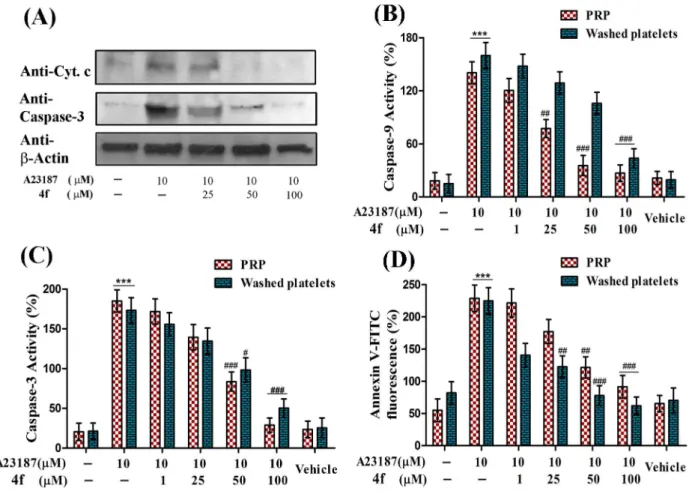
Release of cytochrome c from mitochondria to cytosol is mediated by the formation of MPTP. Altered redox conditions and peroxidation of cardiolipin play critical role in the formation of MPTP and the release of cyt c into the cytosol. A23187 was used as agonist in inducing the release of cyt c to the cytosol. On the other hand, 4f was also able to diminish the expression of cytosolic cyt c induced by A23187 in a dose dependent manner and at 100mM concentration basal levels of cyt c was restored (Fig. 4A).
Inhibition of caspase activity by compound 4f
Intrinsic apoptotic pathway in principally is mediated by caspase-9 which is activated by itself bound to apoptosome complex along with cytosolic cyt c. The activated caspase-9 in turn activates caspase-3 which orchestrates platelet apoptosis. There-fore, in order to investigate the anti-apoptotic properties of 4f, inhibition of caspase-9 and caspase-3 activity is critically impor-tant. Compound 4f which was proved to be effective in ameliorating oxidative stress, was able to significantly inhibit both caspase-9 and caspase-3 activities induced by A23187 in a concentration dependent manner (Fig. 4B& Fig. 4C). Further, inhibition of caspase-3 was confirmed by western blot wherein,
there was significant reduction in activated form of caspase-3 in platelets treated with 4f (Fig. 4A).
Mitigation of PS externalization by compound 4f
Cells undergoing apoptosis are finally marked by the scrambling of PS from inner plasma membrane to outer plasma membrane. A23187 was used as agonist in inducing PS externalization in platelets and its inhibition by 4f was evaluated. As expected, there was dose dependent inhibition of PS externalization by 4f in a concentration dependent fashion and significant inhibition was observed at 100mM concentration in both PRP and washed platelets (Fig. 4D).
Suppression of protein phosphorylation by compound 4f
Recently, it has been revealed that sites for protein phosphor-ylation are exposed by caspase cleavage activity. Therefore, 4f was evaluated for its effects on protein phosphorylation with collagen as agonist. There was significant increase in protein phosphory-lation in platelets treated with collagen whereas, 4f markedly reduced collagen induced protein phosphorylation in a concen-tration dependent manner and at 100mM concentration protein phosphorylation was restored up to basal levels (Fig. 5A).
Figure 4. Effect of compound 4f on A23187 induced (A) Translocation of cytosolic cytochrome C and activation of caspase-3 (B) Caspase-9 and (C) Caspase-3 activities and (D) PS externalization in PRP and washed platelets.Values are presented as mean6SEM (n = 5), expressed as percentage increase in (B & C) caspase activity and (D) Annexin V-FITC fluorescence and expressed as percentage increase in apoptotic platelets expressing PS relative to control. ***p,0.001; significant compared to control.#
p,0.05,##
p,0.01,###
p,0.001; significant compared to A23187.
Inhibition ofc-glutamyl transferase (GGT) activity by compound 4f
GGT is involved in GSH homeostasis and its expression is increased during oxidative stress. In the previous set of experi-ments 4f was proved to be anti-apoptotic by reducing oxidative stress in the platelets. Accordingly, 4f was analyzed for its effects on GGT activity. There was significant increase in GGT activity in platelets treated with A23187 as agonist and as expected there was concentration dependent reduction of GGT activity in agonist activated platelet suggesting that 4f serves as anti-apoptotic molecule basically by reducing oxidative stress in platelets (Fig. 5B).
Cyto-protective effects of compound 4f
In order to investigate whether 4f had cyto-protective effect, MTT assay was performed. A23187 was used as standard agonist, which reduced cell viability from 93% to 21%. However, treating platelets with 4f restored its survivability up to 75% in agonist activated platelets (Fig. 5C). Further, cyto-protective nature of 4f was confirmed by measure the leakage of LDH into the medium. Treatment of platelets with A23187 increased LDH release in to the medium whereas, 4f was able to significantly reduce LDH release indicating its cyto-protective properties (Fig. 5D).
Effect of compound 4f on agonist induced platelet aggregation and platelet adhesion
Further, platelets being the key mediators in maintaining the integrity of endothelium, effect of compound 4f on platelet aggregation and platelet adhesion with collagen was evaluated. In order to stimulate platelet aggregation collagen/ADP/epinephrine were used as agonist. Compound 4f was able to significantly inhibit agonist induced platelet aggregation to varied extent. Of all the agonists tested, the epinephrine-induced aggregation was abolished at 100mM concentration of compound 4f. On the other
hand, compound 4f alone did not exhibit any effect on platelet aggregation up to the tested concentrations (Fig. 6A & Fig. S1– S3).
In order to determine the effect of compound 4f on the platelet receptors for agonists, collagen adhesion was performed using pre-coated collagen microtiter wells. The compound 4f untreated/PBS treated platelet suspension served as control and accounted for 100% adhesion. In contrast, 4f pre-treated platelets did not adhere efficiently to collagen immobilized on the microtiter wells. Compound 4f was able to prevent the binding of platelet with collagen in a concentration dependant fashion in both collagen pre-treated with compound 4f and PRP pre-treated with compound 4f. At 250mM concentration, only 54% adhesion
Figure 5. Effect of compound 4f on (A) Collagen induced protein phosphorylation (B) A23187 induced c-glutamyltransferase
activity (C) MTT cell viability assay (D) LDH release in platelets.(A) Lane I- resting platelets (untreated). Lane II- platelets treated with Collagen (1mg/mL). Lanes III, IV and V- pre-loaded platelets with collagen and incubated with 4f in increasing concentration of 25, 50 and 100mM respectively. Values are presented as mean6SEM (n = 5). ***p,0.001; significant compared to control.#p
,0.05,##p
,0.01,###p
,0.001; significant compared to agonist.
was observed, and thus affecting the collagen binding property of platelets (Fig. 6B).
Discussion
Reduced platelet count or thrombocytopenia is the medical condition where the platelet count drops drastically to 50,000/mL of blood as against the normal count of 150,000 to 450,000 plate-lets/mL of blood. It can be caused by a variety of clinical
conditions including 1) decreased production of platelets or 2) augmented platelet destruction or 3) increased splenic sequestra-tion of platelets. Among these reasons, augmented platelet destruction or apoptosis can be seen in a number of medical and pathological conditions [1,4,35]. The platelet apoptosis is mediated either by immune related or non-immune related causes. The immune thrombocytopenia is a common bleeding disorder characterized by autoantibody-mediated platelet destruction. The auto-antibodies primarily target the membrane receptors and integrin complex of platelets including glycoproteins GPIIb/IIIa and GPIb/IX [36]. In contrast, platelets undergo oxidative stress-mediated apoptosis in non-immune related thrombocytopenia. The adverse effects of thrombocytopenia may lead to morbidity and mortality from severe surgical hemorrhage and delay in the normal process of clotting. Several studies reported oxidative stress or immunologic reaction-induced platelet apoptosis by a wide range of biologicals including antibiotics, anticancer drugs, phytochemicals and hormones [5,8,9]. Therefore, the clinical implication of thrombocytopenia in various human pathologies has strongly demanded the need for small molecular weight natural/synthetic platelet protective molecules. Till date only two phytochemicals (cinnamtannin B1 and crocin) are shown to inhibit platelet apoptosis [17,18]. Hence, there continues the search for potent small molecular weight natural/synthetic molecules to manage thrombocytopenia associated with various human pathol-ogies.
Of late, Ibuprofen, a well known NSAID was reported to interfere with platelet functions and viability thereby, causing thrombocytopenia, nevertheless it is also demonstrated to have anti-cancer, analgesic, anti-pyretic and anti-platelet properties [19,37,38]. Thus, as an attempt to reduce ibuprofen side effects including platelet damage property, in the present study we have synthesized a series of ibuprofen derivatives (4a-f) with improved antioxidant activity. Among the derivatives, compound 4f was a
potent antioxidant and effectively inhibited oxidative stress-induced apoptosis in platelets.
In the first set of experiments, the effect of compound 4f on oxidative stress was assessed. A23187 was used to stimulate the endogenous generation of ROS in platelets. When the platelets were pretreated with 4f prior to A23187 treatment, the ROS generation was found to decrease in a dose-dependent manner. It is reported that it is specifically H2O2that initiates the apoptotic
events in platelets through the intrinsic or mitochondrial pathway. Thus, the results highlight the ability of compound 4f to diminish oxidative stress in platelets and thereby protect them from undergoing an early death. This property may be attributed to the anti-inflammatory and anti-platelet efficacy of the parent compound ibuprofen. Previously it has been reported that ibuprofen treatment decreases the levels of lipid peroxidation, tyrosine nitration, protein oxidation and ROS production in murine model of Alzheimer’s disease [39].
For the next set of experiments, A23187 was used as the agonist to induce the different events of apoptosis. In order to assess the effect of compound 4f on mitochondria, the central players in the intrinsic pathway of apoptosis, changes inDYm were analyzed. A23187 was used as the positive control to induce changes in
DYm. Compound 4f markedly diminished A23187-induced changes inDYm and reinstates the membrane potential. From the result it can be stated that 4f has the capacity to inhibit platelet apoptosis by protecting mitochondria from oxidative stress. In the recent past, a study by Sanz-Blasco et al. [40] demonstrated that ibuprofen prevents mitochondrial Ca2+overload, cyt c release and thus prevent neurotoxicity [40]. Thus, to verify whether the same was true in the case of the effect of 4f on platelets, the levels of intracellular Ca2+and cytosolic cyt c were measured. It was found
that compound 4f could inhibit the increase in intracellular Ca2+ and cytosolic cyt c levels in a dose-dependent manner. Release of cyt c from mitochondrial intermembrane space to the cytosol due to formation of a channel MPTP is a key event that initiates morphological changes during apoptosis. The morphological changes are arbitrated by the activated caspases, finally resulting in PS externalization [7,8,15].
Therefore, the next set of experiments targeted the influence of compound 4f over caspase activity and PS exposure. The results of the study firmly highlight the inhibitory effect of compound 4f on the activities of caspase -9 and -3. Further, it was also shown that compound 4f was to impair PS exposure, the hallmark of an
Figure 6. Effect of compound 4f on (A) Platelet aggregation induced by Collagen/ADP/Epinephrine and (B) Platelet adhesion on immobilized collagen type I with compound 4f pre-treated collagen and PRP pre-treated with compound 4f.Values are presented as mean6SEM (n = 5), expressed as percentage decrease in aggregation and increase in platelet adhesion. **p,0.01, ***p,0.001; significant compared to control.
apoptotic cell. Thus, the current study underscores anti-apoptotic effects of compound 4f on human platelets. It was recently reported that the parent compound possesses chemo-preventive property viathe stimulation of apoptosis [21,41]. Besides, it was also reported that it causes thrombocytopenia, which is a common effect of most of the anti-cancer drugs [21]. Consequently, it would be amazing if compound 4f could wield anti-cancer effect. It has the potential to be developed as an anti-cancer molecule, which has platelet-protective properties.
Furthermore, effect of compound 4f on platelet aggregation was evaluated to delineate its cardio-protective action. Platelet aggregation plays a central role in the perpetuation of CVDs and atherothrombotic disorders. It triggers intraluminal thrombo-sis and thereby promulgates myocardial infarction, stroke and peripheral vascular occlusions [42,43]. Thus, inhibition of platelet aggregation is crucial in prevention of CVDs and associated complications. The results demonstrated that, compound 4f significantly inhibited various agonists (Collagen/ADP/Epineph-rine)-induced platelet aggregation to varied extent. Among the agonists, compound 4f showed better inhibition towards the epinephrine-induced platelet aggregation. The role of compound 4f in blocking epinephrine receptor on platelets is not clear. The receptors for the above-mentioned agonists are high molecular weight glycoproteins and compound 4f might interfere with the binding of agonists to receptors by interacting directly with the membrane proteins or membrane lipids. In continuance, platelet adhesion assay was carried out to verify whether compound 4f prevents the interaction of platelets with collagen. This interaction is crucial for platelet aggregation. The results indicate that compound 4f markedly decreases the collagen-binding efficacy of platelets, suggesting the mechanism through which the compound exerts its antiplatelet effect.
It has been reported that ibuprofen has mild anti-platelet effect, which is very less when compared to the other standard anti-platelet drugs such as aspirin and ketoprofen. However, from the current results the derivative of ibuprofen, compound 4f has been demonstrated to be a very effective anti-platelet agent. Aspirin is the most common anti-platelet agent used in the primary prevention of cardiovascular events due to atherothrombosis. Aspirin inhibits the platelet aggregation by reducing the produc-tion of thromboxane (Tx) A2due to inhibition of cyclooxygenase-1
in platelets [44]. This could be the possible reason for aspirin-associated bleeding during chronic intake of aspirin. In addition, aspirin could also aggravate platelet GP Ib and GPV ectodomain shedding, which is yet another possible mechanism for hemor-rhagic conditions [45]. Recent reports suggested the apoptotic tendency of aspirin toward many cell types including platelets [46]. Zhao et al. [47] demonstrated pro-apoptotic nature of aspirin on platelets and found that aspirin (2.5–20 mM) induced apoptosis in platelets via COX-independent pathway [47]. This could be the possible mechanism for aspirin induced hemorrhage in susceptible patients. In contrast, the compound 4f inhibited the agonist-induced platelet aggregation as well as protected the platelets from oxidative stress-induced apoptosis.
Platelets are known to express the amyloid precursor protein (APP) and exhibit the complete enzymatic machinery to process APP proteins into amyloid-b (Ab) peptides through the same pathway described in the brain. A recent study demonstrated the amyloid-b (Ab) peptide-induced platelet apoptosis by assessing ROS generation, increase of cytosolic Ca2+levels, mitochondrial depolarization, caspase-3 activation, cell shrinkage and cell membrane scrambling. In neurological pathologies, uncontrolled activation of platelets by oxidative stress and other inducers can lead to acute vessel occlusion leading to myocardial infarction and
stroke [3]. In this context, platelet protective and anti-aggregant molecules like compound 4f, crocin and cinnamtannin B1 [17,18] could be used in the treatment regime of neurological disorders as auxiliary therapeutic molecules in order to mitigate the platelet apoptosis and altered hemostasis.
Taken together, the therapeutic drug-induced dysregulated platelet apoptosis is one of the major causes for thrombocytopenia. Furthermore, the formation of MPs during platelet apoptosis might worsen the clinical condition [5]. Platelet-derived MPs (PMPs) are shown to regulate several pathophysiological functions including cell proliferation, differentiation, vascular remodeling, angiogenesis, inflammation and apoptosis. The augmented levels of circulating PMPs have shown to be associated with many pathological conditions including CVDs, thromboembolism, diabetes, rheumatoid arthritis, and cancer [5,16]. Therefore, biologicals with anti-platelet property as well as platelet protective efficacy stand better and thus could receive a lot of attention by the medical practitioners in order to treat thrombolytic disorders. From the results of the present study, it can be noted that compound 4f (100mM) is as effective as cinnamtannin B1 and crocin, the previously reported compounds with platelet protective properties at the same concentration [17,18]. Thus, compound 4f can be taken into account as a potential candidate in the treatment regime of altered platelet function-associated pathologies. In addition, compound 4f can be used as an auxiliary therapeutic agent in order to mitigate the side effects of therapeutic drugs especially platelet apoptosis and microparticle generation. Future studies related to the molecular mechanism of platelet protection and aggregation inhibition of compound 4f is highly exciting.
Supporting Information
Figure S1 Effect of compound 4f on Collagen induced platelet aggregation. Concentration dependent inhibition of collagen induced platelet aggregation by compound 4f: (i) Control (Collagen-2mg/mL), (ii) 50mM and (iii) 100mM compound 4f
respectively and aggregation was performed as described in materials and methods section.
(TIF)
Figure S2 Effect of compound 4f on ADP induced platelet aggregation. Concentration dependent inhibition of ADP induced platelet aggregation by compound 4f: (i) Control (ADP-10mM), (ii) 50mM and (iii) 100mM compound 4f
respectively and aggregation was performed as described in materials and methods section.
(TIF)
Figure S3 Effect of compound 4f on epinephrine induced platelet aggregation. Concentration dependent inhibition of epinephrine induced platelet aggregation by com-pound 4f: (i) Control (Epinephrine-10mM), (ii) 50mM and (iii) 100mM compound 4f respectively and aggregation was performed as described in materials and methods section.
(TIF)
Acknowledgments
Authors thank Mr. SK Naveen Kumar for his kind help during the study and also thank Central Instrumentation Facility, Institute of Excellence (IOE), University of Mysore, Mysore.
Author Contributions
materials/analysis tools: K. S. Rangappa KSG KK MH RMT. Contributed to the writing of the manuscript: K. S. Rangappa KSG MH RMT.
References
1. Chu SG, Becker RC, Berger PB, Bhatt DL, Eikelboom JW, et al. (2010) Mean platelet volume as a predictor of cardiovascular risk: a systematic review and meta-analysis. J Thromb Haemost 8: 148–156.
2. Dindar S, Cinemre H, Sengul E, Annakkaya AN (2013) Mean platelet volume is associated with glycaemic control and retinopathy in patients with Type 2 diabetes mellitus. West Indian Med J 62: 519–523.
3. Gowert NS, Donner L, Chatterjee M, Eisele YS, Towhid ST, et al. (2014) Blood platelets in the progression of Alzheimer’s disease. PLoS One 9: e90523. 4. Tang WH, Stitham J, Jin Y, Liu R, Lee SH, et al. (2014) Aldose
reductase-mediated phosphorylation of p53 leads to mitochondrial dysfunction, and damage in diabetic platelets. Circulation 129: 1598–1609.
5. Thushara RM, Hemshekhar M, Kemparaju K, Rangappa KS, Devaraja S, et al. (2014) Therapeutic drug-induced platelet apoptosis: An overlooked issue in pharmacotoxicology. Arch Toxicol 88: 185–198.
6. Gyulkhandanyan AV, Mutlu A, Freedman J, Leytin V (2013) Selective triggering of platelet apoptosis, platelet activation or both. Br J Haematol 161: 245–254.
7. Leytin V (2012) Apoptosis in the anucleate platelet. Blood Rev 26: 51–53. 8. Thushara RM, Hemshekhar M, Santhosh MS, Devaraja S, Kemparaju K, et al.
(2013) Differential action of phytochemicals on platelet apoptosis: A biological overview Curr Med Chem 20: 1018–1027.
9. Girish KS, Paul M, Thushara RM, Hemshekhar M, Shanmuga Sundaram M, et al. (2013) Melatonin elevates apoptosis in human platelets via ROS mediated mitochondrial damage. Biochem Biophys Res Commun 438: 198–204. 10. Pietraforte D, Vona R, Marchesi A, Tarissi de Jacobis I, Villani A, et al. (2014)
Redox control of platelet functions in physiology and pathophysiology. Antioxid Redox Signal Doi:10.1089/ars.2013.5532.
11. Rukoyatkina N, Mindukshev I, Walter U, Gambaryan S (2013) Dual role of the p38 MAPK/cPLA2 pathway in the regulation of platelet apoptosis induced by ABT-737 and strong platelet agonists. Cell Death Dis 214 e931.
12. Kanbay A, Tutar N, Kaya E, Buyukoglan H, Ozadoqan N, et al. (2013) Mean platelet volume in patients with obstructive sleep apnea syndrome and its relationship with cardiovascular diseases. Blood Coagul Fibrinolysis 24: 532– 536.
13. Ruiz-Argu¨elles GJ, Velazquez-Sanchez-De-Cima S, Zamora-Ortiz G, Hernan-dez-Reyes J, Ruiz-Delqado GJ (2014) Nonalcoholic Fatty Liver Disease May Cause Thrombocytopenia. Acta Haematol 132: 159–162.
14. Li J, Callum JL, Lin Y, Zhou Y, Zhu G, et al. (2014) Severe platelet desialyation in a patient with glycoprotein Ib/IX antibody-mediated immune thrombocy-topenia and fatal pulmonary hemorrhage. Haematologica 99: 61–63. 15. Garcia-Souza LF, Oliveira MF (2014) Mitochondria: Biological roles in platelet
physiology and pathology. Int J Biochem Cell Biol 50: 156–160.
16. Wu ZH, Ji CL, Li H, Qiu GX, Gao CJ, et al. (2013) Membrane microparticles and diseases. Eur Rev Med Pharmacol Sci 17: 2420–2427.
17. A . Bouaziz, C . Romera-Castillo, S . Salido, PJ . Linares-Palomino, J . Altarejos, et al. (2007) Cinnamtannin B-1 from bay wood exhibits antiapoptotic effects in human platelets. Apoptosis 12: 489–498.
18. Thushara RM, Hemshekhar M, Santhosh MS, Jnaneshwari S, Nayaka SC, et al. (2013) Crocin, a dietary additive protects platelets from oxidative stress-induced apoptosis and inhibits platelet aggregation. Mol Cell Biochem 373: 73–83. 19. Saxena A, Balaramnavar VM, Hohlfeld T, Saxena AK (2013) Drug/drug
interaction of common NSAIDs with antiplatelet effect of aspirin in human platelets. Eur J Pharmacol 721: 215–224.
20. Endo H, Yano M, Okumura Y (2014) Ibuprofen enhances the anticancer activity of cisplatin in lung cancer cells by inhibiting the heat shock protein 70. Cell Death Dis 5: e1027.
21. Arnold DM, Kukaswadia S, Nazi I, Esmail A, Dewar L, et al. (2013) A systematic evaluation of laboratory testing for drug-induced immune thrombo-cytopenia. J Thromb Haemost 11: 169–176.
22. Hsieh CL, Yan GC (2000) Antioxidant actions of du-zhong (Eucommia ulmoides Oliv.) toward oxidative damage in biomolecules. Life Sci 66: 1387– 1400.
23. Yamaguchi T, Takamura H, Matoba T, Terao J (1998) HPLC method for evaluation of the free radical-scavenging activity of foods by using1,1,-diphenyl-2-picrylhydrazyl. Biosci Biotechnol Biochem 62: 1201–1204.
24. Kumar MS, Girish KS, Vishwanath BS, Kemparaju K (2011) The metalloprotease, NN-PF3 from Naja naja venom inhibits platelet aggregation primarily by affectinga2b1 integrin. Ann Hematol 90: 569–577.
25. Lopez JJ, Salido GM, Go’mez-Artet E, Rosado JA, Pariente JA (2007) Thrombin induces apoptotic events through the generation of reactive oxygen species in human platelets. J Thromb Haemost 5: 1283–1291.
26. Asai M, Takeuchi K, Uchida S, Urushida T, Katoh H, et al. (2008) Misinterpretation of the effect of amlodipine on cytosolic calcium concentration with fura-2 fluorospectrometry. Naunyn Schmiedebergs Arch Pharmacol 377: 423–427.
27. Ferlini C, Scambia (2007) Assay for apoptosis using the mitochondrial probes, Rhodamine 123 and 10-N-nonyl acridine orange. Nat Protoc 2: 3111–3114. 28. Ronot X, Benel L, Adolphe M, Mounolou JC (1986) Mitochondrial analysis in
living cells: the use of rhodamine 123 and flow cytometry. Biol Cell 57: 1–7. 29. Amor NB, Pariente JA, Salido GM, Rosado JA, Bartegi A (2006).
Thrombin-induced caspases 3 and 9 translocation to the cytoskeleton is independent of changes in cytosolic calcium in human platelets. Blood Cells Mol Dis 36: 392– 401.
30. Rosado JA, Lopez JJ, Gomez-Arteta E, Redondo PC, Salido GM, et al. (2006). Early caspase-3 activation independent of apoptosis is required for cellular function. J Cell Physiol 209: 142–152.
31. Yamazaki Z, Chiba K, Yoshikawa C (2009) Genipin suppresses A23187-induced cytotoxicity in neuro2a cells. Biol Pharm Bull 32: 1043–1046.
32. Sener A, Cevik O, Yanikkaya-Demirel G, Apikoglu-Rabus S, Ozsavci (2012) Influence of plateletc-glutamyltransferase on oxidative stress and apoptosis in the presence of holo-transferrin. Folia Biol 58: 193–202.
33. Bellavite P, Andrioli G, Guzzo P, Arigliano P, Chirumbolo S, et al. (1994) A colorimetric method for the measurement of platelet adhesion in microtiter plates. Anal Biochem 216: 444–450.
34. Lowry OH, Rosebrough NJ, Farr AL, Randall RJ (1951) Protein measurement using folin-phenol reagent. J Biol Chem 193: 265–275.
35. Dimitroulis D, Valsami S, Stamopoulos P, Kouraklis G (2012) Immunological HCV-associated thrombocytopenia: short review. Clin Dev Immunol 2012: 378653.
36. Kashiwagi H, Tomiyama Y (2013) Pathophysiology and management of primary immune thrombocytopenia. Int J Hematol 98: 24–33.
37. Crook J (2010) Fever management: evaluating the use of ibuprofen and paracetamol. Paediatr Nurs 22: 22–26.
38. Rainsford KD (2013) Ibuprofen: from invention to an OTC therapeutic mainstay. Int J Clin Pract Suppl 178: 9–20.
39. Wilkinson BL, Cramer PE, Varvel NH, Reed-Geaghan E, Jian Q, et al. (2012) Ibuprofen attenuates oxidative damage through NOX2 inhibition in Alzheimer’s disease. Neurobiol Aging 197: 21–32.
40. Sanz-Blasco S, Valero RA, Rodrı´guez-Crespo I, Villalobus C, Nunez L (2008) Mitochondrial Ca2+
overload underlies Abeta oligomers neurotoxicity providing an unexpected mechanism of neuroprotection by NSAIDs. PLoS One 3: e2718. 41. Todo M, Horinaka M, Tomosugi M, Tanaka R, Ikawa H (2013) Ibuprofen enhances TRAIL-induced apoptosis through DR5 upregulation. Oncol Rep 30: 2379–2384.
42. Jneid H, Bhatt DL (2003). Advances in antiplatelet therapy. Expert Opin Emerg Drugs 8: 349–363.
43. Fisch AS, Perry CG, Stephens SH, Horenstein RB, Shuldiner AR (2013) Pharmacogenomics of anti-platelet and anti-coagulation therapy. Curr Cardiol Rep 15: 381–393.
44. Schro¨r K (1997) Aspirin and platelets: the antiplatelet action of aspirin and its role in thrombosis treatment and prophylaxis. Semin Thromb Hemost 23: 349– 356.
45. Aktas B, Pozgajova M, Bergmeier W, Sunnarborg S, Offermanns S (2005) Aspirin induces platelet receptor shedding via ADAM17 (TACE). J Biol Chem 280: 39716–39722.
46. Saito T, Tamura D, Asano R (2014) Usefulness of selective COX-2 inhibitors as therapeutic agents against canine mammary tumors. Oncol Rep 31: 1637–1644. 47. Zhao L, Zhang W, Chen M, Zhanq J, Zhanq M, et al. (2013) Aspirin Induces