CLINICAL SCIENCE
I Cardiopulmonary Physiotherapy Laboratory, Nucleus of Research in
Physi-cal Exercise, Physiotherapy Department - Federal University of São Carlos (UFSCar) - São Carlos/SP, Brazil.
II UNINOVE - São Paulo, Brazil.
Email: [email protected] Tel: 55 16 3351.8705
Received for publication on May 29, 2009 Accepted for publicaiton on August 06, 2009
ACUTE APPLICATION OF BILEVEL POSITIVE
AIRWAY PRESSURE INFLUENCES THE CARDIAC
AUTONOMIC NERVOUS SYSTEM
Camila Bianca Falasco Pantoni,I Renata Gonçalves Mendes,I Luciana Di
Thommazo,I Aparecida Maria Catai,I Luciana Maria Malosá Sampaio,II Audrey
Borghi-SilvaI
doi: 10.1590/S1807-59322009001100008
Pantoni CBF, Mendes RG, Di Thommazo L,Catai AM, Sampaio LMM, Borghi-Silva, A. Acute application of bilevel positive airway pressure inluences the cardiac autonomic nervous system. Clinics. 2009;64(11):1085-92.
INTRODUCTION: Noninvasive positive pressure has been used to treat several diseases. However, the physiological response of the cardiac autonomic system during bilevel positive airway pressure (Bilevel) remains unclear.
OBJECTIVE: The aim of this study was to evaluate the heart rate variability (HRV) during Bilevel in young healthy subjects.
METHODS: Twenty men underwent 10-minute R-R interval recordings during sham ventilation (SV), Bilevel of 8-15 cmH2O and Bilevel of 13-20 cmH2O. The HRV was analyzed by means of the parallel R-R interval (mean R-Ri), the standard deviation of all R-Ri (SDNN), the root mean square of the squares of the differences between successive R-Ri (rMSSD), the number of suc-cessive R-Ri pairs that differ by more than 50 milliseconds (NN50), the percentage of sucsuc-cessive R-Ri that differ by more than 50 milliseconds (pNN50), the low frequency (LF), the high frequency (HF) and SD1 and SD2. Additionally, physiological variables, including blood pressure, breathing frequency and end tidal CO2, were collected. Repeated-measures ANOVA and Pearson cor-relation were used to assess the differences between the three studied conditions and the cor-relationships between the delta of Bilevel at 13-20 cmH2O and sham ventilation of the HRV indexes and the physiological variables, respectively.
RESULTS: The R-Ri mean, rMSSD, NN50, pNN50 and SD1 were reduced during Bilevel of 13-20 cmH2O as compared to SV. An R-Ri mean reduction was also observed in Bilevel of 13-20 cmH2O compared to 8-15 cmH2O. Both the R-Ri mean and HF were reduced during Bilevel of 8-15 cmH2O as compared to SV, while the LF increased during application of Bilevel of 8-15 cmH2O as compared to SV. The delta (between Bilevel at 13-20 cmH2O and sham ventilation) of ETCO2 correlated positively with LF, HF, the LF/HF ratio, SDNN, rMSSD and SD1. Acute application of Bilevel was able to alter the cardiac autonomic nervous system, resulting in a reduction in parasympathetic activity and an increase in sympathetic activity and higher level of positive pressure can cause a greater inluence on the cardiovascular and respiratory system.
KEYWORDS: Heart rate; Neural control; Heart rate variability; Noninvasive positive pressure ventilation; Physiological responses.
INTRODUCTION
Noninvasive positive pressure ventilation (NiPPV) has gained widespread acceptance for the support of cardiovascular1-3 and respiratory dysfunction.4 NiPPV is used
to improve gas exchange and the ventilatory pattern, reduce respiratory work and increase tidal volume.3 Some modes of
NiPPV have become standard therapy for treatment in acute care settings,5 by reducing the respiratory effort, the need for
intubation, the length of hospital stay6 and mortality,7 while
improving respiratory mechanics and alveolar ventilation.8
However, the NiPPV application ordinarily used in respiratory interventions may affect intrathoracic hemodynamic and cardiovascular stability by reducing venous return and right ventricular end-diastolic volume.9,10 These
Respiratory phases during normal breathing, left stroke volume and arterial blood pressure variations are sensed by baroreceptors. These baroreceptors provoke parallel R-R interval (R-Ri) changes by means of baroreflex physiology.11,12 In addition, hemodynamic oscillations during
normal (negative pressure) respiration are predominantly due to changes in intrathoracic pressure.13 In this context,
the alterations caused by NiPPV application can also produce physiological responses in cardiac beat intervals and inluence the autonomic neural control of heart rate (HR).
Recent studies14 have demonstrated that short-term
administration of continuous positive airway pressure (CPAP) in normal subjects causes signiicant alterations in R-Ri variability. However, we have not encountered any studies on the effects of bilevel positive airway pressure ventilation (Bilevel), which produces biphasic respiratory cycles (inspiratory and expiratory pressures), nor on the possibility of different pressure levels differentially affecting hemodynamic responses and thus cardiac autonomic control.
Bilevel has commonly been used as an effective means for the management of several diseases.14,15 However,
its physiological responses at different levels remain to be clarified. Therefore, the aim of the present study was to noninvasively evaluate cardiac autonomic neural control by examining heart rate variability (HRV) during Bilevel application and to correlate HRV alterations with physiological measurements. The present study tested the hypothesis that acute Bilevel application can modify HRV in healthy young men and, based on a previous study16, that
physiological variables can modulate HRV changes.
MATERIAL AND METHODS
Study population
A total of 20 non-smoking, healthy young men were evaluated and included in this study. All volunteers underwent anamnesis, physical and clinical examination, a 12-channel electrocardiogram and incremental exercise testing. Subjects with heart rhythm disorders, obstructive or restrictive respiratory disturbances and acute or chronic illness evidence were excluded from the study. None of the subjects were receiving acute or chronic medications. The study protocol was approved by the Federal University of São Carlos ethics committee and all subjects signed a written consent form prior to the initiation of the study.
Design and procedures
This was a prospective, double-blind, randomized controlled trial.
Experimental procedures
Experimental procedures were performed on a single day and always began at the same time of day (7:00 AM) at a room temperature between 22°C and 24°C and a relative air humidity between 50 and 60%. Each subject was instructed to avoid caffeinated and alcoholic beverages for at least 12 hours before the test and to avoid moderate or heavy exercise on the day before the application of the protocol. The experiment was performed by two investigators. One investigator was aware of the NiPPV intervention and operated the device, while the other investigator was responsible for the data physiological collection and data analysis and was not aware of the pressure levels applied.
Heart rate and R-Ri were recorded by the CM5 lead, which was chosen to provide higher R wave and lower T wave curve electrocardiogram (ECG) signal amplitudes. The ECG and HR were obtained from a one-channel heart monitor (Ecaix TC500, São Paulo, Brazil) and processed using an analog-digital converter (PCI7030/640E, National Instruments, Co., Austin, TX, USA), which represents an interface between the ECG monitor and a Pentium III microcomputer. The R-Ri (milliseconds (ms)) was calculated using speciic software.17 Before starting the
experimental procedure, the subjects were asked to remain in a sitting position for 15 minutes to adjust themselves to the environmental conditions.
Data collection was performed during spontaneous breathing (sham ventilation), as well as during ventilatory assistance, delivered by a Bilevel ventilator mode (BiPAP S, Respironics Inc, Murrysville, PA) applied via a nasal mask (chosen previously by volunteers as the more comfortable mask) under the following conditions for 10 minutes each:
1) Sham Ventilation (SV): HR and R-Ri were recorded during simulated ventilation with the same nasal mask and device, but without positive pressure application.
2) Expiratory positive airway pressure (EPAP) of 8 cmH2O and inspiratory positive airway pressure (IPAP) of 15 cmH
2O (hereafter referred to as Bilevel of 8-15): HR and
R-Ri were recorded during Bilevel application with levels titrated to 8 cmH2O and 15 cmH2O of EPAP and IPAP, respectively.
3) EPAP of 13 cmH
2O and IPAP of 20 cmH2O (hereafter
referred to as Bilevel of 13-20): HR and R-Ri were recorded during Bilevel application with levels titrated to 13 cmH2O and 20 cmH2O of EPAP and IPAP, respectively.
A capnometer (BCI 1050, Waukesha, USA) was used to monitor the breathing frequency (BF) and end-tidal of carbon dioxide (ETCO2), while peripheral oxygen saturation (SpO2) was obtained by pulse oximetry (OX-P-10, Emai Transmai, Sao Paulo, Brazil). Systolic blood pressure (SBP) and diastolic blood pressure (DBP) were measured by an indirect method, using a sphygmomanometer, at the end of each studied condition (SV and Bilevel application at both levels applied) in order to investigate the inluence of positive pressure on these variables. For all conditions, the subjects were instructed to breathe calmly while SpO2, BF and ETCO2 were monitored.
Before starting the HR and R-Ri recordings, there was an adaptation period for each of the levels of ventilatory assistance of approximately 10 minutes for each subject, as well as an interval period of 15 minutes between the study conditions, in order for physiological variables to return to baseline values.
HRV analysis
All artifacts were reviewed by visual inspection on the computer display. Only segments with >90% pure sinus beats were included in the inal analysis. The data were entered into Kubios HRV Analysis software (MATLAB, version 2 beta, Kuopio, Finland).
HRV was analyzed with linear statistical measures in time-and frequency-domain. The mean of all R-Ri (R-Ri mean), the standard deviation of all R-Ri in ms (SDNN, which relects all of the cyclic components responsible for variability in the period of recording and is a estimate of overall HRV18), the root mean square of the squares of the
differences between successive R-Ri in ms (rMSSD), the number of successive R-Ri pairs that differ by more than 50 ms (NN50) and the percentage of successive R-Ri pairs that differ by more than 50 milliseconds (pNN50) were computed as time domain measures, with the last three indexes used to express parasympathetic activity.18
In the frequency domain, the power spectral components were reported using Fast Fourier Transform at low frequency (LF) and high frequency (HF) in normalized units. The LF component is representative of sympathetic activity, although it is also inluenced by parasympathetic components.18,19
The HF component is representative of the parasympathetic component18 and the LF/HF ratio represents sympathetic
vagal balance.19
In addition, nonlinear statistical measures by a Poincaré plot were analyzed with the standard deviation of the Poincaré plot perpendicular to the line-of-identity and related to short-term variability that is mainly caused by RSA (SD1) in ms, which represents a measure of parasympathetic
nervous system activity, and the standard deviation of the Poincaré plot along the line-of-identity (SD2), which is representative of the total HRV.20
Statistical analysis
Data were submitted to a frequency distribution analysis test (Shapiro-Wilk´s), and as it presented normality values, the parametric test was applied. Repeated-measures analysis of variance (ANOVA) with the Tukey post-test was utilized in GraphPad InStat for Windows program, version 3.0 (1994-1999). The level of signiicance was set at p<0.05. A Pearson correlation analysis was employed to assess the relationships between the delta of higher Bilevel application (13-20 cmH2O) and the SV of the HRV indexes and of the physiological variables (blood pressure, breathing rate and end-tidal CO2). Employing data from a pilot study, the sample size was determined using a 5% signiicance level, a power of 80% and an expected mean difference between SV and Bilevel for a mean heart rate of approximately 5 beats per minute (bpm) and a variability of 10 bpm. The estimated sample size was determined to be 14 subjects per group. (StatCalc version 6.0.1 2009).
RESULTS
Twenty healthy young men that were referred to the study were eligible. The following characteristics describe the subjects: mean age of 23±2.2 years, mean weight of 75±11.3 kg, mean height of 182±6.7 cm and a mean body mass index of 23±2.4 kg/m2.
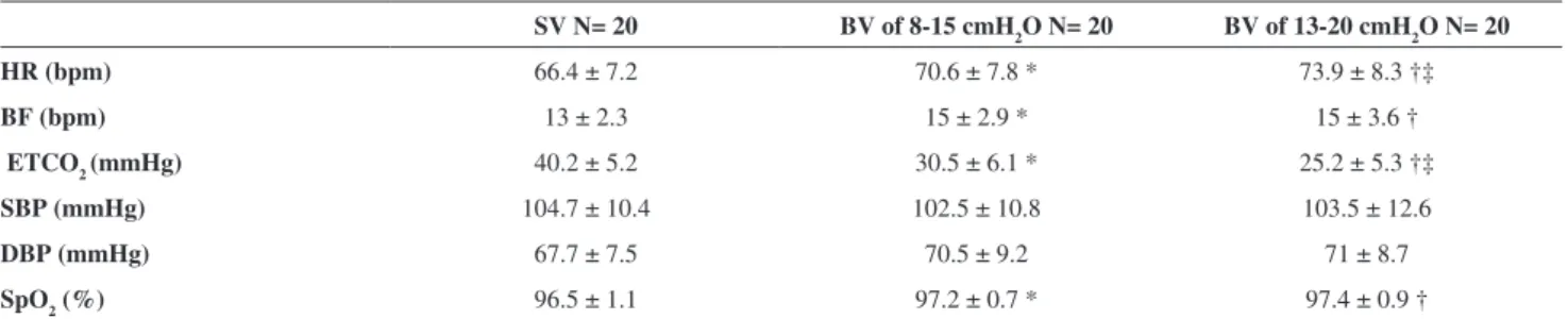
Table 1 shows the mean HR, BF, ETCO2, SBP, DBP and SpO2 of the study subjects. Compared to SV, both Bilevel conditions resulted in a signiicant increase in HR, BF and SpO2 and a decrease in ETCO2. The application of Bilevel of 13-20 cmH2O increased HR and decreased ETCO2, compared to the lower levels of positive pressure applied.
HRV changes: In terms of the HRV variables during SV and Bilevel application at two different levels, the application of 13-20 cmH2O was associated with a reduction in the R-Ri mean, RMSSD, NN50, pNN50 and SD1 as compared to SV. There was also a reduction in the R-Ri mean with the application of 13-20 cmH2O as compared to the 8-15 cmH2O Bilevel application. Compared to SV, the 8-15 cmH2O Bilevel application resulted in signiicantly lower values of the R-Ri mean and HF, while increasing the value of LF (Table 2).
DISCUSSION
Summary of Findings
The present study evaluated the acute effects of NiPPV on HRV and other physiological variables in healthy young men. The main indings were that Bilevel application is able to modify HRV and physiological variables. In addition, we found that a higher level of positive pressure can cause a greater inluence on the cardiovascular and respiratory system. Furthermore, we observed that ETCO2 may modulate the HRV indexes.
HRV Changes during Bilevel Application
The present study shows that Bilevel application, at
different levels, signiicantly modiies HRV in young healthy subjects. A Bilevel application of 8-15 cmH2O was able to increase sympathetic response and decrease parasympathetic activity. The higher level of positive pressure provoked a parasympathetic reduction with alterations of almost all vagal representative indexes, compared to SV. Moreover, when compared to the lower pressure application, the higher level of positive applied pressure caused a greater inluence on the cardiovascular system as seen by a greater reduction in the mean R-Ri.
Previous data have demonstrated that the autonomic nervous system is signiicantly active during the application of positive airway pressure ventilation10. Heindl et al.21
observed an increase in muscle sympathetic nerve activity during short-term application of CPAP at 10 cmH2O in healthy subjects. In contrast to our study using Bilevel, the Table 2 - Heart rate variability of healthy volunteers during sham ventilation (SV) and Bilevel ventilation (BV)
SV N= 20 BV of 8-15 cmH2O N= 20 BV of 13-20 cmH2O N= 20
Time domain
Mean RR (ms) 919.2±109.2 866.4±102.7 * 826.9±99.4 †‡
SDNN (ms) 54.1±23.4 56.5±26.6 49.3±20.6
rMSSD (ms) 50.2±32.5 45.6±29.4 38.9±18.9 †
NN50 (ms) 81±71.2 68.2±60.2 51.1±40.1 †
pNN50 (%) 26.2±22.9 21.7±19.5 17.1±13.3 †
Nonlinear
SD1 (ms) 35.8±23.1 32.6±20.9 27.9±13.5 †
SD2 (ms) 85.4±32.7 93.1±40.3 85.7±31.8
Frequency Domain
LF (nu) 60.1±20.5 72.5±21.7 * 68.1±21.4
HF (nu) 39.9±20.5 27.5±21.7 * 31.9±21.4
LF/HF 3.7±7.5 5.2±4.3 4.2±4
Mean RR= the mean of R-R intervals, in ms; SDNN= standard deviation of all R-R intervals, in ms; rMSSD= root mean square of the squares of the differences between successive R-Ri, in ms; NN50= number of successive R-Ri interval pairs that differ by more than 50 ms; pNN50= percentage of suc-cessive R-Ri pairs that differ by more than 50 ms; SD1= the standard deviation of the Poincaré plot perpendicular to the line-of-identity, in ms; SD2= the standard deviation of the Poincaré plot along the line-of-identity, in ms; LF= low frequency, in normalized units; HF= high frequency, in normalized units; LF/HF= ratio of low frequency to high frequency. * Bilevel of 8-15 cmH2O versus SV; † Bilevel of 13-20 cmH2O versus SV; ‡ Bilevel of 13-20 cmH2O
versus Bilevel of 8-15 cmH2O. Repeated-measures analysis of variance (ANOVA) with the Tukey post-test. Data expressed in mean and standard deviation.
Table 1 - Physiological data of the healthy volunteers during sham ventilation (SV) and Bilevel ventilation (BV)
SV N= 20 BV of 8-15 cmH2O N= 20 BV of 13-20 cmH2O N= 20
HR (bpm) 66.4 ± 7.2 70.6 ± 7.8 * 73.9 ± 8.3 †‡
BF (bpm) 13 ± 2.3 15 ± 2.9 * 15 ± 3.6 †
ETCO2 (mmHg) 40.2 ± 5.2 30.5 ± 6.1 * 25.2 ± 5.3 †‡
SBP (mmHg) 104.7 ± 10.4 102.5 ± 10.8 103.5 ± 12.6
DBP (mmHg) 67.7 ± 7.5 70.5 ± 9.2 71 ± 8.7
SpO2 (%) 96.5 ± 1.1 97.2 ± 0.7 * 97.4 ± 0.9 †
HR= heart rate, presented in beats per minute (bpm); BF= breathing frequency, in breathes per minute (bpm); ETCO2= end-tidal of carbon dioxide, in
millimeters of mercury (mmHg); SBP= systolic blood pressure, in mmHg; DBP= diastolic blood pressure, in mmHg; SpO2:= peripheral oxygen saturation,
majority of previous studies have investigated the effects of CPAP modality on the cardiovascular system.
This is the irst study to compare the effects of two levels of Bilevel on cardiac autonomic nervous control. We observed that the magnitude of cardiovascular, hemodynamic and pulmonary responses can be related to the level of positive pressure applied. Some authors14 have
previouslyobserved a difference in the HRV responses in healthy subjects when different levels of CPAP were applied. A greater modiication was observed when high applied levels (>15 cmH2O) were used. These data suggest, in accordance with our results, that the magnitude of cardiac autonomic responses can be inluenced by differences in the levels of positive pressure applied.
T h e e ff e c t s o f ve n t i l a t i o n m e c h a n i c s o n t h e cardiovascular system resulting from the NiPPV application can explain the HRV alterations. Increased intrathoracic pressure application can produce signiicant hemodynamic alterations, such as a reduction in venous return and stroke volume with a tendency to decrease cardiac output.9,22 These
hemodynamic alterations are then sensed by the carotid sinus and aortic baroreceptors, and the luctuations in the cardiac illing are sensed by cardiac receptors.13 Cardiopulmonary
receptors represent another group of baroreceptors that regulate sympathetic outlow.23 Therefore, a reduction in
venous return and cardiac illing pressures can unload all of these cardiopulmonary baroreceptors. Unloading of these baroreceptors results in cardiac autonomic adjustments in healthy subjects, as physiological responses resulting from hemodynamic alterations,24 which was observed in our study.
Besides the acute use of NiPPV in some eventual clinical conditions, this intervention has been routinely applied to treat chronic diseases, such as obstructive sleep apnea syndrome,25 chronic obstructive pulmonary disease
(COPD)26,27 and congestive heart failure.22 Studies in
patients with several different diseases that were submitted to short-term and long-term NiPPV have demonstrated different HRV responses. Kaye et al.22 found a reduction in
cardiac sympathetic tone in patients with congestive heart failure submitted to a short-term nasal CPAP of 10 cmH2O. Maser et al.28 observed an improvement in vagal tone and
cardiovascular autonomic function in patients with sleep-disordered breathing after six weeks of treatment with CPAP. On the other hand, Borghi-Silva et al.29 found a reduction
in parasympathetic activity and an increase in sympathetic activity during Bilevel ventilation in COPD patients that were submitted to similar pressure levels used in our study.
We know that it is impossible to extend the results of our study to patients with disease whose HRV responses may differ from those with normal clinical conditions (as demonstrated in the literature). However, our study is
of interest in clarifying the HRV physiologic responses resulting from the influence on the hemodynamic and cardiac autonomic nervous system produced by the application of this modality. The present study is especially important considering the lack of studies performed with Bilevel and the applicability of this kind of intervention for some chronic diseases (like COPD) and in acute clinical conditions with subjects who are mainly without some previous disease or instability.
Moreover, we reinforce the importance of the applied level of positive airway pressure. Considering the impact of pressure levels on the cardiac autonomic nervous system, care should be applied when selecting the level of pressure to apply. The level of applied pressure should be selected according to the necessity required by each clinical condition. Higher or lower levels of pressure must be chosen in order to provide the best functionality of the cardiovascular system.
Physiological Variables during Bilevel Application
As demonstrated in Table 1, physiological parameters were modiied by Bilevel application at both applied levels as compared to SV. The application of Bilevel at a higher level (13-20 cmH2O) also increased HR and decreased ETCO2 as compared to the low level (8-15 cmH2O). As observed, both pressure levels applied were strong enough to improve the gas exchange, as relected by an increase in oxygen saturation and a decrease in ETCO2. These data relect a ventilation improvement.
The HR behavior is directly related to cardiac autonomic responses provoked by the effect of positive airway pressure, as previously explained. Modiications of the cardiovascular system are required in order to maintain the cardiac output, which were expressed in our results by an increase of HR and a decrease in the mean R-Ri.
Some authors16 have reported that the autonomic neural
system areas that control breathing and heart function are located close to each other in the brainstem. Thus, CO2 alterations can mediate, through chemoreceptors, the cardiac autonomic control system. Therefore, we can speculate that ETCO2 alterations, as well as BF alterations, also contribute to HRV responses. This effect can be explained by the driving effect of CO2 on ventilation. As a result, we cannot exclude the inluence of respiratory variables on cardiac autonomic modulation, besides the mechanical effects previously explained.
Importantly, we must emphasize that our results suggest a greater inluence of a higher level of positive pressure on respiratory variables, as occurred in HRV responses.
Relationship between HRV and Physiological Data
In regards to the moderate correlations found between the delta of the HRV variables and the physiological data, we observed that the higher the ETCO2 variation, the greater the inluence on cardiac autonomic control. Changes in breathing patterns due to altered CO2 are known to interact with the autonomic cardio-respiratory control system.30 CO
2
concentrations in the blood may affect the iring rate of the autonomic nervous system to the respiratory muscles and the cardio-respiratory control network. This change in iring rate may in turn inluence the HRV spectrum.28 Similar to
our results, a decrease in HF power was found to be related to low CO2 breathing. In this context, our study suggests that ETCO2 variation can modulate the autonomic nervous system as well as HRV responses.
Study Implications
Understanding the physiological effects of NiPPV and its influence on the cardiac autonomic nervous system in healthy volunteers is important to clarify how positive pressure, applied at different levels, can alter HRV in a healthy cardiovascular system. While it is dificult to extend the results of our study to patients affected with disease, our indings presented here should be considered when choosing the best and safest therapeutic treatment of patients with cardiopulmonary conditions, as well as those with eventual acute clinical situations without previous disease or instability. Our data suggest that care should be taken when determining the levels of positive pressure to apply, according to the physiological adjustments and autonomic response.
Study Limitations
Due to the noninvasive nature of our study, several
methodological aspects need further clariication. Although it was not possible to directly measure the tidal volume in this study, we believe that this variable could have an important role in the cardiac autonomic control alterations that we observed.
Secondly, we measured the cardiac autonomic nervous system control of HR by HRV, a noninvasive measurement. Nonetheless, this important tool has been validated by previous pharmacologic blockage.18,19
Another important issue is the time period of Bilevel application used in the present study. Our study results were limited to short-term application of positive pressure, which allowed us to evaluate only the acute effects of this ventilatory modality. Such short-term NiPPV is already routinely applied during respiratory therapy intervention sessions for inpatients and outpatients with acute and chronic dysfunctions in care settings.31,32 However, it would be
interesting to investigate the long-term effects of different levels of positive pressure on the cardiac autonomic nervous system. We hypothesize that the long-term effects could be different from those found in the present study.
Moreover, we only studied healthy subjects. We cannot completely extrapolate our results to real-life patients with disease, whose HRV responses may be different from those with normal clinical conditions. Nevertheless, our study intended to clarify the HRV physiologic responses resulted from the application of Bilevel, considering its impact on cardiac autonomic nervous system, particularly during higher levels. In this context, we wanted to reinforce the importance of the level applied and the care required for each clinical condition.
CONCLUSION
In summary,the acute application of Bilevel was able to alter cardiac autonomic nervous system function, as well as physiological variables, in young healthy subjects. Compared to a low level of positive pressure, a higher level of positive pressure causes a greater inluence on the cardiovascular and respiratory system. Moreover, ETCO2 strongly modulates HRV of these subjects.
ACKNOWLEDGEMENTS
REFERENCES
1. Artz M, Schulz M, Wensel R, Montalvàn S, Blumberg FC, Riegger GAJ, et al. Nocturnal Continuous Positive Airway Pressure Improves Ventilatory Eficiency During Exercise in Patients With Chronic Heart Failure. Chest. 2007;127:794-802.
2. Chadda K, Annane D, Hart N, Gajdos P, Raphaël JC, Lofaso F. Cardiac and respiratory effects of continuous positive airway pressure and noninvasive ventilation in acute cardiac pulmonary edema. Crit Care Med. 2002;30:2457-61.
3. Vitacca M, Nava S, Confalonieri M, Bianchi L, Porta R, Clini E, et al.The appropriate setting of noninvasive pressure support ventilation in stable COPD patients. Am Rev Respir Dis. 2000;118:1286-93. 4. Phua J, Kong K, Lee KH, Shen L, Lim TK. Noninvasive ventilation
in hypercapnic acute respiratory failure due to chronic obstructive pulmonary disease vs. other conditions: effectiveness and predictors of failure. Intensive Care Med. 2005;31:533-9.
5. Curtis JR, Cook DJ, Sinuff T, White DB, Hill N, Keenan SP, et al. Society of Critical Care Medicine Palliative Noninvasive Positive Ventilation Task Force. Noninvasive positive pressure ventilation in critical and palliative care settings: understanding the goals of therapy. Crit Care Med. 2007;35:932-9.
6. Carrera M, Marín JM, Antón A, Chiner E, Alonso ML, Masa JF, et al. A controlled trial of noninvasive ventilation for chronic obstructive pulmonary disease exacerbations. J Crit Care. 2009;24:473.e7-14. 7. Tomii K, Seo R, Tachikawa R, Harada Y, Murase K, Kaii R, et al.
Impact of noninvasive ventilation (NIV) Trial for various types of acute respiratory failure in the emergency department; decreased mortality and use of the ICU. Respir Med. 2009;103:67-73.
8. Matte P, Jacquet L, Van Dyck, Goenen M. Effects of conventional physiotherapy, continuous positive airway pressure and non-invasive ventilatory support with bilevel positive airway pressure after coronary artery bypass grafting. Acta Anaesthesiol Scand. 2000;44:75-81. 9. Barbas CSV, Bueno MAS, Amato MBP, Hoelz C, Rodrigues-Junior M.
Interação cardiopulmonar durante a ventilação mecânica. Revista da Sociedade de Cardiologia do Estado de São Paulo. 1998;8:406-19. 10. Frazier SK, Moser DK, Stone KS. Heart rate variability and
hemodynamic alterations in canines with normal cardiac function during exposure to pressure support, continuous positive airway pressure, and a combination of pressure support and continuous positive airway pressure. Biol Res Nurs. 2001;2:167-74.
11. Lanfranchi PA, Somers VK. Arterial baroreflex function and cardiovascular variability: interactions and implications. Am J Physiol Regulatory Integrative Comp Physiol. 2002;283:815-26.
12. Longo A, Ferreira D, Correia MJ. Variabilidade da freqüência cardíaca. Rev Port Cardiol 1995;14:241-62.
13. Looga R. Relex cardiovascular responses to lung inlation: a review. Respir Physiol. 1997;109:95-106.
14. Valipour A, Schneider F, Koessler W, Saliba S, Burghuber OC. Heart rate variability and spontaneous barorelex sequences in supine healthy volunteers subjected to nasal positive airway pressure. J Appl Physiol. 2005;99:2137-43.
15. Moster WG, Reier CE, Gardier RW, Hamelberg W. Cardiac output and postganglionic sympathetic activity during acute respiratory alkalosis. Anesthesiology. 2005;31:29-35.
16. Pöyhönen M, Syväoja S, Hartikainen J, Ruokonen E, Takala J. The effect of carbon dioxide, respiratory rate and tidal volume on human heart rate variability. Acta Anaesthesiol Scand. 2004;48:93-101. 17. Silva E, Catai AM, Trevelin LC, Guimaraes JO, Silva Junior LP, Silva
LMP, et al. Design of computerized system to evaluate the cardiac function during dynamic exercise. Physics in Medicine and Biology. 1994;1:409 (Abstract).
18. Task Force, Heart rate variability: standards of measurement, physiological interpretation and clinical use. Task Force os the European Society of Cardiology and the North American Society of Pacing and Electrophysiology. Circulation. 1996;93:1043-65.
19. Malliani A, Pagani M, Lombardi F, Cerutti S. Cardiovascular neural regulation explored in the frequency domain. Circulation. 1991;84:1482-92.
20. Guzik P, Piskorski J, Krauze T, Schneider R, Wesseling KH, Wykretowicz A, et al. Correlations between the Poincaré plot and conventional heart rate variability parameters assessed during paced breathing. J Physiol Sci. 2007;57:63-71.
21. Heindl S, Dodt C, Krahwinkel M, Hasenfuss G, Andreas S. Short term effect of continuous positive airway pressure on muscle sympathetic nerve activity in patients with chronic heart failure. Heart. 2001;85:185-90.
22. Kaye DM, Mansield D, Aggarwal A, Naughton MT, Esler MD. Acute effects of continuous positive airway pressure on cardiac sympathetic tone in congestive heart failure. Circulation. 2001;103:2336-38. 23. Mitchell JH, Victor RG. Neural control of the cardiovascular system:
insights from muscle sympathetic nerve recordings in humans. Med Sci Sports Exerc. 1996;28:S60-9.
24. Sundlöf G, Wallin BG. Effect of lower body negative pressure on human muscle nerve sympathetic activity. J Physiol. 1978;278:525-32. 25. Arias MA, García-Río F, Alonso-Fernández A, Mediano O, Martínez
I, Villamor J. Obstructive sleep apnea syndrome affects left ventricular diastolic function: effects of nasal continuous positive airway pressure in men. Circulation 2005;112:375-83.
26. Sin DD, Wong E, Mayers I, Lien DC, Feeny D, Cheung H, et al. Effects of nocturnal noninvasive mechanical ventilation on heart rate variability of patients with advanced COPD. Chest. 2007;131:156-63.
27. Borghi-Silva A, Di Thommazo L, Pantoni CBF, Mendes RG, Salvini TF, Costa D. Non-invasive Ventilation Improves Peripheral Oxygen Saturation and Reduces Fatigability of Quadriceps in Patients with COPD. Respirology. 2009; 14:537-44.
28. Maser RE, Lenhard MJ, Rizzo AA, Vasile AA. Continuous positive airway pressure therapy improves cardiovascular autonomic function for persons with sleep-disordered breathing. Chest. 2008;133;86-91. 29. Borghi-Silva A, Reis MS, Mendes RG, Pantoni CBF, Simões RP,
30. Brown TE, Beightol LA, Koh J, Eckberg DL. Important inluence of respiration on human RR interval power spectra is largely ignored. J Appl Physiol. 1993;75:2310-7.
31. Borghi-Silva A, Mendes RG, Costa FS, Di Lourenzo VAP, Oliveira CR, Luzzi S. The influences of positive end expiratory pressure (PEEP) associated with physiotherapy intervention in phase I cardiac rehabilitation. Clinics. 2005;60:465-72.