ROOT HAIR DEFECTIVE SIX-LIKE Class I
Genes Promote Root Hair Development in the
Grass
Brachypodium distachyon
Chul Min Kim1,2, Liam Dolan1,2*
1Department of Plant Sciences, University of Oxford, Oxford, United Kingdom,2Oxford Martin School, University of Oxford, Oxford, United Kingdom
Abstract
Genes encoding ROOT HAIR DEFECTIVE SIX-LIKE (RSL) class I basic helix loop helix proteins are expressed in future root hair cells of theArabidopsis thalianaroot meristem where they positively regulate root hair cell development. Here we show that there are three RSL class I protein coding genes in theBrachypodium distachyongenome,BdRSL1,
BdRSL2andBdRSL3, and each is expressed in developing root hair cells after the asym-metric cell division that forms root hair cells and hairless epidermal cells. Expression of BdRSL class I genes is sufficient for root hair cell development: ectopic overexpression of any of the three RSL class I genes induces the development of root hairs in every cell of the root epidermis. Expression of BdRSL class I genes in root hairlessArabidopsis thaliana root hair defective6 (Atrhd6) Atrsl1double mutants, devoid of RSL class I function, restores root hair development indicating that the function of these proteins has been conserved. However, neither AtRSL nor BdRSL class I genes is sufficient for root hair development in
A.thaliana. These data demonstrate that the spatial pattern of class I RSL activity can account for the pattern of root hair cell differentiation inB.distachyon. However, the spatial pattern of class I RSL activity cannot account for the spatial pattern of root hair cells inA.
thaliana. Taken together these data indicate that that the functions of RSL class I proteins have been conserved among most angiosperms—monocots and eudicots—despite the dramatically different patterns of root hair cell development.
Author Summary
Root hairs are tubular extensions that extend from specialized cells in the root surface. They take up nutrients and water from the soil and tether the root to its substrate. The dif-ferentiation of root hair cells in the cress family is controlled by a group of regulators called RSL class I transcription factors. The spatial arrangement of root hair cells in grasses is very different from cresses likeArabidopsis thaliana. Root hair cells form in discrete longi-tudinal files in cresses: there are stripes of root hair cells that alternate with stripes of hair-less epidermal cells. Root hair cells alternate with hairhair-less epidermal cells in a chessboard a11111
OPEN ACCESS
Citation:Kim CM, Dolan L (2016) ROOT HAIR DEFECTIVE SIX-LIKE Class I Genes Promote Root Hair Development in the GrassBrachypodium distachyon. PLoS Genet 12(8): e1006211. doi:10.1371/journal.pgen.1006211
Editor:Li-Jia Qu, Peking University, CHINA
Received:July 19, 2015
Accepted:July 5, 2016
Published:August 5, 2016
Copyright:© 2016 Kim, Dolan. This is an open access article distributed under the terms of the Creative Commons Attribution License, which permits unrestricted use, distribution, and reproduction in any medium, provided the original author and source are credited.
Data Availability Statement:All relevant data are within the paper and its Supporting Information files.
Funding:This research was funded by a fellowship from the National Research Foundation of Korea (Grant 352-2006-2-F00001) to CMK, a grant from the Oxford Martin School and a European Research Council Advanced Grant (EVO500 contract number 25028) to LD. None of these agencies had any role in the execution of the research or in the preparation of this manuscript.
pattern in the root epidermis of grasses. We show that the pattern of RSL class I gene expression defines the pattern of root hair cell differentiation in the root epidermis of the grassBrachypodium distachyonbut not in the cressArabidopsis thaliana; ectopic expres-sion of RSL genes can transform every cell into a root hair cell in the grass but not in the cress. Despite these differences in development we also show that the function of RSL class I genes has been conserved since these genes last shared a common ancestor approxi-mately 200 million years ago.
Introduction
Root hairs are filamentous extensions of epidermal cells that extend the absorbing surface of roots into the surrounding soil. They play essential functions in nutrient acquisition and are particularly important for the uptake of nutrients with limited soil mobility such as phosphate [1,2]. The spatial pattern of root hair cell and hairless epidermal cell differentiation varies among angiosperms [3–6]. In many taxa—including the grass family, the Poaceae—root hair cells alternate with hairless epidermal cells along every epidermal cell file [4,7]. In other taxa—
including the cress family, the Brassicaeae—cell files comprising only root hair cells are flanked by two or more files that contain only hairless epidermal cells [8]. In Brassiceae files of root hair cells are located between a pair of underling cortical cells while hairless epidermal cell files develop over single cortical cells [9,10]. In the Poaceae the two epidermal cell types develop in any position relative to underlying cortical cells [4,7].
The differentiation of root hair cells inA.thalianais positively regulated by the activity of ROOT HAIR DEFECTIVE SIX-LIKE (RSL) class I basic helix transcription factors in future root hair cells [11]. The striped pattern of epidermal cell types that develops results, in part, from RSL class I transcription and translation in cells overlying longitudinal cortical cell junc-tions and the transcriptional repression of these genes in the epidermal cells overlying cortical anticlinal walls. The transcriptional repressor GLABRA2 accumulates in the future non-hair cells and represses RSL transcription; class I RSL genes are expressed in root epidermal cells in whichGL2is not expressed [12]. The spatial pattern ofGL2expression is determined by a sig-naling system, which produces a transcriptionally active complex—containing the WERE-WOLF (WER) Myb transcriptional activator—in the future hairless epidermal cell files that promotesGL2expression and an inactive complex (containing the CAPRICE Myb transcrip-tional repressor) in the future hair cell files [13–15].
A.thalianaRSL class I genes are expressed in future root hair cells located in the meristem [11]. The expression of RSL class I in the future hair cells positively regulate the expression of RSL class II genes in the elongation zone and these genes promote root hair initiation and elon-gation. A key RSL class II gene isAtRSL4, which is sufficient for root hair elongation; loss of
AtRSL4function results in the development of fewer and shorter root hairs while constitutive expression results in the constitutive elongation of root hair cells [16].
hair development inLotus japonicusandA.thaliana[20,21]. It is likely thatOsRHL1promotes the expression of genes required for the growth or root hairs.B.distachyon TRYPTOPHAN AMINOTRANSFERASE OF ARABIDOPSIS RELATED2(BdTAR2)is required for auxin bio-synthesis in the root and for root hair elongation;Bdtar2loss of function mutants develop shorter root hairs than wild type [22].
Given the central regulatory role played by RSL class I genes during root hair development
in A.thalianawe tested the hypothesis thatRSLgenes positively regulate root hair develop-ment in the grassB.distachyon. We show here that class I RSL genes promote root hair devel-opment and expression is sufficient for root hair develdevel-opment. This suggests that the function of RSL genes in promoting root hair cell differentiation is conserved among monocots and eudicots,
Results
RSL class I genes are present in grass and cereal genomes
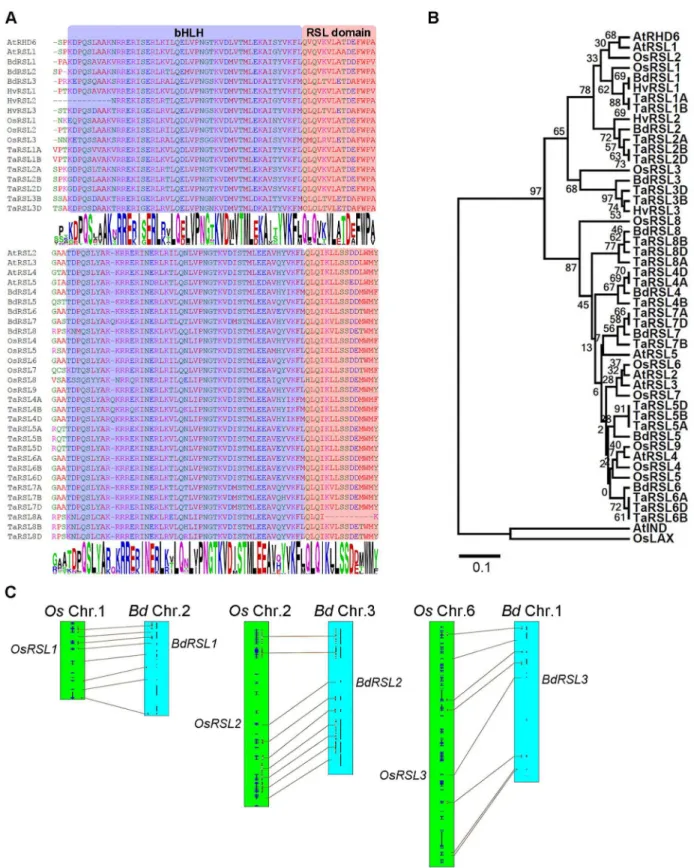
To determine if RSL class I genes control the development of root hair cells inB.distachyonwe searched for similar genes in the genomes of members of the grass family (Poaceae) (S1A Fig). We discovered genes encoding proteins with the conserved RSL domain next to the bHLH domain (Fig 1A). The topology of gene trees constructed using alignments of the basic helix-loop-helix domain and conserved RSL motif from these proteins showed that threeB. distach-yongenes (BdRSL1,BdRSL2andBdRSL3) (Fig 1BandS1B Fig) and threeO.sativagenes–
(OsRSL1,OsRSL2andOsRSL3) were most closely related to the previously characterized RSL class I genesAtRHD6andAtRSL1ofA.thaliana. BdRSL1, BdRSL2 and BdRSL3 are respec-tively 84%, 89% and 73% identical to AtRHD6 in the bHLH-RSL domain (S1 Table) but there is no conservation outside these conserved regions. Gene order (synteny) indicates that
OsRSL1andBdRSL1,OsRSL2andBdRSL2, andOsRSL3andBdRSL3are orthologous gene pairs (Fig 1C). RSL class I genes were also identified in genomes of other members of the grass family includingHordeum vulgareandTriticum aestivum. The RSL class I clade was sister to a clade that contained theA.thalianaRSL class II proteins, AtRSL2, AtRSl3, AtRSL4 and AtRSL5. FiveB.distachyonproteins, sixO.sativaand 15T.aestivumproteins were identified that belonged to the RSL class II clade. Taken together these data indicate that RSL class I and RSL class II genes are present in the genomes of members of the grass family.
RSL class I genes are expressed in developing epidermal cells of
Brachypodium distachyon
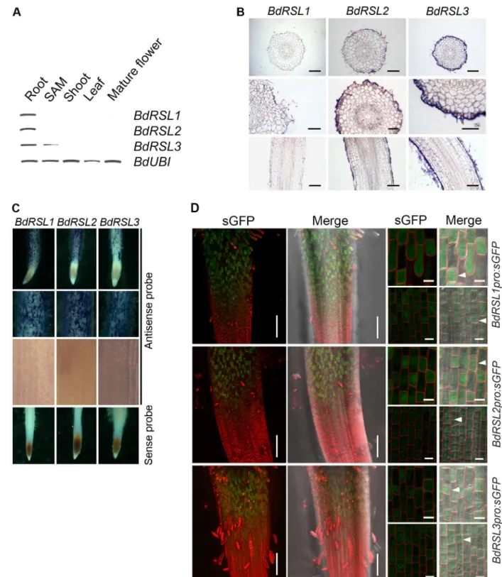
RSL class I genes are expressed in theA.thalianaroot meristem and mRNA disappears from cells before root hair initiation. We set out to determine if this expression pattern is conserved inB.distachyon. First, using RT-PCR we detectedBdRSL1,BdRSL2mRNA only in roots while
Fig 1. Genes encoding RSL class I proteins are present in the genomes of members of the grass family. (A)Alignment of conserved regions ofBrachypodium distachyon(Bd),Oryza sativa(Os),Triticum aestivum(Ta),Hordeum vulgare(Hv) and
cells and continue to be expressed during root hair morphogenesis. To verify independently that RSL class I genes were expressed in developing root hair cells in the elongation and differ-entiation zones, we identified the cells in which the RSL class I promoters were active. We used plants transformed with gene constructs in which the RSL class I promoter controlled expres-sion of the synthetic Green Fluorescent Protein (sGFP) [23]. GFP fluorescence was not detected in the meristem ofBdRSL1pro:sGFP,BdRSL2pro:sGFP or BdRSL3pro:sGFP trans-formed plants, confirming the conclusion that these genes are not expressed in the dividing cells of the root (Fig 2D). However, GFP fluorescence was detected in developing root hair cells in the elongation zone, and in the differentiation zone where the root hairs actively elongated (Fig 2DandS2 Fig). The earliest detectable fluorescence was found in cells at the beginning of the elongation zone (after the completion of asymmetric mitosis). While the promoters of each of the three RSL class I genes was preferentially expressed in the smaller daughter cells (Fig 2D) they were occasionally active in the large daughter cells. InBdRSL1pro:sGFProots shown inFig 2D, seven of the eight small cells in the field of view expressed GFP and none of the seven long expressed GFP (Fig 2D). InBdRSL2pro:sGFProots all 14 of the short cells in the field of view expressed GFP while one of the 13 long cells expressed GFP (Fig 2D). InBdRSL3pro:sGFP
roots all 16 of the short cells in the field of view expressed GFP while two of the 17 long cells expressed GFP. These data indicate that the RSL class I genes are preferentially expressed in the smaller daughter cell that forms from the asymmetric mitosis that forms a root hair cell and a hairless epidermal cell pair. GFP fluorescence was detected later in development in cells with elongating root hairs (S2 Fig). These data indicate that RSL class I genes are preferentially expressed in developing root hair cells, from at or just after the formative asymmetric cell divi-sion and continues through root hair elongation. This expresdivi-sion pattern is different from that observed inA.thalianawhere RSL class I genes are expressed in the meristem and expression is not detectable in cells with growing root hairs. Despite the differences in expression pattern, these data are consistent with the hypothesis that RSL class I genes positively regulate root hair cell development inB.distachyon.
RSL class I genes are sufficient for root hair cell development in
Brachypodium distachyon
To determine if RSL class I proteins are sufficient for root hair development in the root epider-mis, the relative numbers of root hair cells and hairless epidermal cells on wild type and plants constitutively expressingBdRSL1,BdRSL2orBdRSL3were compared. To overexpress each RSL class I gene constitutively,B.distachyonwas transformed with constructs in which each gene was placed under the control of the promoter and first intron of theZea mays Ubiquitin1
gene (ZmUBIpro). Plants lines in which each of the RSL class I genes was overexpressed were
identified (S3A Fig). Almost every cell in the root epidermis developed root hairs in each of the overexpressing lines (Fig 3A). While 51% of epidermal cells develop as root hair cells in wild type, 95%, 96% and 96% of epidermal cells developed root hairs inZmUBIproBdRSl1,ZmUBI -proBdRSL2andZmUBIproBdRSL3lines respectively (Fig 3B). Furthermore, root hairs were lon-ger in the RSL class I overexpressing lines than in wild type controls (S3B Fig). This
demonstrates not only that RSL class I proteins positively regulate the specification of the relationship between RSL class I and class II proteins fromBrachypodium distachyon(Bd),Oryza sativa(Os),Triticum aestivum
(Ta),Hordeum vulgare(Hv) andArabidopsis thaliana(At) based on the protein sequence of bHLH and RSL domains. AtbHLH040 (AtIND) and OsbHLH123 (OsLAX) were used as out groups. Numbers below branches indicate bootstrap percentages.(C)Synteny between RSL class I genes ofB.distachyonandO.sativa. Connecting lines between linkage groups define chromosome regions with collinear orthologous genes. The locations ofRSL1,RSL2andRSL3orthlogs are indicated.
class I gene promoters. Two left-hand columns: root tips and white arrows highlight appearance of GFP fluorescence. Two right-hand columns: there are four images for each reporter construct. The top row is a representative pair of images of the root epidermis at the root hair initiation stage. White arrowheads indicate GFP fluorescence in the short cells bearing from which root hairs develop. The lower pair of images show the root epidermis at the transition between meristem and elongation zone where the asymmetric cell division has occurred. White arrowheads indicate GFP fluorescence in a smaller daughter cell resulting from an asymmetric cell division. Scale bars 50μm (two left-hand columns), 10μm (two right-hand columns).
doi:10.1371/journal.pgen.1006211.g002
Fig 3. RSL class I expression is sufficient for root hair cell development inB.distachyon. (A)Plants transformed withZmUBIpro:
BdRSL1,ZmUBIpro:BdRSL2orZmUBIpro:BdRSL3develop root hairs on almost every root epidermal cell. Scale bar 100μm.(B)
Percentage root hair cells (grey bar) and percentage hairless epidermal cells (white bar).(C)Asymmetric mitoses occur in plants that overexpress RSL class I genes. White arrows indicate the location of the new cell wall formed after asymmetric division forms a relatively small cell and a relatively large cell. Scale bar 10μm.
epidermal cell identity inB.distachyonbut also that these genes are sufficient for root hair cell development.
The expression pattern of RSL class I genes suggests thatBdRSL1,BdRSL2andBdRSL3act in the elongation zone after the asymmetric cell division that generates root hair cells. To test if these genes act after the formative asymmetric cell division, the positioning of new transverse cell walls was determined in plants transformed withZmUBIproBdRSL1,ZmUBIproBdRSL2or
ZmUBIproBdRSL3gene constructs. In wild type, the last cell division in a file is accompanied by an asymmetric mitosis forming a small daughter cell with root hair cell fate and a larger cell with hairless epidermal cell fate (Fig 3C). Asymmetric cell divisions were observed in all cell files of the RSL class I overexpressing plants and were indistinguishable from wild type; asym-metric mitoses formed a cell pair in which one cell was larger than the other (Fig 3C). In the overexpressing plants, both daughter cells developed root hairs, in contrast to wild type where only the smaller daughter cell formed a root hair. Taken together these data indicate that RSL class I proteins are sufficient for root hair cell development in the root epidermis ofB. distach-yonand they likely act after the asymmetric cell division that generates the root hair cell. We conclude that the spatial pattern of root hair cell differentiation in theB.distachyonroot epi-dermis is largely dictated by the pattern of RSL class I gene expression.
RSL class I genes positively regulate root hair growth and likely act after
the asymmetric cell division that forms root hair cells
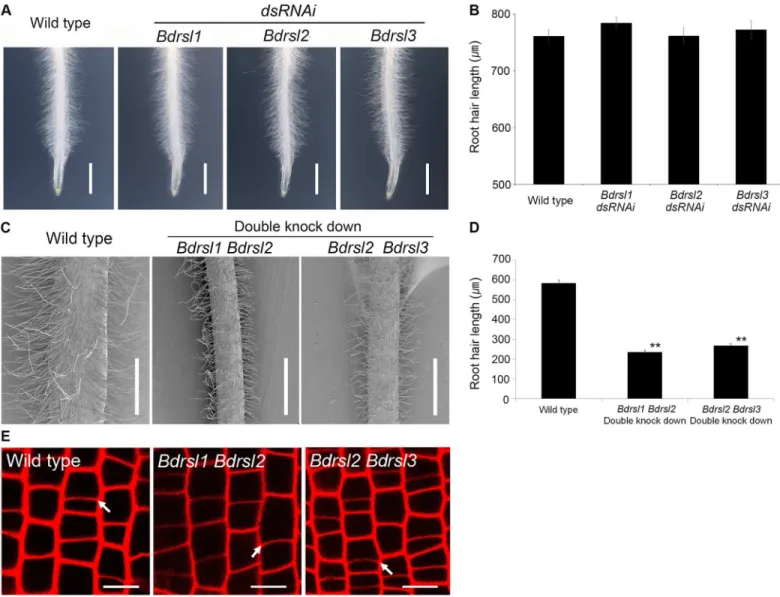
To verify independently when RSL genes act during root hair cell development we tested if the asymmetric formative cell division occurred in transgenic lines with reduced RSL class I func-tion. Reduced expression of individualBdRSL1,BdRSL2andBdRSL3genes had no impact on root hair cell development (Fig 4A and 4B), Therefore, lines with decreased expression of two RSL class I genes were constructed. Steady state levels ofBdRSL1andBdRSL3mRNA were reduced by more than 50% in one set of lines while steady state levels ofBdRSL2andBdRSL3
mRNA were reduced by more 50% in the other set of lines (S4 Fig). Imaging of the cross walls on propidium iodide stained roots revealed that the asymmetric cell divisions occurred in
BdRSL1 BdRSL2andBdRSL2 BdRSL3knock down plants and were indistinguishable from wild type (Fig 4E). These data are consistent with the hypothesis that RSL gene function does not regulate asymmetric cell division, although we cannot rule out the possibility that there may be some residual RSL1 class I activity in the double knockdown plants that is sufficient for division asymmetry. Furthermore, root hairs of theBdRSL1 BdRSL2double knockdown plants were 50% shorter than wild type, and root hairs onBdRSL2 BdRSL3 doubleknockdown plants were 54.0% shorter than wild type (Fig 4C and 4D). This indicates that the expression of RSL class I genes promotes root hair cell morphogenesis and differentiation. Taken together these data suggest that RSL genes positively regulate root hair development and act after the asym-metric cell division that forms hair cells inB.distachyon.
The function of RSL class I proteins is conserved between
B
.
distachyon
and
A
.
thaliana
Given the conservation of the bHLH and RSL domains of RSL class I proteins between B. dis-tachyonandA.thaliana, we predicted that their function would be at least partially conserved (S1 Table). If the function of RSL class I genes was conserved sinceB.distachyonandA. thali-analast shared a common ancestor, we predicted that expression of RSL class I genes fromB.
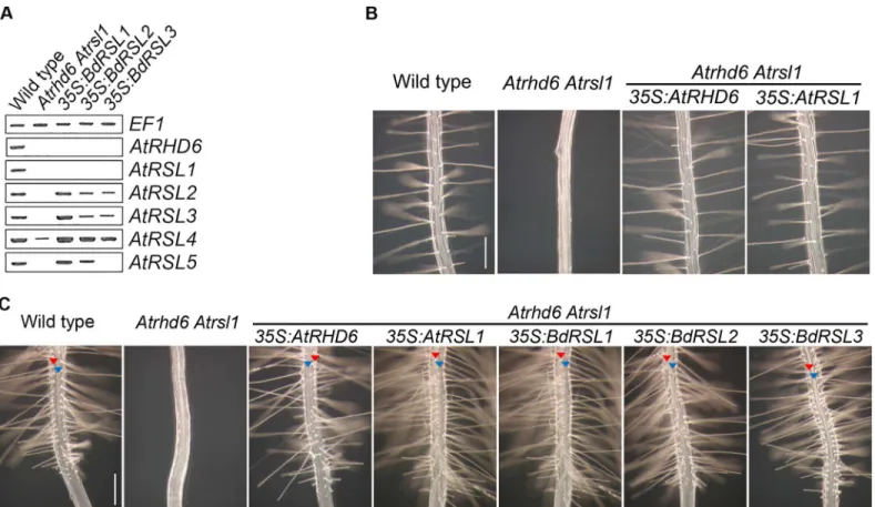
there was no detectableAtRSL2,AtRSL3, orAtRSL5expression andAtRSL4was reduced in the
Atrhd6 Atrsl1double mutant (Fig 5A) [16]. However, expression ofAtRSL2,AtRSL3,AtRSL4
andAtRSL5was restored inAtrsl1 Atrhd6double mutants transformed with35S:BdRSL1or
35S:BdRSL2gene constructs (Fig 5A). The35S:BdRSL3transgene partly restoredAtRSL2,
AtRSL3,AtRSL4expression (Fig 5A). To test if restoration of RSL class II gene expression byB.
distachyonRSL class I genes also restored root hair elongation, we characterized root hair growth inAtrhd6 Atrsl1double mutants expressing theB.distachyonRSL class I genes. While root hairs do not develop onAtrhd6 Atrsl1double mutants, root hairs formed on the double mutants transformed with35S:BdRSL1,35S:BdRSL2or35S:BdRSL3gene constructs (Fig 5B); root hair development was indistinguishable fromAtrhd6 Atrsl1 35S:RHD6plants. This indi-cates that expression of theBdRSL1,BdRSL2andBdRSL3genes can replace the missing RSL class I gene function in theA.thaliana Athd6 Atrsl1double mutant.
Fig 4. RSL class I genes act after the asymmetric cell division that forms root hair cells inB.distachyon. (A, B)Root hair length inBdrsl1,Bdrsl2, andBdrsl3dsRNAi single knock down lines is indistinguishable from wild type. Scale bar 10 mm. Error bar is SD.(C, D)Root hairs are shorter inBdrsl1 Bdrsl2andBdrsl2 Bdrsl3double knock down lines than that of wild type. Scale bar 500μm. Asterisks in(D)indicate statistically significant differences between knock down lines and wild type at**P<0.01 (t-test). Error bar indicates SD.(E)Asymmetric mitoses occur in all cell files of the RSL class I double knock down plants. White arrows indicate positions of new transverse cell walls formed after asymmetric mitosis. Scale bar 10μm.
Since RSL class I expression was sufficient for root hair development inB.distachyon, we tested if RSL class I expression was sufficient for root hair development inA.thaliana. First, we characterized the pattern of epidermal cell differentiation inAtrhd6 Atrsl1double mutants transformed with35S:RHD6or35S:RSL1gene constructs. Root hair cells developed in longitu-dinal cell files and were flanked by files of hairless epidermal cells in the transformed plants—
the same pattern observed in wild type—while theAtrhd6 Atrsl1mutants were root hairless (Fig 5C). These data indicate that constitutive expression ofA.thalianaRSL class I genes did not induce ectopic root hair cell development inA.thaliana, unlikeB.distachyonwhere ectopic RSL class I gene expression resulted in ectopic root hair cell development (Fig 3A).
If the function of RSL class I genes has been conserved sinceA.thalianaandB.distachyon
last shared a common ancestor, we predicted that the phenotype ofA.thalianaplants that ectopically expressB.distachyonRSL class I genes would be the same as the phenotype of plants that ectopically expressA.thalianaRSL class I genes.Atrhd6 Atrsl1double mutants were transformed with35S:BdRSL1,35S:BdRSL2and35S:BdRSL3transgenes. Files of root hair cells and hairless epidermal cells formed on theAtrhd6 Atrsl1double mutants expressing indi-vidual RSL class I genes; ectopic hair cells did not develop (Fig 5C). These plants were morpho-logically indistinguishable from theAtrhd6 Atrsl1double mutants transformed with35S:
Fig 5. The functionB.distachyonandA.thalianaRSL class I genes is conserved. (A)Steady state levels ofAtRSLclass I and class II mRNA in
Atrhd6 Atrsl1double mutants, and inAtrhd6 Atrsl1double mutants transformed BdRSL class I genes under the control of the35Spromoter. Lane 1: wild type, Lane2:Atrhd6 Atrsl1double mutant, Lane3:Atrhd6 Atrsl1 35S:BdRSL1, Lane4:Atrhd6 Atrsl1 35S:BdRSL2, Lane5:Atrhd6 Atrsl1 35S:BdRSL3.(B) Phenotype of wild type;Atrhd6 Atrsl1double mutant;Atrhd6 Atrsl1 35S:AtRHD6;Atrhd6 Atrsl1 35S:AtRSL1. Scale bar 200μm.(C)Files of root hair cells and hairless epidermal cells form in wild-type,Atrhd6 Atrsl1 35S:AtRHD6,Atrhd6 Atrsl1 35S:AtRSL1,Atrhd6 Atrsl1 35S:BdRSL1,Atrhd6 Atrsl1 35S:
BdRSL2andAtrhd6 Atrsl1 35S:BdRSL3.Atrhd6 Atrsl1double mutants do not develop root hairs. Red arrowhead indicates a hair cell file, blue arrowhead indicates the position of a hairless epidermal cell file. Scale bar 200μm.
AtRHD6orAtRSL1(Fig 5CandS5B Fig). These data indicate that neither the expression ofB.
distachyonnorA.thalianaRSL class I genes is sufficient for root hair cell development inA.
thaliana. To independently verify thatB.distachyonRSL class I expression was not sufficient for root hair development inA.thalianawe ectopically overexpressed these genes in wild type plants. Root hair cells developed in longitudinal cell files and were flanked by files of hairless epidermal cells in wild type transformed with35S:BdRSL1,35S:BdRSL2or35S:BdRSL3(S5A and S5B Fig). These data suggest that the pattern of root hair cell differentiation in theA. thali-anaroot epidermis cannot be dictated by the spatial pattern of RSL class I gene expression alone.
The different phenotypes that result from the ectopic expression of RSL class I genes inB.
distachyonandA.thalianademonstrate that while the pattern of RSL class I gene expression determines the pattern of hair cell differentiation inB.distachyon, the pattern of expression in
A.thalianaalone cannot account for the pattern of epidermal differentiation.
Discussion
The function of RSL class I genes as positive regulators of root hair cell development is con-served betweenB.distachyonandA.thaliana. We conclude that RSL class I genes controlled root hair cell development in the common ancestor of grasses and brassicas which existed some time before 200 million years ago [24]. This demonstrates that RSL class I genes are likely to promote root hair cell differentiation in monocots and eudicots even though different spatial patterns of epidermal development can exist among these taxa.
Despite the conservation of RSL class I function betweenB.distachyonandA.thaliana, the different phenotypic consequences of ectopic RSL class I gene expression in these species high-lights a major divergence in the mechanism controlling root epidermal development in each. RSL class I expression is sufficient for hair cell development inB.distachyon–ectopic overex-pression of any of the three RSL class I genes induces the development of root hairs in every cell of the root epidermis. This demonstrates that RSL class I proteins can initiate the root hair cell developmental program in the grass. By contrast, ectopic overexpression of AtRSL class I genes inA.thalianadoes not induce the development of ectopic root hair cells in the root epi-dermis. This suggests that whileA.thalianaRSL class I proteins promote root hair cell develop-ment, their expression alone is not sufficient for root hair cell development in other cell types of the root epidermis. These observations could be explained by either (i)B.distachyonRSL class I genes being functionally different fromA.thalianaRSL class I genes, or (ii) the presence of an activity that represses root hair cell development inA.thalianadespite the high levels of RSL class I gene activity in the experimental transgenic plants. It was possible to rule out the former because while expression ofB.distachyonRSL class I genes in theA.thaliana Atrhd6 Atrsl1double mutants was sufficient to restore root hair development in root hair cells, their expression was not sufficient for root hair development in the cells in the hairless epidermal cell position [9]. This suggests that the differences in the ability of RSL class I genes to induce root hair development is due to factors that repress hair development inA.thalianain develop-ing hairless epidermal cells that are missdevelop-ing fromB.distachyon. Taken together these data sug-gest that the mechanism controlling root hair cell differentiation is conserved betweenB.
distachyonandA.thaliana, but the mechanism that controls the spatial patterning of the two cells types in the root epidermis is different between the two species.
While RSL class I genes positively regulate root hair cell development in bothB.distachyon
differentiation zone, but there is no expression in the meristem. By contrast,AtRHD6and
AtRSL1are expressed in the meristem and no expression of these RSL class I genes is detected in the developing root hair cells in the elongation or differentiation zones [11]. This different timing of RSL class I expression mirrors the morphological differentiation of the two cell types in these species. Epidermal cells become morphologically different relatively late development in the grass—after the last meristematic cell division—while differentiation is morphologically detectable in the meristem ofA.thaliana, where files of relatively short cells are destined to develop root hairs and relatively long cells are destined to become hairless epidermal cells. Therefore, morphological differentiation is first visible in the meristem inA.thalianaand in the elongation zone ofB.distachyonand this mirrors the patterns of RSL class I gene expres-sion in the two species. Consequently we can reconcile the different expresexpres-sion patterns if RSL class I genes are expressed in future hair cells as soon as they become morphologically different from neighbouring future hairless epidermal cells. Since this event occurs in the meristem ofA.
thaliana, RSL class I genes are expressed in the meristem and since this event occurs after the last cell division inB.distachyon, the genes are in the elongation zone. However this does not explain why the RSL class I genes continue to be expressed during root hair elongation in the grass. It is possible that the continued expression ofBdRSL1,BdRSL2andBdRSL3in these cells maintains the growth program active.
We conclude that RSL class I genes positively regulate the development of root hair cells in
B.distachyonwhich demonstrates that the mechanism of RSL-regulated root hair differentia-tion has been conserved between Brassicaceae and Poaceae which last shared a common ances-tor some time around 200 million years ago [24]. We also present evidence that suggests that the expression patterns RSL class I genes can account for the pattern of root hair cell differenti-ation found in members of the grass family.
Materials and Methods
Plant material and growth
Brachypodium distachyon(line Bd21) caryopses (grain) were sterilized in 10% NaClO and 0.1% Triton X-100 with gentle shaking for 15 minutes, then washed with sterilized water 5 times. Grain were then placed on wet filter paper in a Petri dish sealed with cling film and incu-bated for 2 days in the dark at 5°C (B.distachyon) to ensure that all grain germinated at the same time. Grain were placed in Petri dishes containing media as in described previously [25] but modified to contain 1% sucrose and 0.5% phytagel (Sigma, UK). Plants were grown verti-cally in a growth chamber with a 16 hour light period at 23°C for 5 days.Arabidopsis thaliana
wild type plants of the ecotype Columbia-0 (Col0) were used.Arabidopsis thalianaseeds were sterilized in 5% NaClO for 5 min and washed with sterilized water for 5 times. Seeds were sown and germinated in Petri dishes containing sterilized media as in Maet al. [28] modified to con-tain 1% sucrose and 0.5% phytagel (Sigma, UK). The plants were grown vertically in the growth chamber with a 16 hour light period at 23°C for 5 days.
Alignment and phylogenetic analysis
(http://www.atgc-montpellier.fr/phyml/) and thMaximum Likelihood method with the follow-ing parameters: SPR &NNI, aLRT SH-like. The constructed tree file was visualized usfollow-ing MEGA [27] (version 6.0;http://www.megasoftware.net/index.html). Logos of amino acid sequence alignments were created with WebLogo (http://weblogo.berkeley.edu/). The multiple alignment to score similarity of amino acid was carried out Mview program (http://www.ebi. ac.uk/Tools/msa/mview/).
SyntenyB.distachyonandO.sativagenomes were analysed using SyMAP v4.2 software (http://www.symapdb.org/projects/poaceae/), and the ORFs in bothO.sativaandB.distachyon
genome were identified using a BLAST filtering approach (BLASTP, e-value<0.01). All
pre-dicted gene sequences were searched against all available databases of fully sequenced genomes (O.sativa;http://signal.salk.edu/cgi-bin/RiceGE,B.distachyon;http://jbrowse.brachypodium. org/JBrowse.html).
Generation of transgenic plants
B.distachyonoverexpression constructs were based on pVec8- GFP [28] (a gift from Philippe Vain, John Innes Centre, Norwich, UK). The GFP sequences were removed and a unique KpnI site was generated betweenZmUBIpromoter andNosterminator to make the pVec8-overex-pression vector. For the constitutive expVec8-overex-pression of RSL class I genes inB.distachyon, the coding sequences of RSL class I were amplified from root cDNA with primer pairs that added a KpnI recognition site at both ends:BdRSL1F (KpnI): 5’-CGGTACCCTCATGGCAAGCAGGCAC G-3’, BdRSL1 R (KpnI): 5’-CGGTACCGCATCATGCATGGCCTAGG-3’forBdRSL1(836bp); BdRSL2 F (KpnI): 5’-CGGTACCCTGATCATGGCATTAGTGCG-3’,BdRSL2R (KpnI): 5’ -CGGTACCGGTACGTGTTTCCTTCTAGC-3’forBdRSL2(951bp); BdRSL3 F (KpnI): 5’ -CGGTACCGCCATGGCTCTAGTGGGTC-3’,BdRSL3R (KpnI): 5’-CGGTACCGCTATATA CTGCTAGCTCC-3’forBdRSL3(1157bp) (the KpnI site is underlined). The PCR products were subcloned into the pGEM-T Easy Vector (Promega), and sequenced using SP6 and T7 primers to verify the PCR product. Cloned PCR fragments were digested with KpnI and ligated into KpnI-digested pVec8-overexpression plasmid. Sense orientations were confirmed by sequencing with the forward primer 50-TGATGGCATATGCAGCAGC-3’from the ZmUBI
promoter.
The dsRNAi site of pANDA vector [29], containing attR cassettes at both sites of the GUS linker in the sense and antisense orientations were introduced to pVec8-overexpression KpnI site betweenZmUBIpromoter andNosterminator. RNA silencing constructs carrying the 3’
region of each class I RSL gene were made using the modified pVec8dsRNAivector to silence the endogenous class I RSL gene. 50 ng cDNA was used for PCR amplification with each of the following gene specific primer sets (underlines are attB site to create entry clones using the BP recombination reaction) BdRSL1 dsRNA F: GGGGACAAGTTTGTACAAAAAAGCAGGCT TCGAATCAATCCTAGGCCATG, BdRSL1 dsRNA R: GGGGACCACTTTGTACAAGAAA GCTGGGTCAGTGACCAGAACTCCGAAC for BdRSL1 dsRNAi (239bp); BdRSL2 dsRNA F: GGGGACAAGTTTGTACAAAAAAGCAGGCTTCGGCAATTCAACTGCTCCAG,BdRSL2
was amplified with each of the following gene specific primer sets (attB site underlined to create entry clones using the BP recombination reaction) and cloned into pDONR207. BdRSL1pro F: GGGGACAAGTTTGTACAAAAAAGCAGGCTTC GTTACTATGGGCTACTGGG,
BdRSL1pro R:GGGGACCACTTTGTACAAGAAAGCTGGGTC GAGCTAGGCAGCTATAA CAC, for BdRSL1 promoter, BdR2pro F:GGGGACAAGTTTGTACAAAAAAGCAGGCTTC GCAACATGATCGAGGATCC, BdR2pro R:GGGGACCACTTTGTACAAGAAAGCTGG GTC GATCAGTTGATCACGTATCAATC, for BdRSL2 promoter, BdR3pro F:GGGGACAA GTTTGTACAAAAAAGCAGGCTTC CGTATCCGGTGAAAACGAC, BdR3pro R:
GGGGACCACTTTGTACAAGAAAGCTGGGTC GGCGGTGGGACTAAGCGG, for BdRSL3 promoter. After sequencing, to confirm the insertion, a GATEWAY LR reaction was then performed with pVEC8-R1-CmR-ccdB-R2:sGFP vector.
TransgenicB.distachyonplants were generated usingAgrobacterium tumefaciensmediated transformation method as described previously [33]. Immature embryonic calli were induced from immature embryos cultured on MSB3 + Cu0.6 medium for 6 weeks to produce callus, and then co-cultivated with Agrobacterium strain AGL1 harboring the pVec8 binary vector including target gene, before being transferred to selective medium supplemented with 50μg/L
hygromycin. Hygromycin resistant calli were subsequently regenerated on regeneration media [30]. The transgenic plantlets were then transferred to the greenhouse in pots of John Innes No. 1 compost with a 16-hour light period at 23°C to grow and produce seeds. T3 seeds that were homozygous for the transgene were harvested and several lines tested for gene expression levels and used for further analysis.
For overexpression inA.thaliana,BdRSL1,BdRSL2, andBdRLS3cDNAs were used and subcloned into the pGEM-T Easy Vector (Promega) (see above). cDNA was inserted into the pCAMBIA1300 binary vector. All binary vectors were transferred into Agrobacterium strain GV3101.Arabidopsisplants were transformed using a modified version of the floral dip method [31]. Agrobacterium transformed with a binary vector was grown overnight at 28°C in LB medium with antibiotic (Kanamycin 50μg/ml and Rifampicin 125μg/ml) for plasmid
selec-tion. 2 ml of this culture was used to inoculate 500 ml of fresh LB media, which contained anti-biotics, and incubated for 24 hours at 28°C. Cells were pelleted by centrifugation and then resuspended in an equal volume of infiltration medium (MS salts, 5% (w/v) sucrose, 0.05% (v/v) Silwet L-77 (Vac-in-Stuff, Lehle Seeds)) and 0.15 mM acetosyringone.Arabidopsisplants were dipped into the bacterial suspension for 60–90 seconds and covered with a plastic bag for 24 hour to maintain humidity. After removing the bag, plants were returned to normal green-house conditions for seed harvesting. Transformants were selected on kanamycin (50μg/ml).
Root hair measurements
Images of root hairs of 5 day old seedling were visualized using a Nikon Eclipse E600 epifluor-escence light microscope (Nikon, Japan) with Nikon Plan Apo objectives and bright field optics, and a Leica M165 FC stereo microscope and images captured with a Leica DFC310 FX camera. Images were postprocessed with ImageJ (http://rsb.info.nih.gov/ij/) and Adobe Photo-shop CS4.
Gene expression analysis
Super Script III First-Strand Synthesis System for RT-PCR using oligo dT primer (Invitrogen). Semi quantitative RT-PCR was routinely carried out for 30 to 35 cycles, depending on the lin-ear range of PCR amplification for each gene. Each PCR cycle included incubations at 98°C for 10 s, 58°C for 20 s, and 72°C for 30 s. Specific primers were designed to generate RT-PCR prod-ucts:BdRSL1RT: 5’-GAAGAAGCAGTGTGGAGGAAG-3’and 5’-GACCATGTCAACCTTT GTGCC-3’;BdRSL2RT: 5’-GACGTTGCAGGAGATGGTG-3’and 5’-CGAGCACCTTGAC TTGCAG-3’;BdRSL3RT: 5’-CAAGCTGCCAAGAGCCGTC-3’and 5’-CTGTCTCGAGCAC CGTGAG-3’;BdUBIRT: 5’-CAATGTCAAGGCGAAGATCC-3’and 5’-GGTCTTCACGAAG ATCTGC-3’;AtRHD6RT: 5’-CCTAAATCCGCTGGAAACAA-3’and 5’-CTCTTCGATTCTT GGCTGCT-3’;AtRSL1RT: 5’-CCCTAAACTGGCTGGCAATA-3’and 5’-TCTTGGCTGCTA GGCTTTGT-3’;AtRSL2RT: 5’-TCCCCAATGGAACAAAGGTC-3’and 5’-TCTCGGTGAGC TGAGACCAA-3’;AtRSL3: 5’-GGAGCCAGAAATGCGTAGAG-3’and 5’-GTCTCCACCGT TTGATTCGT-3’;AtRSL4RT: 5’-GTGCCAAACGGGACAAAAGT-3’and 5’-TTGTGATGGA ACCCCATGTC-3’;AtRSL5RT: 5’-GCAGGAACTTCACGTAATGGA-3’and 5’-TATACGCT AGGAAACGAAGAGAAA-3’;EF1α: RT: 5’-GGTGGTGGCATCCATCTTGTTACA-3’and 5’-TGAGCACGCTCTTCTTGCTTTCA-3’
Confocal microscopy
Roots were mounted in 10 ng/ml propidium iodide (Sigma) solution. Confocal images of root epidermal cells were obtained by Leica TCS SP5 confocal laser scanning microscopes using 543-nM and 490-nM laser line for excitation of PI and GFP, respectively. Images were post-processed with ImageJ (http://rsb.info.nih.gov/ij/) and Adobe Photoshop CS4.
Cryo-scanning electron microscopy
For cryo-scanning electron microscopy, seedlings were placed on moist nitrocellulose paper, mounted on a stub and immersed in liquid nitrogen slush. Roots were transferred to a cold stage. After removal of water by sublimation, roots were sputter coated with gold and observed with a Philips XL30 FEG scanning electron microscope equipped with an Oxford Instruments CT1500 HF cryo-stage at -147°C. Digital images were captured with the maximum pixel den-sity. Image J (http://rsb.info.nih.gov/ij/) and Adobe Photoshop CS4 were used to process the images.
Whole mount in situ hybridization
In situ hybridization was used to determine the cells in which RSL class I mRNA accumulated. Antisense probes for the class IRSLgene were made by amplifying the full coding sequence including the 5’UTR and 3’UTR by PCR using full length cDNA primers (see above). PCR fragments were cloned into pGEM-T Easy Vector (Promega). To generate specific antisense probes, full length cDNA was transcribed from the terminal SP6 or T7 RNA polymerase prim-ing sites of these vectors, then treated DNase and precipitated RNA by ethanol. Full length RNA was hydrolyzed by carbonate buffer (200 mM, pH10) to create 150 bp fragments for hybridization.In vitrotranscription, digoxigenin labeling of RNA probes, tissue preparation, and in situ hybridization were carried out as described in Dreaet al. [32] and Derbyshireet al. [33].
Supporting Information
genes that were used as outgroupsAtINDandOsLAX. (B)Alignment of RSL class I and class II gene sequences used for phylogenetic analysis.
(TIF)
S2 Fig. TheBdRSL1promoter is active in root hair cells.Fluorescence produced from the activity of theBdRSL1pro:sGFPtransgene in root hair cells. Scale bar 50μm.
(TIF)
S3 Fig. Vector constructs used for the overexpression ofBdRSL1,BdRSL2,BdRSL3. (A)
BdRSLclass I cDNAs (BdRSL1,BdRSL2, andBdRSL3) were inserted between theZmUBI pro-moter and theNosterminator. Arrows indicate restriction sites and the names of the respective restriction enzymes.(B)Root hairs are longer in lines transformed withZmUBIpro:BdRSL1,
ZmUBIpro:BdRSL2andZmUBIpro:BdRSL3than wild type. Asterisks indicate statistically signifi-cant differences between overexpression lines and wild type atP<0.01 (t-test). Error bar
indi-cates SD. (TIF)
S4 Fig. Steady state mRNA levels are reduced inBdrsl1 Bdrsl2,and Bdrsl2 Bdrsl3double knock down lines ofB.distachyon.Steady state levels ofBdRSL1andBdRSL2mRNA are lower inBdrsl1 Bdrsl2double knockdown lines than in wild type. Steady state levels ofBdRSL2
andBdRSL3mRNA are lower inBdrsl1 Bdrsl2double knockdown lines than in wild type. Error bar is SD.
(TIF)
S5 Fig. Expression ofB.distachyonRSL class I genes in wild typeA.thalianadoes not induce ectopic root hair cell development. (A)Wild-type (Col-0)A.thalianatransformed with35S:BdRSL1,35S:BdRSL2and35S:BdRSL3. Scale bar 500μm.(B)Mean root hair number
mm-1(± SD) in lines transformed with35S:BdRSL1,35S:BdRSL2and35S:BdRSL3is indistin-guishable from wild-type.
(TIF)
S1 Table. Percentage amino acid identity between the bHLH and RSL domain ofA. thali-anaandB.distachyonRSL class I proteins.
(PDF)
Author Contributions
Conceived and designed the experiments: CMK LD. Performed the experiments: CMK. Ana-lyzed the data: CMK LD. Contributed reagents/materials/analysis tools: CMK LD. Wrote the paper: LD CMK.
References
1. Jungk A. Root hairs and the acquisition of plant nutrients from soil. J Plant Nutr Soil Sci. 2001; 164: 121–129.
2. Gahoonia TS, Nielsen NE. Variation in root hairs of barley cultivars doubled soil phosphorus uptake. Euphytica. 1997; 98: 177–182.
3. Leavitt RG. Trichomes of the root in vascular cryptogams and angiosperms. Proc Bost Soc Nat Hist. 1904; 31: 273–313.
7. Kim CM, Dolan L. Root hair development involves asymmetric cell division in Brachypodium distachyon and symmetric division inOryza sativa. New Phytol. 2011; 192: 601–610. doi:10.1111/j.1469-8137. 2011.03839.xPMID:21848982
8. Cormack RGH. Investigations on the development of root hairs. New Phytol. 1935; 34: 30–54. 9. Dolan L, Duckett CM, Grierson C, Linstead P, Schneider K, Lawson E, et al. Clonal relationships and
cell patterning in the root epidermis of Arabidopsis. Development. 1994; 2474: 2465–2474.
10. Dolan L, Costa S. Evolution and genetics of root hair stripes in the root epidermis. J Exp Bot. 2001; 52: 413–417. PMID:11326047
11. Menand B, Yi K, Jouannic S, Hoffmann L, Ryan E, Linstead P, et al. An ancient mechanism controls the development of cells with a rooting function in land plants. Science. 2007; 316: 1477–1480. PMID: 17556585
12. Masucci JD, Schiefelbein JW. Hormones act downstream of TTG and GL2 to promote root hair out-growth during epidermis development in the Arabidopsis root. Plant Cell. 1996; 8: 1505–1517. PMID: 8837505
13. Ryu KH, Kang YH, Park Y, Hwang I, Schiefelbein J, Lee MM. The WEREWOLF MYB protein directly regulates CAPRICE transcription during cell fate specification in the Arabidopsis root epidermis. Devel-opment. 2005; 132: 4765–4775. PMID:16207757
14. Tominaga R, Iwata M, Okada K, Wada T. Functional analysis of the epidermal-specific MYB genes CAPRICE and WEREWOLF in Arabidopsis. Plant Cell. 2007; 19: 2264–2277. PMID:17644729 15. Lee MM, Schiefelbein J. WEREWOLF, a MYB-related protein in Arabidopsis, is a position-dependent
regulator of epidermal cell patterning. Cell. 1999; 99: 473–483. PMID:10589676
16. Yi K, Menand B, Bell E, Dolan L. A basic helix-loop-helix transcription factor controls cell growth and size in root hairs. Nat Genet. Nature Publishing Group; 2010; 42: 264–267.
17. Marzec M, Melzer M, Szarejko I. Root hair development in the grasses: what we already know and what we still need to know. Plant Physiol. 2015; 168: 407–414. doi:10.1104/pp.15.00158PMID: 25873551
18. Marzec M, Melzer M, Szarejko I. The evolutionary context of root epidermis cell patterning in grasses (Poaceae). Plant Signal Behav. 2014; 9: 1–5.
19. Ding W, Yu Z, Tong Y, Huang W, Chen H, Wu P. A transcription factor with a bHLH domain regulates root hair development in rice. Cell Res. 2009; 19: 1309–1311. doi:10.1038/cr.2009.109PMID: 19752888
20. Bruex A, Kainkaryam RM, Wieckowski Y, Kang YH, Bernhardt C, Xia Y, et al. A gene regulatory net-work for root epidermis cell differentiation in Arabidopsis. PLoS Genet. 2012; 8: e1002446. doi:10. 1371/journal.pgen.1002446PMID:22253603
21. Karas B, Amyot L, Johansen C, Sato S, Tabata S, Kawaguchi M, et al. Conservation of lotus and Arabi-dopsis basic helix-loop-helix proteins reveals new players in root hair development. Plant Physiol. 2009; 151: 1175–1185. doi:10.1104/pp.109.143867PMID:19675148
22. Pacheco-Villalobos D, Sankar M, Ljung K, Hardtke CS. Disturbed local auxin homeostasis enhances cellular anisotropy and reveals alternative wiring of auxin-ethylene crosstalk inBrachypodium distach-yonseminal roots. PLoS Genet. 2013; 9: e1003564. doi:10.1371/journal.pgen.1003564PMID:
23840182
23. Chiu Wan-Ling, Niwa Y, Zeng W, Hirano T, Kobayashi H, Sheen J. Engineered GFP as a vital reporter in plants. Curr Biol. 1996; 6: 325–330. PMID:8805250
24. Wolfe KH, Gouy M, Yang YW, Sharp PM, Li WH. Date of the monocot-dicot divergence estimated from chloroplast DNA sequence data. Proc Natl Acad Sci U S A. 1989; 86: 6201–6205. PMID:2762323 25. Ma Z, Bielenberg DG, Brown KM, Lynch JP. Regulation of root hair density by phosphorus availability
in Arabidopsis thaliana. Plant, Cell Environ. 2001; 24: 459–467.
26. Abascal F, Zardoya R, Posada D. ProtTest: Selection of best-fit models of protein evolution. Bioinfor-matics. 2005; 21: 2104–2105. PMID:15647292
27. Tamura K, Peterson D, Peterson N, Stecher G, Nei M, Kumar S. MEGA5: Molecular evolutionary genet-ics analysis using maximum likelihood, evolutionary distance, and maximum parsimony methods. Mol Biol Evol. 2011; 28: 2731–2739. doi:10.1093/molbev/msr121PMID:21546353
28. Holme IB, Brinch-Pedersen H, Lange M, Holm PB. Transformation of barley (Hordeum vulgare L.) by
Agrobacterium tumefaciensinfection of in vitro cultured ovules. Plant Cell Rep. 2006; 25: 1325–1335. PMID:16832622
30. Alves SC, Worland B, Thole V, Snape JW, Bevan MW, Vain P. A protocol for Agrobacterium-mediated transformation ofBrachypodium distachyoncommunity standard line Bd21. Nat Protoc. 2009; 4: 638– 649. doi:10.1038/nprot.2009.30PMID:19360019
31. Clough SJ, Bent AF. Floral dip: A simplified method for Agrobacterium-mediated transformation of Ara-bidopsis thaliana. Plant J. 1998; 16: 735–743. PMID:10069079
32. Drea S, Leader DJ, Arnold BC, Shaw P, Dolan L, Doonan JH. Systematic spatial analysis of gene expression during wheat caryopsis development. Plant Cell. 2005; 17: 2172–2185. PMID:16006577 33. Derbyshire P, Drea S, Shaw PJ, Doonan JH, Dolan L. Proximal-distal patterns of transcription factor