TREM2
mRNA Expression in Leukocytes Is
Increased in Alzheimer
’
s Disease and
Schizophrenia
Yoko Mori1*, Yuta Yoshino1, Shinichiro Ochi1, Kiyohiro Yamazaki1, Kentaro Kawabe1, Masao Abe1, Tomoji Kitano1, Yuki Ozaki1, Taku Yoshida1, Shusuke Numata2,
Takaaki Mori1, Junichi Iga1, Norio Kuroda3, Tetsuro Ohmori2, Shu-ichi Ueno1
1Department of Neuropsychiatry, Molecules and Function, Ehime University Graduate School of Medicine, Toon, Ehime, Japan,2Department of Psychiatry, Course of Integrated Brain Sciences, Medical Informatics, Institute of Health Biosciences, The University of Tokushima Graduate School, Kuramoto-cho, Tokushima, Japan,3Kuroda Hospital, Masaki-cho, Ehime, Japan
Abstract
TREM2andTYROBPare causal genes for Nasu–Hakola disease (NHD), a rare autosomal recessive disease characterized by bone lesions and early-onset progressive dementia. TREM2forms a receptor signaling complex withTYROBP, which triggers the activation of
immune responses in macrophages and dendritic cells, and the functional polymorphism of TREM2is reported to be associated with neurodegenerative disorders such as Alzheimer’s disease (AD). The objective of this study was to reveal the involvement ofTYROBPand TREM2in the pathophysiology of AD and schizophrenia.Methods: We investigated the
mRNA expression level of the 2 genes in leukocytes of 26 patients with AD and 24 with schizophrenia in comparison with age-matched controls. Moreover, we performed gene association analysis between these 2 genes and schizophrenia.Results: No differences were found inTYROBPmRNA expression in patients with AD and schizophrenia; however, TREM2mRNA expression was increased in patients with AD and schizophrenia compared
with controls (P<0.001). There were no genetic associations of either gene with schizo-phrenia in Japanese patients.Conclusion:TREM2expression in leukocytes is elevated not only in AD but also in schizophrenia. Inflammatory processes involvingTREM2may occur in schizophrenia, as observed in neurocognitive disorders such as AD.TREM2expression in leukocytes may be a novel biomarker for neurological and psychiatric disorders.
Introduction
Nasu–Hakola disease (NHD), also called polycystic lipomembranous osteodysplasia with scle-rosing leukoencephalopathy (PLOSL), is an extremely rare autosomal recessive disease charac-terized by bone lesions and early-onset progressive neurocognitive disorders [1]. NHD is caused by mutations in the triggering receptor expressed on myeloid cell 2 (TREM2) on OPEN ACCESS
Citation:Mori Y, Yoshino Y, Ochi S, Yamazaki K,
Kawabe K, Abe M, et al. (2015)TREM2mRNA
Expression in Leukocytes Is Increased in Alzheimer’s Disease and Schizophrenia. PLoS ONE 10(9): e0136835. doi:10.1371/journal.pone.0136835
Editor:David R Borchelt, University of Florida, UNITED STATES
Received:May 20, 2015
Accepted:August 10, 2015
Published:September 2, 2015
Copyright:© 2015 Mori et al. This is an open access article distributed under the terms of theCreative Commons Attribution License, which permits unrestricted use, distribution, and reproduction in any medium, provided the original author and source are credited.
Data Availability Statement:All relevant data are within the paper.
Funding:This work was partially supported by a Health and Labor Science Research Grant from the Japanese Ministry of Health, Labour and Welfare; a Grant-in-Aid for Scientific Research from the Japanese Ministry of Education, Culture, Sports, Science and Technology; and the Ehime Graduate University of Medicine’s Good Practice Fund.
chromosome 6p21.1 or TYRO protein tyrosine kinase binding protein(TYROBP)on chromo-some 19q13.1 [2]. These genes encode different domains of the same receptor signaling protein in the activation of immune response, called the TREM2/TYROBP signaling cascade. Several studies have shown that TREM2/TYROBP signaling is essential for the development of osteo-clasts and dendritic cells under homeostatic conditions [3] and that synaptogenesis is deregu-lated in TYROBP-deficient mice [4,5]. However, the absence of immunological derangement in NHD remains enigmatic.
TREM2is also known to be associated with Alzheimer’s disease (AD) and other neurode-generative diseases. A functional single nucleotide polymorphism (SNP) inTREM2
(rs75932628>T, p.R47H) is associated with AD [6], Parkinson’s disease [7], frontotemporal dementia [8], and amyotrophic lateral sclerosis [9]. Moreover, Lue et al. [10] reported that
TREM2expression is upregulated in the brain of patients with AD. Although a vast amount of clinical data has strongly implicated the role of inflammatory and degenerative processes in the pathophysiology of schizophrenia,TREM2expression in schizophrenia has not yet been exam-ined. In the present study, we report gene expression and association analyses of bothTYROBP
andTREM2in patients with AD and schizophrenia.
Methods
Subjects
Descriptive data for each group of participants are shown inTable 1. All participants in this study were of Japanese origin and unrelated to each other.
Participants in AD analysis. We recruited 26 patients with AD [8 males and 18 females, mean age ± standard deviation (SD) = 79.6 ± 4.0 years] from Ehime University Hospital and related community hospitals. AD was diagnosed according to criteria established by the National Institute on Aging and the Alzheimer’s Association and classified as probable AD dementia [11] with bilateral hippocampal atrophy using brain CT or brain MRI findings. Sub-jects were also assessed by the Mini Mental State Examination (MMSE) [12] and Clinical
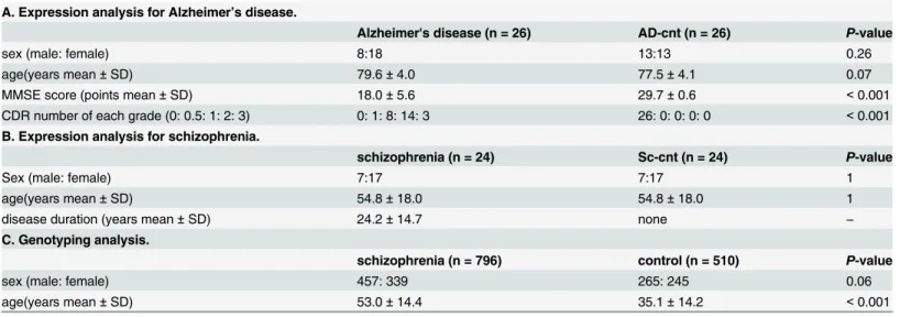
Table 1. Demographic data and clinical characteristics of each group.
A. Expression analysis for Alzheimer’s disease.
Alzheimer's disease (n = 26) AD-cnt (n = 26) P-value
sex (male: female) 8:18 13:13 0.26
age(years mean±SD) 79.6±4.0 77.5±4.1 0.07
MMSE score (points mean±SD) 18.0±5.6 29.7±0.6 <0.001
CDR number of each grade (0: 0.5: 1: 2: 3) 0: 1: 8: 14: 3 26: 0: 0: 0: 0 <0.001
B. Expression analysis for schizophrenia.
schizophrenia (n = 24) Sc-cnt (n = 24) P-value
Sex (male: female) 7:17 7:17 1
age(years mean±SD) 54.8±18.0 54.8±18.0 1
disease duration (years mean±SD) 24.2±14.7 none ―
C. Genotyping analysis.
schizophrenia (n = 796) control (n = 510) P-value
sex (male: female) 457: 339 265: 245 0.06
age(years mean±SD) 53.0±14.4 35.1±14.2 <0.001
AD-cnt, the controls against Alzheimer’s disease in expression analysis; MMSE, Mini Mental State Examination; CDR, Clinical Dementia Rating. The score of 0–3 shows classification of dementia (0 = none, 0.5 = questionable, 1 = mild, 2 = moderate, 3 = severe); Sc-cnt, the controls against schizophrenia in expression analysis. The p-value was calculated by student T test, Chi-squired test, and Fisher’s exact test.
Dementia Rating (CDR) [13]. The AD control group (AD-cnt) included 22 age-matched elderly participants with normal cognitive function (13 males and 13 females, mean
age ± SD = 77.5 ± 4.1 years) who agreed to participate in this study. For inclusion, subjects had to be capable of living independently, have MMSE scores over 28, and be free of cognitive impairment or morphological brain abnormality.
Participants in schizophrenia analysis. We recruited 24 patients with schizophrenia (7 males and 17 females, mean age ± SD = 54.8 ± 18.0 years, disease duration at blood draw = 24.2 ± 14.7 years) as well as 24 age-matched controls (Sc-cnt; 7 males and 17 females, mean age ± SD = 54.8 ± 18.0 years) from Ehime University Hospital and related community hospitals. In addition, we enrolled 796 patients with schizophrenia (457 males, 339 females, age = 53.0 ± 3.4 years, 34 patients did not indicate age) who visited Ehime or Tokushima Uni-versity Hospitals for a gene association study. Schizophrenia was diagnosed according to the Diagnostic and Statistical Manual of Mental Disorders IV criteria by at least 2 certified psychia-trists on the basis of clinical interviews and review of medical records. The comparison group included 510 healthy volunteers (265 males, 245 females, mean age ± SD = 35.1 ± 14.2 years) without psychiatric signs, psychiatric family history, or past history of mental disorders.
NHD participant. A 42-year-old male NHD patient homozygous for aTYROBPmutation (TYROBPc.141delG;manuscript in preparation) was included as a reference. We obtained his RNA twice, at intervals of 30 months, and confirmed that both samples expressed equivalent levels ofTYROBP. We also analyzed his father and mother (aged 72 and 68 years, respectively), both of whom were heterozygous for theTYROBPc.141delG mutation.
Ethical issues
All procedures followed were in accordance with the ethical standards of the responsible com-mittee on human experimentation (institutional and national) and with the Helsinki Declara-tion of 1964 and later revision. This study was approved by the instituDeclara-tional ethics committees of The Ethics Review Committee for Human Genome/Gene Analysis Research in Ehime Uni-versity Graduate School of Medicine and the UniUni-versity of Tokushima Graduate School as “Genetic Studies on Neuropsychiatric Diseases”(registry number; 25-K4), and written
informed consent was confirmed by all participants or their guardians before the acquisition of blood samples.
Blood sample collection
Whole peripheral blood samples were collected for the extraction of total RNA and genomic DNA, according to the standard protocol, in PaxGene Blood RNA Systems tubes (BD, Tokyo, Japan) and potassium EDTA tubes, respectively. Absorption spectrophotometry, using Nano-Drop-1000 (Thermo Fisher scientific, Yokohama, Japan), was used to determine RNA concen-trations and purity (260/280 ratio above 1.8 was used). RNA (1μg per sample) was reverse-transcribed using the High-Capacity cDNA Reverse Transcription Kit (Applied Biosystems, CA, USA), in a total reaction volume of 40μL. Genomic DNA was isolated from whole blood leukocytes using the QIAamp DNA Blood Mini Kit (Qiagen, Tokyo, Japan), according to the manufacturer’s protocol.
Expression analyses
For a quantitative estimate ofTYROBPandTREM2mRNA levels, the StepOnePlus Real-Time PCR System (Applied Biosystems) was used. Specific TaqMan probes were employed (Assay ID:TYROBP; Hs00182426_m1,TREM2; Hs00219132_m1), and the relative expression level of
GAPDH[14–16], serving as an internal standard (all Taqman probes from Applied Biosys-tems). The final volume reaction was 10μL using the TaqMan Universal Master Mix (Applied Biosystems). Relative mRNA levels were calculated via the 2−ΔΔCTmethod [17] using StepOne software (Applied Biosystems). We averaged the fold changes from three wells for each sample (triplicate) and used this average value for statistical analyses. The NHD patient was used for calibration in all experiments to correct the observational error.
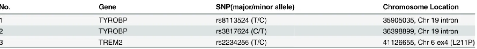
SNP analysis
Three SNPs, rs8113524 (NG_009304.1; assay ID: C___2604899_10), rs3817624 (NG_ 009304.1; assay ID: C__25603557_20), and rs2234256 (NG_011561.1; assay ID: C__ 15948232_10) (Table 2) were selected using HaploView v4.2 (Cambridge, MA) as tag SNPs, with squared correlation coefficient (r2) between 2 SNPs>0.8 and minor allele frequency (MAF)>0.01, on the basis of the current International HapMap project database (http:// hapmap.ncbi.nlm.nih.gov/index.html.en). These were analyzed for association study with schizophrenia. Additional SNPs, rs429358 (NG_007084.2; assay ID: C___3084793_20) and rs7412 (NG_007084.2; assay ID: C____904973_10) were used to determine apolipoprotein E (APOE) isoforms using a real-time SNP genotyping system (TaqMan Assays, Applied Biosys-tems). Following this, 1× TaqMan PCR Master Mix, 1× TaqMan SNP genotyping assay, 10 ng genomic DNA, and ultrapure water to a final reaction volume of 6μL were mixed in each well of an optical plate. Allelic discrimination was performed using StepOnePlus and analyzed using its software.
Statistics
Statistical analyses were performed using SPSS v22 (IBM, Tokyo, Japan). Expression levels of
TYROBPandTREM2were compared using the Mann–Whitney U-test and Student’st-test. Correlations of gene expression with age, disease duration, and MMSE score were analyzed using the Spearman test. Fisher’s exact test with simulatedP-values was used to compare
TREM2genotype distributions between patients with schizophrenia and controls. Linkage dis-equilibrium (LD),TYROBPhaplotypes, genotype distributions, minor allele frequencies, and Hardy–Weinberg equilibrium (HWE) were determined using HaploView v4.2. Statistical power was calculated with GPower 3 (http://www.gpower.hhu.de/), and statistical significance was defined at the 95% confidence level (P= 0.05). The ratio of successful genotyping was over 99%, and we extrapolated the missing values for genotype analysis. Bonferroni corrections were applied to maintain an overall type I error rate of 0.05, taking multiple comparisons into account.
Results
TYROBP
and
TREM2
expression in NHD
The NHD patient homozygous for theTYROBPc.141delG mutation exhibited the lowest level ofTYROBPexpression among the study participants, while his parents, heterozygous for the
Table 2. Characteristics of the selected two tagging SNPs inTYROBPand one tagging SNP inTREM2.
No. Gene SNP(major/minor allele) Chromosome Location
1 TYROBP rs8113524 (T/C) 35905035, Chr 19 intron
2 TYROBP rs3817624 (C/T) 36398899, Chr 19 intron
3 TREM2 rs2234256 (T/C) 41126655, Chr 6 ex4 (L211P)
mutation, showed no difference from other groups. In contrast,TREM2was expressed in the NHD patient and his parents at the same levels as that in control subjects.
TYROBP
and
TREM2
expression in AD and schizophrenia
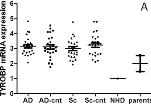
Fig 1showsTYROBPandTREM2expression in leukocytes of patients with AD or schizophre-nia and their controls.TYROBPexpression in patients with AD and schizophrenia was similar to that in their respective control groups (P= 0.44 andP= 0.13, respectively). In contrast,
TREM2expression was significantly higher in patients with AD and schizophrenia compared to that in their respective controls (P<0.001 in both cases). This was also confirmed after Bon-ferroni corrections. There was no correlation between TREM2 expression and other clinical variables such as age (r = 0.08,P= 0.71), MMSE (r = 0.01,P= 0.95) in AD, and disease dura-tion (r = 0.28,P= 0.19) in schizophrenia.
TREM2
expression in AD with
APOE
ε
4
AD patients were typed for theAPOEε4 allele (Table 3). There were no differences in age, sex, MMSE score, or CDR between the 14 participants (all wereε3/ε4) in theAPOEε4-positive AD group [ε4(+) AD] and the 12 participants (all wereε3/ε3) in theAPOEε4-negative AD group [ε4(−) AD]. In the AD control group (n= 26), only 2 subjects were positive for the
APOEε4 allele. For these 3 groups,TYROBPandTREM2mRNA expression levels in leuko-cytes are shown inFig 2. Inε4(+) AD,TREM2expression was significantly higher than that in AD-cnt (P<0.001). Although there were no significant differences betweenε4(+) andε4(−) AD (P= 0.07) or betweenε4(−) AD and AD-cnt (P= 0.07), the relative expression of both genes tended to decrease across groups, in the orderε4(+) AD,ε4(−)AD, and AD-cnt.
Case
–
control association studies in schizophrenia
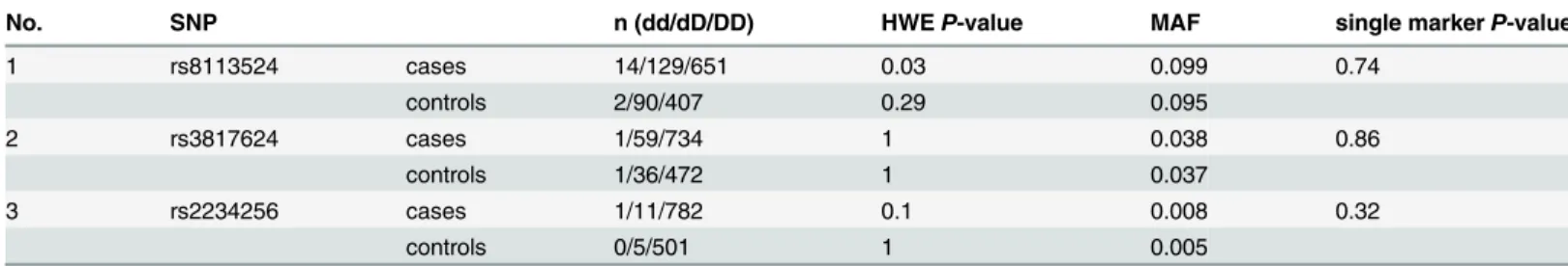
We performed genotyping of 3 tag SNPs (rs8113524 and rs3817624 inTYROBPand rs2234256 inTREM2) in 796 patients with schizophrenia and 510 controls (unrelated Japanese partici-pants). The results are shown inTable 4. The result of haplotype analysis betweenTYROBP
and schizophrenia is shown inTable 5. In schizophrenia, rs8113524 was not in HWE; however, rs3817624 and rs2234256 were in HWE. No associations between eitherTYROBPorTREM2
and schizophrenia were revealed by single marker or haplotype analyses.
Discussion
In NHD, the TREM2/TYROBP signaling pathway is damaged by the mutations in either TYR-OBPorTREM2, and this has been suggested to cause defects in microglial activity [18]. It is reported thatTYROBPexpression is normal in the brain of NHD individuals with theTREM2
mutation [2] and thatTREM2mRNA extracted from leukocytes is severely reduced in NHD individuals with this mutation [19]. Our results, showing decreased levels ofTYROBPmRNA and normal levels ofTREM2mRNA in leukocytes of an NHD patient with theTYROBP muta-tion, are consistent with previous reports.
In the present study, we found thatTREM2was more highly expressed in leukocytes of patients with AD and schizophrenia, which were of different disease and age groups, whereas
Fig 1. Expression of TYOBP (1A) and TREM2 (1B) in each group.TYROBP expression was the lowest in the NHD patient and showed no difference in other groups.TREM2 expression was higher in schizophrenic and AD patients than in the respective controls.P<0.01 is indicated by**when thePvalues were calculated by Mann–Whitney U-test.
TREM2is highly expressed in microglia of the AD brain [10], andTrem2expression is particu-larly high in the amyloid plaques of AD model mice [22]. HigherTREM2mRNA levels in
APOEε4-positive individuals may suggests thatTREM2expression in leukocytes reflects the AD pathology. Although, unexpectedly, we found no differences between theε4(+) AD andε4 (−) AD groups or between theε4(−) AD and AD-cnt groups, it is possible that this is a
false-negative result and that differences in expression may be confirmed by increasing the number of subjects.
Hu et al. [16] reported thatTREM2mRNA and protein levels were significantly higher in monocytes of AD and that the expression was inversely associated with the MMSE score.
TREM2mRNA was significantly increased in leukocytes of AD in the present study, and we found the tendency to inverse correlation betweenTREM2mRNA expression in leukocytes and the MMSE score in APOEε4-negative AD patients (P= 0.08, r =−0.53). We suggest that elevatedTREM2expression in leukocytes can be utilized as a novel biological marker for AD and may associate with severity in APOEε4-negative AD patients.
Surprisingly,TREM2mRNA expression was also elevated in patients with schizophrenia. To date, this is the first report showingTREM2upregulation in schizophrenia. Although schizophrenia has been thought of as a non-neurodegenerative disorder, the evidence from sev-eral recent reports suggests that schizophrenia may actually be neurodegenerative [23]. Com-pared to brain tissues,TREM2is highly expressed in microglia [24] and reduced microglial activity is reported in Trem2-deficient mice [25]. Although we did not examine inflammatory markers such as C-reactive protein and IL-6, it is well known that C-reactive protein [26,27] and IL-6 [28] are elevated in schizophrenic patients who are free of overt inflammation. Inter-estingly, increased microglial activity in schizophrenia was also confirmed by both electron microscopy [29] and positron emission tomography [30]. HigherTREM2mRNA levels in leu-kocytes of schizophrenia may be associated with peripheral inflammation or microglial involvement. Because inflammation in glial cells may lead to neuronal changes in schizophre-nia [31],TREM2expression in brain samples of patients with schizophrenia should be studied in the future.
TYROBPexpression was not changed in either AD or schizophrenia. TREM2 in extracellu-lar regions acts as a receptor to bind the ligand [32], whereas TYROBP is present as a second transmembrane receptor and transmits signal [33]. The difference in the role of each domain in TYROBP/TREM2 signaling may explain the different expression patterns between TYROBP and TREM2 in leukocytes. Further studies of the TREM2/TYROBP signaling should be per-formed to elucidate its role in neurological and psychiatric diseases.
We could not find significant association between TYROBP and TREM2 gene polymor-phisms and schizophrenia. In addition, the allele rs8113524 was not present in HWE in schizophrenic patients; this may be due to small population size and low allele frequency [34]. Recently, an association study of 2190 Japanese patients with late-onset AD revealed that
Table 3. Characteristics of APOEε4 positive and negative group in AD.
ε4-positive (n = 14) ε4-negative (n = 12) P-value
sex (male: female) 5:09 3:09 0.68
age(years mean±SD) 80.1±4.3 78.9±3.7 0.45
MMSE score (points mean±SD) 17.7±7.1 18.2±3.6 0.84
CDR number of each grade (0: 0.5: 1: 2: 3) 0: 0: 5: 7: 2 0: 1: 3: 7: 1 0.64
AD, Alzheimer’s Disease; MMSE, Mini Mental State Examination; CDR, Clinical Dementia Rating. The score of 0–3 shows classification of dementia (0 = none, 0.5 = questionable, 1 = mild, 2 = moderate, 3 = severe). TheP-value was calculated by Studentttest, Chi-squired test, and Fisher’s exact test.
Fig 2. Expression of TYOBP (2A) and TREM2 (2B) in APOEε4-positive AD [ε4(+) AD], APOE ε4-negative AD [ε4(
−) AD], and AD-cnt groups.TYROBP expression showed no difference. TREM2
expression was significantly higher inε4-positive AD group than in AD-cnt (including 2ε4-positive subjects)
group. The expression levels tended to decline in the following order:ε4(+) AD,ε4(
−) AD, and AD-cnt. P<0.001 after Bonferroni correction is indicated by* *when thePvalues were calculated by Mann–Whitney
U-test.
TREM2variants, including R47H and L211P, were not associated with the disease [35]. There-fore, higher level ofTREM2mRNA in AD and schizophrenia in this study may not be caused by gene polymorphisms but by other factors such as epigenetic modification.
Our study has several limitations that should be considered. All patients with schizophrenia were taking antipsychotics and all patients with AD were taking cholinesterase inhibitors at the time of the blood sampling. Although our NHD patient was also taking antipsychotics and his
TREM2mRNA levels were similar to those of controls, we could not exclude the possibility that these pharmacological treatments affected the expression of both genes. Influences of food or physical condition (e.g., body mass index, smoking, infection, and inflammation) on gene expression in leukocytes were not examined. Moreover, it remains to be confirmed whether
TREM2expression in the brain is in good agreement with that in leukocytes. In addition, future studies should assess the correlation between the severity of inflammation or its markers in central spinal fluid (such as neopterin) and expression of the 2 genes.
In conclusion, we report for the first time thatTREM2expression in leukocytes is elevated not only in AD but also in schizophrenia and may be a clinical biomarker for these diseases. We believe that our study is an important first step in understanding the role ofTREM2in the pathophysiology of AD and schizophrenia.
Acknowledgments
We thank Miss Takako Muneta for her technical assistance and Dr. Win Thiri Kyaw for her proofreading and encouragement. This work was partially supported by a Health and Labor Science Research Grant from the Japanese Ministry of Health, Labour and Welfare; a Grant-in-Aid for Scientific Research from the Japanese Ministry of Education, Culture, Sports, Science and Technology; and the Ehime Graduate University of Medicine’s Good Practice Fund.
Table 4. Genotyping results of the selected two tagging SNPs inTYROBPand one tagging SNP inTREM2.
No. SNP n (dd/dD/DD) HWEP-value MAF single markerP-value
1 rs8113524 cases 14/129/651 0.03 0.099 0.74
controls 2/90/407 0.29 0.095
2 rs3817624 cases 1/59/734 1 0.038 0.86
controls 1/36/472 1 0.037
3 rs2234256 cases 1/11/782 0.1 0.008 0.32
controls 0/5/501 1 0.005
d, minor allele; HWE, Hardy‐Weinberg equilibrium; MAF, Minor Allele Frequency
The single marker p-value is calculated by Chi-squared test with omission the missing values.
doi:10.1371/journal.pone.0136835.t004
Table 5. Result of association study in the TYROBP gene.
Haplotype Frequencies Ratio Counts Frequencies P-Value
case control case control
TC 0.87 1370.0: 218.0 890.7: 135.3 0.863 0.868 0.69
CC 0.1 157.0: 1431.0 97.3: 928.7 0.099 0.095 0.74
TT 0.04 61.0: 1527.0 38.0: 988.0 0.038 0.037 0.86
P-value is calculated by Chi-squared test with omission the missing values.
Author Contributions
Conceived and designed the experiments: YM SU. Performed the experiments: YM YY KY TK. Analyzed the data: YM. Contributed reagents/materials/analysis tools: YM SO KK KY MA YO TY SN TM NK TO. Wrote the paper: YM JI SU.
References
1. Hakola HP. Neuropsychiatric and genetic aspects of a new hereditary disease characterized by pro-gressive dementia and lipomembranous polycystic osteodysplasia. Acta Psychiatr Scand Suppl 1972; 232: 1–173. PMID:4509294
2. Paloneva J, Manninen T, Christman G, Hovanes K, Mandelin J, Adolfsson R, et al. Mutations in two genes encoding different subunits of a receptor signaling complex result in an identical disease pheno-type. Am J Hum Genet 2002; 71: 656–662. PMID:12080485
3. Paradowska-Gorycka A, Jurkowska M. Structure, expression pattern and biological activity of molecu-lar complex TREM-2/DAP12. Human Immunology 2013; 74: 730–737. doi:10.1016/j.humimm.2013. 02.003PMID:23459077
4. Colonna M. DAP12 signaling: from immune cells to bone modeling and brain myelination. Journal of Clinical Investigation 2003; 111: 313–314. PMID:12569153
5. Kaifu T, Nakahara J, Inui M, Mishima K, Momiyama T, Kaji M, et al. Osteopetrosis and thalamic hypo-myelinosis with synaptic degeneration in DAP12-deficient mice. Journal of Clinical Investigation 2003; 111: 323–332. PMID:12569157
6. Guerreiro R, Wojtas A, Bras J, Carrasquillo M, Rogaeva E, Majounie E, et al. TREM2 variants in Alzhei-mer’s disease. The New England Journal of Medicine 2013; 368: 117–127. doi:10.1056/
NEJMoa1211851PMID:23150934
7. Rayaprolu S, Mullen B, Baker M, Lynch T, Finger E, Seeley W, et al. TREM2 in neurodegeneration: evi-dence for association of the p.R47H variant with frontotemporal dementia and Parkinson’s disease. Molecular Neurodegeneration 2013; 8: 19. doi:10.1186/1750-1326-8-19PMID:23800361 8. Giraldo M, Lopera F, Siniard AL, Corneveaux JJ, Schrauwen I, Carvajal J, et al. Variants in triggering
receptor expressed on myeloid cells 2 are associated with both behavioral variant frontotemporal lobar degeneration and Alzheimer’s disease. Neurobiol Aging 2013; 34: 2077.e11–18.
9. Cady J, Koval ED, Benitez BA, Zaidman C, Jockel-Balsarotti J, Allred P, et al. TREM2 variant p.R47H as a risk factor for sporadic amyotrophic lateral sclerosis. JAMA Neurol 2014; 71: 449–453. doi:10. 1001/jamaneurol.2013.6237PMID:24535663
10. Lue L, Schmitz C, Sorrano G, Sue L, Beach T, Walker D. TREM2 protein expression changes correlate with Alzheimer’s disease neurodegenerative pathologies in postmortem temporal cortices. Brain Pathol. 2015; 25(4): 469–480 doi:10.1111/bpa.12190PMID:25186950
11. McKhann G, Knopman D, Chertkow H, Hyman B, Jack C, Kawas C, et al. The diagnosis of dementia due to Alzheimer’s disease: recommendations from the National Institute on Aging-Alzheimer's Associ-ation workgroups on diagnostic guidelines for Alzheimer's disease. Alzheimer’s & Dementia: The Jour-nal of the Alzheimer's Association 2011; 7: 263–269.
12. Folstein MF, Folstein SE, McHugh PR.“Mini-mental state”: A practical method for grading the cognitive state of patients for the clinician. J Psychiatr Res 1975; 12: 189–198. PMID:1202204
13. Morris JC. The Clinical Dementia Rating (CDR): current version and scoring rules. Neurology 1993; 43: 2412–2414.
14. Carrol ED, Salway F, Pepper SD, Saunders E, Mankhambo LA, Ollier WE, et al. Successful down-stream application of the Paxgene Blood RNA system from small blood samples in paediatric patients for quantitative PCR analysis. BMC Immunol. 2007; 12: 8:20. PMID:17850649
15. Watanabe S, Iga J, Ishii K, Numata S, Shimodera S, Fujita H, et al. Biological tests for major depressive disorder that involve leukocyte gene expression assays. J Psychiatr Res. 2015; 66–67: 1–6. doi:10. 1016/j.jpsychires.2015.03.004PMID:25943949
16. Hu N, Tan MS, Yu JT, Sun L, Tan L, Wang YL, et al. Increased expression of TREM2 in peripheral blood of Alzheimer’s disease patients. J Alzheimers Dis 2014; 38: 497–501. doi:10.3233/JAD-130854 PMID:24002183
17. Livak KJ, Schmittgen TD. Analysis of Relative Gene Expression Data Using Real-Time Quantitative PCR and the 2(-Delta Delta C(T)) Method. Methods 2001; 25: 402–408. PMID:11846609
19. Chouery E, Delague V, Bergougnoux A, Koussa S, Serre JL, Mégarbané A. Mutations in TREM2 lead to pure early-onset dementia without bone cysts. Human Mutation 2008; 29: E194–204. doi:10.1002/ humu.20836PMID:18546367
20. Neumann H, Takahashi K. Essential role of the microglial triggering receptor expressed on myeloid cells-2 (TREM2) for central nervous tissue immune homeostasis. J Neuroimmunol 2007; 184: 92–99. PMID:17239445
21. Kiialainen A, Hovanes K, Paloneva J, Kopra O, Peltonen L. Dap12 and Trem2, molecules involved in innate immunity and neurodegeneration, are co-expressed in the CNS. Neurobiology of Disease 2005; 18: 314–322. PMID:15686960
22. Frank S, Burbach G, Bonin M, Walter M, Streit W, Bechmann I, et al. TREM2 is upregulated in amyloid plaque‐associated microglia in aged APP23 transgenic mice. Glia 2008; 56: 1438–1447. doi:10.1002/ glia.20710PMID:18551625
23. Monji A, Kato T, Mizoguchi Y, Horikawa H, Seki Y, Kasai M, et al. Neuroinflammation in schizophrenia especially focused on the role of microglia. Progress in Neuro-Psychopharmacology & Biological Psy-chiatry 2013; 42: 115–121.
24. Hickman S, Khoury J. TREM2 and the neuroimmunology of Alzheimer’s disease. Biochem Pharmacol. 2014; 88(4): 495–498. doi:10.1016/j.bcp.2013.11.021PMID:24355566
25. Cantoni C, Bollman B, Licastro D, Xie M, Mikesell R, Schmidt R, et al. TREM2 regulates microglial cell activation in response to demyelination in vivo. Acta Neuropathol 2015; 129: 429–447. doi:10.1007/ s00401-015-1388-1PMID:25631124
26. Avramopoulos D, Pearce BD, McGrath J, Wolyniec P, Wang R, Eckart N, et al. Infection and inflamma-tion in schizophrenia and bipolar disorder: a genome wide study for interacinflamma-tions with genetic variainflamma-tion. PloS one. 2015; doi:10.1371/journal.pone.0116696PMID:25781172
27. Dickerson F, Stallings C, Origoni A, Vaughan C, Khushalani S, Yang S, et al. C-reactive protein is ele-vated in schizophrenia. Schizophr Res. 2013; 143(1): 198–202. doi:10.1016/j.schres.2012.10.041 PMID:23218564
28. Al-Hakeim HK, Al-Rammahi AD, Al-Dujaili AH. IL-6, IL-18, sIL-2R, and TNFαproinflammatory markers
in depression and schizophrenia patients who are free of overt inflammation. J Affect Disord. 2015; doi: 10.1016/j.jad.2015.04.044PMID:25985379
29. Uranova NA, Vikhreva OV, Rachmanova VI, Orlovskaya DD. Ultrastructural alterations of myelinated fibers and oligodendrocytes in the prefrontal cortex in schizophrenia: a postmortem morphometric study. Schizophr Res Treatment 2011; 2011: 325789. doi:10.1155/2011/325789PMID:22937264 30. Doorduin J, Vries E, Willemsen A, Groot J, Dierckx R, Klein H. Neuroinflammation in schizophrenia-related psychosis: a PET study. Journal of Nuclear Medicine: Official Publication, Society of Nuclear Medicine 2009; 50: 1801–1807.
31. Takahashi N, Sakurai T. Roles of glial cells in schizophrenia: possible targets for therapeutic approaches. Neurobiology of Disease 2013; 53: 49–60. doi:10.1016/j.nbd.2012.11.001PMID: 23146995
32. Daws M, Lanier L, Seaman W, Ryan J. Cloning and characterization of a novel mouse myeloid DAP12‐ associated receptor family. Eur J Immunol 2001; 31: 783–791. PMID:11241283
33. Lanier LL, Corliss BC, Wu J, Leong C, Phillips JH. Immunoreceptor DAP12 bearing a tyrosine-based activation motif is involved in activating NK cells. Nature 1998; 391: 703–707. PMID:9490415 34. Masel J. Genetic drift. Curr Biol. 2011; 21(20): R837–838. doi:10.1016/j.cub.2011.08.007PMID:
22032182

![Fig 2. Expression of TYOBP (2A) and TREM2 (2B) in APOE ε 4-positive AD [ ε 4(+) AD], APOE ε 4-negative AD [ ε 4( − ) AD], and AD-cnt groups](https://thumb-eu.123doks.com/thumbv2/123dok_br/18362964.354387/8.918.299.853.107.917/expression-tyobp-trem-apoe-positive-apoe-negative-groups.webp)