Mechanisms of TSC-mediated Control of Synapse
Assembly and Axon Guidance
Sarah Knox1., Hong Ge1., Brian D. Dimitroff1,2
, Yi Ren1,2, Katie A. Howe1,2, Andrew M. Arsham2, Mathew C. Easterday1,2, Thomas P. Neufeld2, Michael B. O’Connor2, Scott B. Selleck1,2
*
1The Developmental Biology Center, Department of Pediatrics, The University of Minnesota, Minneapolis, Minnesota, United States of America,2The Developmental Biology Center, Department of Genetics, Cell Biology and Development, The University of Minnesota, Minneapolis, Minnesota, United States of America
Tuberous sclerosis complex is a dominant genetic disorder produced by mutations in either of two tumor suppressor genes,
TSC1 and TSC2; it is characterized by hamartomatous tumors, and is associated with severe neurological and behavioral disturbances. Mutations inTSC1orTSC2deregulate a conserved growth control pathway that includes Ras homolog enriched in brain (Rheb) and Target of Rapamycin (TOR). To understand the function of this pathway in neural development, we have examined the contributions of multiple components of this pathway in both neuromuscular junction assembly and photoreceptor axon guidance in Drosophila. Expression of Rheb in the motoneuron, but not the muscle of the larval neuromuscular junction produced synaptic overgrowth and enhanced synaptic function, while reductions in Rhebfunction compromised synapse development. Synapse growth produced by Rheb is insensitive to rapamycin, an inhibitor of Tor complex 1, and requireswishful thinking,a bone morphogenetic protein receptor critical for functional synapse expansion. In the visual system, loss ofTsc1in the developing retina disrupted axon guidance independently of cellular growth. Inhibiting Tor complex 1 with rapamycin or eliminating the Tor complex 1 effector, S6 kinase (S6k), did not rescue axon guidance abnormalities ofTsc1mosaics, while reductions in Tor function suppressed those phenotypes. These findings show that Tsc-mediated control of axon guidance and synapse assembly occurs via growth-independent signaling mechanisms, and suggest that Tor complex 2, a regulator of actin organization, is critical in these aspects of neuronal development.
Citation: Knox S, Ge H, Dimitroff BD, Ren Y, Howe KA, et al (2007) Mechanisms of TSC-mediated Control of Synapse Assembly and Axon Guidance. PLoS ONE 2(4): e375. doi:10.1371/journal.pone.0000375
INTRODUCTION
Mutations inTSC1orTSC2result in tuberous sclerosis, a human
syndrome characterized by formation of benign tumors, or hamar-tomas, and a range of neurological and behavioral anomalies, including epilepsy and autism. While neurological dysfunction in patients with tuberous sclerosis is clearly linked to structural brain abnormalities in the central nervous system [1], recent work has provided evidence that TSC1/2 may affect neural development by altering neuronal morphology and function. Loss of TSC function produces changes in dendritic architecture of hippocampal neurons and altered synaptic properties [2]. Rats heterozygous
forTSC2 mutations show disruption of hippocampal physiology,
including long term potentiation, a measure of synaptic plasticity
[3]. Mutations in theDrosophilaortholog ofTSC2, gigas,have also
been shown to produce ectopic axon terminations in addition to the normal projections of sensory neurons [4,5]. It is unclear to what degree neurological deficits associated with tuberous sclerosis complex result from disruptions of cytoarchitecture in specific brain regions or alterations in synaptic function directly.
TSC1andTSC2encoded proteins form a complex that regulates
a small GTP-binding protein, Ras homolog enriched in brain (Rheb), promoting its endogenous GTPase activity and thereby limiting Rheb signaling. Rheb in turn controls the activity of Target of Rapamycin (TOR), a serine-threonine kinase. The TSC-Rheb-TOR pathway is a critical determinant of growth during development, regulating a number of cellular functions including translation, mRNA turnover, protein stability, and actin organization [6]. It is responsive to growth factors, such as insulin and insulin-like growth factors (ILGFs), and also serves as a nutrient sensor, thus integrating numerous signals related to cell and tissue growth. TOR plays a pivotal role in this signaling pathway, receiving regulatory inputs from Rheb and affecting downstream targets via two distinct molecular complexes. Tor
complex 1 (TORC1) includes Raptor and mLST8, and regulates translation via phosphorylation of S6 kinase (S6K) and 4E-binding protein (4EBP). Tor complex 2 (TORC2) includes Rictor in addition to Tor and mLST8; in both yeast and mammalian cells TORC2 influences the actin cytoskeleton. Tor complex 1, but not Tor complex 2, is inhibited by the anti-proliferative and immuno-suppressant compound rapamycin, emphasizing that TORC1 and 2 are pharmacologically separable entities. The distinct molecular outputs of TORC1 and 2 have also suggested that TORC2 may be the primary regulator of cell polarity and morphology. It is not known which functions of TSC-Rheb-TOR in the nervous system are mediated by either or both of the two Tor kinase-containing complexes, and if pharmacological intervention in tuberous sclerosis complex patients should best be directed at TORC1, with agents such as rapamycin, or if TORC2-specific agents will also be important.
Academic Editor:Hugo J. Bellen, Baylor College of Medicine, United States of America
ReceivedNovember 29, 2006;AcceptedMarch 19, 2007;PublishedApril 18, 2007
Copyright:ß2007 Knox et al. This is an open-access article distributed under the terms of the Creative Commons Attribution License, which permits unrestricted use, distribution, and reproduction in any medium, provided the original author and source are credited.
Funding:This work was supported by NIH contract grant number GM54832-09 to SBS, the Martin Lenz Harrison Endowment to SBS, and NIH grant RO1 GMO62509 to TN. MBO is an investigator with the Howard Hughes Medical Institute.
Competing Interests:The authors have declared that no competing interests exist.
* To whom correspondence should be addressed.E-mail: [email protected]
The fruit flyDrosophilahas proven to be an important system for understanding the molecular mechanisms of Tsc-Rheb-Tor signaling during development [7]. As in vertebrates, this signaling cascade is a critical regulator of growth. All of the principal
elements of this pathway are represented in Drosophila, including
molecular components upstream of Tsc, such as phosphatidyino-sitol-3 kinase (Pi3K), Akt, Pten and the insulin receptor ortholog, InR. Likewise, molecules that convey the signal downstream of Tsc, including Rheb, Tor, and S6k serve critical roles in the fruit fly. Mutations affecting all these genes have been identified in Drosophila, as well as transgenes that can convey gain-of-function effects. We have used these molecular and genetic tools to explore the function of Tsc-Rheb-Tor signaling in two fundamental processes essential to nervous system development, synapse formation and axon guidance.
TheDrosophilaneuromuscular junction has served as a powerful model for identifying the molecular components required for assembly and plasticity of a defined synapse [8]. This glutamater-gic synapse must respond to greater than a 100-fold increase in the size of the muscle target from first to third instar larval stages. Physiological responses of this synapse are well-characterized using single-cell recording techniques, and morphological development with specific molecular markers has been extensively described. We have used this synapse to determine the role of gain or loss of Tsc-Rheb-Tor signaling on synapse assembly and function.
The visual system ofDrosophilais equally well described in both
molecular and genetic terms [9]. Photoreceptors show stereotyped projections to the brain, and genes required for photoreceptor axon projection and termination have been identified in numerous screens. Methods for making somatic cell mosaics have proven particularly powerful in determining what molecules are required in photoreceptors or in cells along their trajectory into the brain. Previous studies have shown that retinal clones mutant for the DrosophilaTsc2 orthologgigasgenerated enlarged ommatidia with increased numbers of synaptic contact sites in the optic lamina [10]. We have taken advantage of this system to examine Tsc-Rheb-Tor requirements for photoreceptor axon guidance and formation of functional synaptic contacts in the brain.
Our results establish that either gain or loss of signaling via the Tsc-Rheb-Tor pathway affects synapse development at the DrosophilaNMJ. Ectopic activation of the Tsc-Rheb-Tor signaling pathway produced profound synaptic overgrowth with commen-surate increases in synaptic function. We show that Rheb-mediated enhancement of synaptic function depends upon bone
morphoge-netic protein (BMP) signaling mediated by wishful thinking (wit),
a type II receptor. In the visual system, increased Tsc-Rheb-Tor signaling produced cell autonomous defects in photoreceptor axon guidance. Both genetic and pharmacological evidence suggest that TORC2 serves critical functions in both synapse development and
axon guidance inDrosophila. Axon guidance phenotypes produced
by null mutations inPtenandTsc1are distinct, demonstrating that
regulation of signaling by these two tumor suppressor genes are not functionally equivalent in the nervous system.
RESULTS
Activation of Tor signaling produces synaptic
growth and enhanced synaptic function
Tsc1/2 affect growth by inhibiting Rheb, a small GTP-binding protein that in turn governs Tor activity. Overexpression of Rheb
activates the pathway independent ofTscgene function [11–13].
We have used theGal4-UASsystem to overexpressRhebin either
the motoneuron or the muscle of theDrosophilathird instar larval
neuromuscular junction (NMJ), a well-characterized glutamatergic
synapse [8] that shows dynamic growth during larval
develop-ment. Ectopic expression ofRhebin the motoneuron of the third
instar larval NMJ using a pan-neuronal Gal4 line (elav-Gal4)
resulted in more than a doubling of synapse size, measured by the number of synaptic boutons/muscle area (Figure 1A–C, quantified
in D). Similar results were seen using a motoneuron-specific
OK6-Gal4line (data not shown). We found no evidence of motoneuron
axon misrouting at this level of Rheb activation; the motoneuron axon follows the normal trajectory and synapses at the correct
location on muscle 6 and 7(data not shown). Indeed,
elav-Gal4.UAS-Rheb animals are viable, indicating this degree of
pathway activation is considerably more mild than loss ofTsc1(see
below). Expression of Rheb selectively in muscle (G14-Gal4.
UAS-Rheb), while producing enlargement of muscle cells, did not
increase the proportional size of the synapse (bouton number/ muscle area, Figure 1D). Activation of Tor by overexpression of Pi3K in the motoneuron also produced an enlarged synapse, but to a lesser degree than overexpression of Rheb (Figure 1C, D).
Enlargement of the NMJ inDrosophilais not always associated
with an electrophysiologically competent synapse. For example, highwiremutants display large NMJs but markedly compromised synaptic function [14,15]. We therefore assessed the electrophys-iological behavior of the NMJ in animals overexpressing Rheb in the motoneuron. This synapse showed nearly a doubling of the quantal content, a measure of the number of synaptic vesicles released per motoneuron firing (Fig 1I). The amplitude of the excitatory junctional potential (EJP), the voltage change in the muscle elicited by a suprathreshold stimulation of the motoneuron,
also increased significantly compared to control synapses
(Figure 1E, F). Mini-excitatory junctional potentials (mEJPs) are depolarizations of the muscle that result from spontaneous neurotransmitter release and provide a measure of vesicular
fusion. While the mEJP frequencies ofRheboverexpressing animals
showed no significant change (Figure 1G), the mEJP amplitudes were lower than matched controls (Figure 1H). In all, activation of Tor signaling via overexpression of Rheb produced an expanded synapse that was fully functional.
Reduction of Tor signaling produces a small synapse
with compromised function
To determine if reduced Tsc-Rheb-Tor signaling compromises synapse growth and function we overexpressed Tsc1 and Tsc2 in
the motoneuron, or compromisedRhebactivity using a
combina-tion of hypomorphic Rheb alleles previously shown to cause
reductions in cell size and number as well as S6k activity [11].
Overexpression ofUAS-Tsc1/Tsc2has been shown to limit growth
mediated by Rheb [11–13], and we observed that Tsc1 and 2 overexpression in the motoneuron reduced synapse size compared to controls (Figure 2A, B, quantified in D). Consistent with this
finding,Rheb hypomorphic mutant larvae showed a significantly
reduced number of boutons per unit muscle area compared to heterozygous controls (Fig 2C, D). The NMJs of these animals also revealed significant changes in synaptic function. mEJP
frequen-cies inRhebmutant animals were half that of controls (Fig 2E, G),
and EJP amplitudes were significantly reduced (Figure 2F). We
also saw a reduction in the quantal content of Rheb mutants
(Figure 2I), while mEJP amplitude showed no significant change (Figure 2H). Thus, reducing Tor activity by either of two mechanisms, overexpression of Tsc1/2 or partial loss-of-function
mutations inRheb, compromised synapse morphological
develop-ment and function. Electrophysiology of hypomorphicTormutants
Rapamycin does not inhibit synapse growth
mediated by overexpression of Rheb
To evaluate if Rheb overexpression-mediated synapse expansion takes place via known growth regulatory pathways, we grew larvae
from hatching to the third instar larval stage on rapamycin-containing food. Rapamycin has been shown to block growth
mediated by TORC1 inDrosophila, and we used a concentration
that produced clear developmental delay [16]. Culturing larvae
bearingelav-Gal4andUAS-Rheb+
transgenes on rapamycin reduced Figure 1. Activation of the Tor pathway produces synaptic growth and enhanced physiological function.The morphology of the third instar larval NMJ was visualized with the presynaptic marker anti-cysteine string protein (CSP) and confocal microscopy. Images shown are stacks of 20 or more optical sections. Neuronal (elav-Gal4) expression of eitherRheb(B) orPi3K(C) increased the size of the synapse compared to control animals bearing theelav-Gal4transgene alone (A). Numbers of synaptic boutons/muscle area are quantified in D. Expression of eitherUAS-Rheb(n = 41) orUAS-Pi3K (n = 41) produced a significant increase in the number of boutons/muscle area compared to controls (n = 44), while expression ofUAS-Rhebin the muscle (driven byG14-Gal4, n = 18) produced a reduction. Neuron-specific expression ofRhebalso produced electrophysiological changes at the NMJ, determined by intracellular recordings from abdominal muscle 6 in third instar larvae. The amplitude of the EJP was significantly increased in animals expressingUAS-Rheb(n = 21) compared to controls withelav-Gal4alone (n = 12). Examples of EJP voltage traces are shown in E, and mean EJP values are quantified in F. Quantal content, a measure of the number of synaptic quanta released in a single firing of the motoneuron, was nearly doubled by neuron-directed expression ofRhebcompared to controls (I). Mini-EJP amplitude was decreased in these animals (H), while mEJP frequency showed no significant change (G). In this and all subsequent figures, *** denotes p-values less than 0.00005 using a student t-test comparison with controls, ** denotes p-values less than 0.005, and * denotes p-values less than 0.05. The scale bar is 50 microns in panel A. doi:10.1371/journal.pone.0000375.g001
overall growth, including muscle size, but did not suppress the synaptic enlargement measured either by the number of synaptic boutons/muscle area or the number of motoneuron branches (Figure 3A–C, quantified in D, E). These findings show that Rheb-mediated synaptic growth did not require TORC1 activity, implicating TORC2 and its regulation of the actin cytoskeleton as serving critical functions in synaptic growth control.
In-terestingly, culturing elav-Gal4 control larvae (without the
UAS-Rhebtransgene) on rapamycin produced both an increase in the
number of boutons per unit muscle area and an in increase in
motoneuron branching (Figure 3D, E). This raises the possibility that blocking the activity of TORC1 with rapamycin indirectly influences the activity of other Tor complexes.
animals bearing a null mutation in S6k. S6k mutants are small, with a reduced muscle surface area compared to controls. The synapse size, however, as measured by the number of boutons per unit muscle area, is not reduced (data not shown). These findings
contrast with the effects of Rheb mutations or Tsc1 and Tsc2
overexpression (Figure 2), but are consistent with the finding that rapamycin does not suppress synapse overgrowth (Figure 3). Together, these results suggest that TORC1 and S6k do not contribute significantly to the proportional growth of the NMJ.
Rheb-mediated changes in synapse function require
the BMP-signaling receptor encoded by
wishful
thinking
Growth factor-mediated signaling, including both Wingless (Wg) and
BMP pathways, is important for normal NMJ growth inDrosophila.
Animals bearing mutations inwishful thinking (wit), a gene encoding
a BMP type II receptor, show a very small NMJ with dramatically compromised synaptic function [17–19]. To determine the relation-ship between Rheb-regulated synaptic growth and BMP-mediated synapse assembly we tested the ability of Rheb overexpression to
rescue the synaptic growth defect of wit mutants. While Rheb+
expression in the motoneuron was able to restore the number of
synaptic boutons to wild-type levels inwitmutant larvae, the number
of boutons was significantly less than with Rheb overexpression alone (Figure 4A–E). Rheb overexpression modestly increased mini
EJP frequency ofwitmutants (Figure 4G), but showed no capacity to
rescue either quantal content or EJP amplitudes (Figure 4F–I),
thereforewitis clearly required for most Rheb-directed effects on
synapse function. While these results do not establish the nature of the communication between Tor-Tsc signaling and the BMP pathway, it does demonstrate that an intact BMP system is necessary for Rheb-directed changes in synapse function.
Tsc-Rheb-Tor signaling is critical for axon guidance
in the visual system
Another fundamental aspect of neural development is the correct specification of axon pathfinding and synapse formation with the
correct targets. The Drosophila visual system offers a powerful
experimental model for assessing the function of a signaling system in axon guidance. To evaluate the function of the Tsc-Rheb-Tor signaling pathway in axon guidance we generated genetic mosaic animals where mutant photoreceptor neurons project to a
pheno-typically wild-type brain. In the fruit fly Drosophila, each retinal
sensory unit, or ommatidium, is comprised of eight photorecep-tors, R1-8. In the third instar larval brain, R1-6 project to the first optic ganglion, the lamina, and terminate to form a discrete plexus where synapses will form later in development (Figure 5A). R7 and R8 project to a deeper level in the brain, the medulla, forming a discrete set of projections seen in both larval and pupal brains. In 40h pupae the R7 and R8 projections terminate in distinct layers Figure 3. Rapamycin does not blockRheb-mediated synapse growth.Panels A–C show anti-CSP staining of NMJ synaptic boutons, demonstrating that the TORC1 inhibitor rapamycin does not block synapse growth in control animals or in larvae with neuron-directed expression ofRheb( elav-Gal4.UAS-Rheb). Panels D and E provide quantification of bouton numbers/muscle area and numbers of motoneuron branches, respectively, for elav-gal4controls (n = 44), animals with neuronal expression ofRheb(n = 41), control animals treated with rapamycin (n = 26), andRhebexpressing animals treated with rapamycin (n = 29). The scale bar is 50 microns.
doi:10.1371/journal.pone.0000375.g003
in the medulla, producing a highly regular and stereotyped pattern
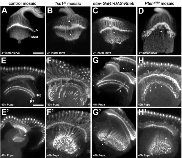
(Figure 5E). Loss of Tsc1 function in the retina produces an
enlarged eye disc with an increased number of photoreceptors
[20–22]. Axon projections fromTsc1mutant photoreceptors in the
brains of third instar larvae and pupae showed severe axon
guidance abnormalities (Figure 5B, F, F9, quantified in Table 1). In
third instar larvae, R1-6 termination at the lamina plexus is disorganized, producing an irregular termination zone (compare Figure 5A to 5B). R7/R8 terminations within the larval medulla are also abnormal (Figure 5B, arrowhead). At the 40 hr pupal
stage we observed gaps in the R7/R8 layers with large axon bundles, or fascicles, that projected past their appropriate
termination points (Figure 5F, F9).
To evaluate the degree of pathway activation mediated by
pan-neuronal expression ofUAS-Rheb, which we used above to evaluate
the role of Tor signaling at the NMJ, we examined photoreceptor
pathfinding in elav-Gal4.UAS-Rheb larvae and pupae. These
animals survive to adulthood and show disruptions in photore-ceptor projections, but to a significantly lesser extent than found in
Tsc1mosaic animals (see Table 1). Inelav-Gal4.UAS-Rhebpupal
Figure 4.Rheb-mediated synapse expansion and physiological function is BMP-signaling dependent.Anti-CSP staining of synaptic boutons (panels A–C) shows the effects ofwiton synapse growth (B), and the effects of neuron-directed expression ofRhebonwitmutant NMJs (C) compared to wild-type (A). Synapse size, measured by either the number of boutons/muscle area (D) or the number of motoneuron branches (E), is dramatically reduced inwitmutants (n = 20) compared to wild-type (n = 12), and is partially rescued by neuron-directed expression ofRheb(elav-Gal4.UAS-Rheb, n = 24). Reductions in EJP amplitudes (F), mini-EJP amplitudes (H), and quantal content (I) mediated by loss ofwit(n = 8) are not rescued by neuron-directed expression ofRheb(n = 16) (n = 10 for wild-type).The decrease in mini-EJP frequency ofwitmutants, a measure of spontaneous vesicle release, is rescued to a significant degree by expression of Rheb in the motoneuron (G). The scale bar represents 50 microns.
brains, abnormal bundles of axons that penetrate into deeper brain structures were found, but this phenotype was markedly less
severe than inTsc1mosaic animals (Figure 5G, G9, Table 1). Close
inspection of R7 and R8 endings in the medulla revealed individual photoreceptor axons growing past the correct termina-tion site (Figure 5G inset). R1-6 endings in the larval brain also
show irregularities, but the lamina plexus is less disrupted than in
Tsc1mosaics (Figure 5C). These findings indicate that the degree
of pathway activation achieved with elav-Gal4.UAS-Rheb is
markedly less than produced by loss of Tsc1. Moreover, these
results suggest that there is a continuum of axon pathfinding abnormalities with different levels of pathway activation. Figure 5. Photoreceptor axon projection defects associated with increased Tor signaling.(A–D) Dorsal-posterior views of third instar optic lobes stained with MAb24B10 to visualize photoreceptor projections. (A) Mitotic clones in anFRT82Bcontrol background show proper termination of photoreceptor axons R1-6 at the lamina plexus (LP), and termination of photoreceptors R7 and R8 in the medulla (Med). (B)Tsc129mutant axons terminate at incorrect positions above and below the lamina (arrowheads) and produce a broadened lamina plexus. (C) Neuronal expression of Rheb creates axon termination defects similar to those seen inTsc1mosaics (D)Ptendj189mutant photoreceptors leave gaps and holes (arrowhead) in the lamina plexus, which is broader and noticeably ‘‘peaked.’’ The medulla contains axon projections which are thicker and much longer than in controls (arrow). (E–H9) Dorsal view of optic lobes from 40h pupae stained with MAb24B10. E9–H9are lower optical planes of the optic lobes shown in E–H, respectively. (E, E9) Control photoreceptors R7 and R8 show two distinct layers of termination in the medulla (labels), and are arranged in a highly regular pattern (arrowhead). (F) Animals withTsc129mutant photoreceptors show severe disruption of the R7 and R8 termination layers. Instead of terminating at the correct positions, the axons fail to de-fasciculate, forming dense bundles (arrowheads) that project beyond the medulla. (G, G9) Neuron-directed expression of Rheb causes axon bundles to project beyond the medulla in a fashion similar toTsc1mosaics (arrowheads), but the phenotype is much less severe. (G, inset) Individual Rheb-overexpressing axons show an intermediate termination defect, stopping several microns beyond their normal targets (arrowheads in inset). (H)Ptendj189
mutant axons exhibit gaps and collapses in the R7/R8 termination zone (arrowhead). Thick axon bundles can be seen that bypass their usual stopping points and then loop back to terminate at other locations in the R7/R8 layers (arrows). (H9, F9) Axon bundles inPtendj189
mosaics are not as densely packed as those ofTsc129
mosaics (arrowheads), but are still disorganized. All scale bars are 50 microns.
doi:10.1371/journal.pone.0000375.g005
Pten, another negative regulator of cell growth and proliferation, encodes a phosphatase that converts the lipid signaling molecule
phosphatidylinositol 3,4,5 triphosphate (PIP3) to PIP2, an inactive
form, thus antagonizing PI3K activation of the TOR pathway. Like Tsc1, Pten retinal mosaics show eye overgrowth and precocious
differentiation [23–27]. To determine if disruptions ofPtenfunction
affect axon guidance, we generated mosaic animals. Ptenmutant
photoreceptor projections showed disorganization of axon termini in the third instar larval brain and were notable for a misshapen and concave lamina plexus with a large number of gaps (Figure 5D,
quantified in Table 1). At the pupal stage,Ptenmutant projections
showed significantly less severe defects than photoreceptors bearing
a Tsc1 null mutation (Figure 5H, H9 and Table 1), with fewer
projections failing to stop at the normal medulla termination sites. The penetrance of pathfinding, defasciculation, and termination
defects in 40h pupae was lower in Ptenthan inTsc1null mutant
photoreceptors projecting to a wild-type CNS (Table 1). In sum,Pten
andTsc1mutant photoreceptor projections show distinct patterns of
photoreceptor axon guidance defects, despite the fact that these two inhibitors of Tsc-Rheb-Tor signaling have similar influences on cell size, growth, and differentiation [20–22,24–26,28].
We also observed distinct effects ofTsc1andPtenretinal mosaics
on the differentiation of lamina neurons and visual system glia, detected with anti-Dachshund and anti-Repo antibodies,
re-spectively (Figure S1). Pten mutant retinal projections produced
an abnormally large lamina not seen inTsc1mosaics (Figure S1A–
C). In bothPtenandTsc1mosaics visual system glia were found in
the brain in roughly normal positions (Figure S1D–F), although
some disorganization was evident in brains receivingTsc1mutant
photoreceptor projections. It is possible that this disruption of glial architecture may partially contribute to the axon projection defects
observed inTsc1mutants.
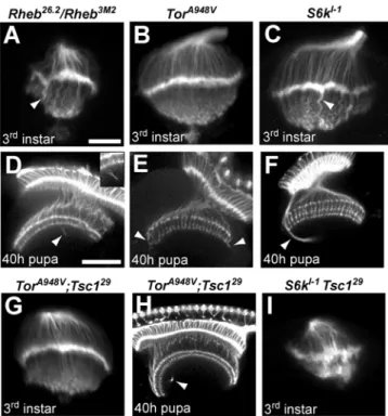
To evaluate the effects of reduced Tor signaling, we examined
axon guidance in animals bearing hypomorphic mutations inTor
andRheb, as well as a null allele ofS6k, a key downstream target of
TORC1. In all three of these mutants, mild axon projection defects were observed (Figure 6A–F, Table 1). Third instar larvae had irregular laminas and abnormally thick projections to the medulla (Figure 6A–C, arrowheads). In 40 h pupae, R7 and R8 terminations were largely normal, but there were projections which misrouted and failed to terminate correctly (Figure 6D–F,
Table 1). Genetic mosaic analysis ofRhebmutant photoreceptor
projections showed the same phenotypes, demonstrating that normal levels of Tor-Tsc signaling in the retina are required for proper photoreceptor targeting (data not shown). These findings establish that reductions in Tor-Tsc signaling also produce axon guidance defects, although quite mild in comparison to activation
of the pathway achieved by loss ofTsc1function. However, only
the S6k mutants are null in these experiments, and we cannot
therefore fully assess the contributions of Tor or Rheb to axon
guidance compared toTsc1.
To determine if the functional relationships critical for growth control are also in effect for axon guidance, we conducted genetic
epistasis experiments between Tsc1 and bothTor and S6k. Tsc1
mosaic pupae show severe axon guidance abnormalities andTsc1
mutant animals do not survive to the pupal stage; in contrast,
animals bearing both a Tsc1 mutation and a hypomorphic Tor
allele survived to pupal stages and showed only modest axon guidance abnormalities in larval and pupal brains (Figure 6G, H, Table 1). The gross disruptions of R7/R8 terminations in the
medullas of 40h Tsc1 mosaic pupae were almost completely
rescued by the presence of a hypomorphic allele ofTor. Genetic
mosaics withTsc1 Rhebdouble mutant chromosomes also showed
dramatic rescue of photoreceptor axon guidance defects (data not
Table 1.Axon guidance defects in animals with altered Tsc-Rheb-Tor signaling
. . . .
(Percent of optic lobes affected)
Incorrect Terminations
3rdinstar larvae ThickLP Gapsin LP LPPeaked LongR7/8 Gapsin Med. AboveLP BelowLP In Med.
Tsc129(n = 58) 70 41 7 5 31 45 52 72
Ptendj189
(n = 38) 24 100 79 63 29 58 68 95
Rheb3M2/26.2
(n = 22) 68 55 0 0 41 32 18 36
TorA948V(n = 12) 17 17 0 8 0 0 17 33
S6kl-1
(n = 49) 29 41 2 0 2 10 14 4
TorA948V Tsc129
(n = 14) 21 29 0 0 7 7 0 0
S6kl-1
Tsc129(n = 23) 65 83 0 0 70 43 17 43
wild-type+Rap (n = 80) 19 13 0 0 9 21 14 13
Tsc129
+Rap (n = 60) 76 54 2 6 59 46 71 63
40-hour pupae Pathfinding
Defects De-fasciculation Defects Termination Layer Defects Tsc129
(n = 60) 100 100 100
elav-Gal4.UAS-Rheb(n = 23) 83 43 39
Ptendj189(n = 73) 25 30 8
Rheb26.2
(n = 80) 36 3 19
S6kl-1
(n = 32) 28 9 25
TorA948V(n = 20) 35 0 10
TorA948V Tsc129
(n = 25) 40 4 16
*Tsc129 , Ptendj189
, andRheb26.2
shown). In contrast,S6knull mutations did not ameliorate theTsc1 axon projection defects in the larval brain, and both the lamina plexus and medulla projections were highly disordered (Figure 6I, Table 1). These findings demonstrate that Tor and Rheb, but not S6k, are critical components of the photoreceptor axon guidance signaling system downstream of Tsc1.
In order to evaluate if the growth control functions of Tsc-Rheb-Tor signaling are important for axon guidance, we used rapamycin to inhibit the abnormal growth produced by loss of
Tsc1function. Feeding animals with rapamycin between hatching
and the third instar larval stage blocked the retinal cell growth and
proliferation defects ofTsc1mutant photoreceptor mosaics. This
was evident in both the overall size of the developing retina and the size of the photoreceptor cell bodies (Figure 7A–C). While the
growth defects of Tsc1 mosaics were rescued by rapamycin
treatment, photoreceptors from these animals still showed severe axon guidance abnormalities in the third instar larval brain, with an irregular and disrupted lamina plexus, as well as disorganized projections to the medulla (Figure 7E, Table 1). Treatment of
wild-type controls with rapamycin produced only mild defects in the lamina plexus (Figure 7D, Table 1) supporting the hypothesis that Tsc1-mediated regulation of axon guidance operates largely via a rapamycin-insensitive function of Tor. We noted that the
excessive growth of Pten mutant retinas was not rescued by
rapamycin treatment, in contrast to the effects of this TORC1
inhibitor on Tsc1mosaics. While the growth and differentiation
phenotypes ofPtenandTsc1 mutant retinas are comparable, the
difference in their rapamycin responses highlights how disruption of signaling by these two regulators is distinct.
DISCUSSION
Tsc-Rheb-Tor signaling in neural development
The Tsc-Rheb-Tor pathway is critical for integrating a variety of signals that govern cellular and organismal growth. Inappropriate activation of the pathway also leads to severe neurological and behavioral abnormalities, including mental retardation, autism,and epilepsy [1,6]. WhileTSCmutations produce hamartomatous
growths in the brain, recent evidence has suggested that these benign tumors may not be solely responsible for the nervous system dysfunction that is a hallmark of tuberous sclerosis
complex. Loss ofTSC2in hippocampal neurons produces changes
in neuronal morphology and synaptic transmission [2].
Heterozy-gosity for TSC2 in the rat compromises several measures of
hippocampal long term potentiation [3]. Loss ofPten, an important
upstream regulator of Tsc-Rheb-Tor signaling, in a limited set of neurons also affects neuronal morphology and socialization Figure 6. Effects of mutations that downregulate the Tor pathway on
photoreceptor axon guidance, and genetic epistasis withTsc1. Optic lobes from third instar larvae (A–C) and 40h pupae (D–F) stained with MAb24B10. (A) Larvae heteroallelic for a hypomorphic combination of Rheballeles show abnormal photoreceptor patterning and contain thick axon bundles that extend into the medulla (arrowhead). (D) At the 40 h pupal stage, Rheb mutants display axons that bypass their normal targets in the R7/R8 termination zones (arrowhead). (B) Larvae homozygous for a hypomorphic Tor allele show fairly normal photoreceptor patterning, but at the pupal stage (E) misrouted axons can be seen in the medulla (arrowheads). (C) S6knull homozygous larvae show thick axon bundles projecting past the lamina (arrowhead), whileS6kpupae (F) display misrouted axons that initially bypass the R7/ R8 termination zone (arrowhead). (G, H) Animals doubly mutant forTor andTsc1 do not show the severe photoreceptor defects seen when axons are mutant forTsc1alone (compare to Figure 5B, F, F9), although mild defects similar to those in Tor mutants are still apparent (arrowhead). (I)S6k-Tsc1double homozygous mutants display a severe phenotype dissimilar to mutants for eitherS6korTsc1alone. The scale bar is 25 microns in panel A, 50 microns in panel D.
doi:10.1371/journal.pone.0000375.g006
Figure 7. Axon guidance defects inTsc1mosaics are not suppressed by blocking growth.(A–C) Third instar eye discs from wild type and Tsc1mosaic larvae raised with or without rapamycin (rap). Ommatidial units, comprised of eight photoreceptors, were visualized with phalloidin (red) that detects F-actin, and MAb24B10 (green). Phalloidin staining is strongest at the perimeter of each ommatidium, outlining each sensory unit. Rapamycin treatment ofTsc1mosaic eye discs (C) restored eye disk size and cell size compared to wild type (A). (D and E) Rapamycin treated third instar larval brains stained with MAb24B10. Rapamycin treatment blocked abnormal growth of the retina and the increase in photoreceptor cell size, but did not ameliorate the abnormal axon projections also characteristic to untreatedTsc129mosaics. The scale bars in panel A represent 50 microns in the left image, 10 microns in the right image. The scale bar is 50 microns in panel D.
doi:10.1371/journal.pone.0000375.g007
behavior [29]. These findings collectively provide evidence that Tsc-Rheb-Tor signaling is critical for the morphological and functional development of the nervous system. It is not clear, however, if the entire Tsc-Rheb-Tor signaling network is critical for nervous system development, or if neural function is strictly a consequence of altered growth regulation. It is also not known if loss of signaling is as detrimental to neuronal development as inappropriately elevated
signaling, such as occurs with loss ofTSCfunction. We have taken
advantage of the genetic and molecular tools available in the fruit fly Drosophilato address these questions. Our findings demonstrate that appropriate levels of Tsc-Rheb-Tor signaling are critical for both NMJ development and for axon guidance in the visual system. In both these contexts, effects are independent of growth, implicating TORC2 rather than TORC1 as the complex mediating Tsc-Rheb-Tor signaling influences in the nervous system.
Tsc-Rheb-Tor effects on neural development are
independent of growth regulation
Given the importance of Tsc-Rheb-Tor signaling in regulating cellular and tissue growth, it was important to determine if disruption of this pathway affects neural development via its effects on growth or through signaling components independent of those that govern cellular size and growth. To address this issue we used both pharmacological and genetic methods to block the increased growth produced by pathway activation. The immunosuppressant rapamycin is a TORC1-specific inhibitor that prevents activation
of S6k and blocks growth mediated by loss ofTsc1. Rapamycin
treatment retarded growth in larvae with pan-neuronal expression
ofRheb, but failed to reduce the synapse expansion characteristic of
these animals. Similarly, while rapamycin effectively reduced the
retinal overgrowth ofTsc1mosaic animals, it failed to suppress the
photoreceptor axon guidance defects seen in the visual system.
Loss ofS6kfunction also failed to ameliorate axon guidance defects
inTsc1mosaic animals. This contrasts with effects ofTorpartial
loss-of-function mutations, which effectively rescued axon
guid-ance defects ofTsc1mutants. Collectively, these findings
demon-strate that the role of Tsc-Rheb-Tor signaling in synapse assembly and axon guidance is largely independent of TORC1, S6k, and their effects on growth. Indeed, while animals bearing null alleles
ofS6khave some axon pathfinding defects, the effects are relatively
modest compared to Tsc1mosaics, indicating that S6k does not
provide the critical outputs affecting axon guidance.
Our findings parallel recent work in the mouse, where neuronal
hypertrophy produced by loss of Pten in granule neurons of the
cerebellum and dentate gyrus was not rescued by loss ofS6k1[30].
It is also of note that some but not allTsc1/2-mediated changes in
dendritic morphology of hippocampal neurons in organotypic cultures were suppressed by rapamycin treatment [2]. Our findings suggest that inhibition of growth regulatory components in tuberous sclerosis patients, such as achieved with rapamycin and related agents, may not affect all processes that are deranged in the nervous system.
Recent studies of Pi3 kinase, Akt and InR inDrosophila have
shown that activation of signaling upstream of Tsc1/2 also produces increases in synapse size, both at the NMJ as well as central synapses [31]. Expression of these components in adult neurons demonstrated that Pi3 kinase-mediated synaptogenesis was age-independent, and therefore not a developmentally re-stricted phenomenon. In agreement with studies reported here, the expanded NMJs produced by activation of Pi3 kinase were functional, with increased stimulus-induced EJPs. Overexpression
of theDrosophilaortholog of the epidermal growth factor receptor
(EgfR) in central neurons increased neuronal cell size, without an
increase in synapse number. These results are consistent with those reported here where we have been able to directly suppress growth mediated by Tsc-Rheb-Tor pathway activation without altering effects on synapse formation or axon guidance.
Recent studies have also demonstrated a link betweenTsc1/Tsc2
andhighwire, a gene known to effect synapse size and functionality in Drosophila[32]. ThehighwireorthologPamwas shown to bind Tsc2 in pull-down assays, and it has been suggested that Pam may function as an E3 ubiquitin ligase to regulate the intracellular levels of the Tsc1/Tsc2 complex. This concept of Highwire as a negative
regulator of Tsc levels is consistent with our findings, sincehighwire
mutants have been shown to possess enlarged NMJs similar to what we see for Rheb overexpression [14]. Despite this, the enlarged
synapses ofhighwiremutants display compromised synaptic function
which is contrary to what we found when overexpressing Rheb, so Highwire is likely to have multiple functions at the synapse besides simply the regulation of Tsc.
Contributions of TORC1 versus TORC2 in synapse
assembly and axon guidance
Tor has a number of molecular outputs that influence many cellular processes; notable among these are cellular growth and cellular morphology. TORC1, which contains Raptor and is sensitive to the anti-proliferative agent rapamycin, is a major contributor to the regulation of cellular growth, in large measure due to its effects on protein synthesis. TORC2, which includes Rictor, is implicated in the control of cell morphology mediated by regulation of the actin cytoskeleton [33]. Both pharmacological and genetic studies presented here argue in favor of Tor complex 2 providing an essential regulatory component of both synapse
growth and axon guidance inDrosophila. Our results support recent
work showing that changes in dendritic morphology of
hippo-campal neurons produced by loss ofTsc1 required regulation of
the actin-depolymerizing factor Cofilin [2], implicating TORC2-mediated processes. There is a considerable body of work demonstrating that control of the actin cytoskeleton is critical for NMJ growth and function [34–36] and TORC2 may provide an important component of that control. Regulation of actin is also essential for axon guidance in the visual system (reviewed in [37,38]), and disruption of Tor-mediated control of actin may be
the underlying molecular deficit inTsc1mosaics.
Either gain or loss of Rheb signaling compromises
neuromuscular junction assembly and axon
guidance
A number of studies have suggested that TOR activation produced
by loss of TSC1/2 affects neuronal morphology and synaptic
function. Our findings support these observations; elevated Rheb activity produces synaptic enlargement and enhanced physiological
function at theDrosophilaNMJ. However, it was not evident from
earlier studies whether loss of signaling through Rheb and Tor is also important for neural development. We provide evidence that this is
the case. Partial loss-of-function mutations inRhebcompromise NMJ
growth and function, as well as photoreceptor axon targeting in the
visual system. Overexpression ofTsc1andTsc2in the motoneuron
also limited synaptic growth, supporting the conclusion that depressed levels of Rheb activity compromise synapse development.
Rheb-mediated synaptic development is dependent
on a functional BMP signaling system
these effects are dependent on signaling systems known to be critical for synapse development. At the Drosophila NMJ, BMP signaling is critical for normal growth and function. Mutations in
wit, a gene encoding a type II BMP receptor, produce a small and
poorly functioning NMJ [17,19]. These deficits can be rescued by
motoneuron expression ofwit+
, demonstrating that BMP signaling in the motoneuron is critical for synaptic expansion during larval growth. To determine if Rheb-mediated synaptic growth required
BMP signaling, we placedelav-Gal4andUAS-Rhebtransgenes into
awitmutant background. While overexpression of Rheb and the
accompanying activation of the Tor pathway partially rescued the
defect in synapse growth produced by loss ofwitfunction, it was
unable to restore a normal EJP response or rescue quantal content. These findings establish that Tsc-Rheb-Tor mediated effects on synapse morphology are partially dependent on BMP signaling, and are fully dependent on BMP activity for a physiologically competent synapse. Our findings also establish that the functional
deficits inwitmutants are not simply the result of reduced synapse
size, since restoration of synapse size by expression ofUAS-Rheb
does not restore physiological function. Intersection of BMP, and Akt/PTEN/TOR signaling has been noted for other systems, and our results indicate the relationship between these pathways is important for synapse growth and plasticity as well [39].
Loss of function mutations in
Tsc1
and
Pten
have
different effects on axon guidance
Previous analysis ofgigas/Tsc2mutants demonstrated that loss of
this gene in mechanoreceptors affects axon targeting, producing projections to novel areas in the CNS in addition to innervation of normal targets [5]. We have used genetic mosaics to evaluate the function of Tsc-Rheb-Tor signaling in photoreceptor axon
guidance. Animals homozygous for Tsc1 in the retina showed
grossly aberrant photoreceptor projections to both the lamina and medulla. R7 and R8 projections to the medulla in 40h pupae failed to terminate correctly and projected beyond normal targets to inappropriate regions within the brain. Somatic mosaics bearing
retinal neurons mutant forPten also showed photoreceptor axon
guidance defects, but to a notably lesser degree. Since bothTsc1
and Pten alleles used for this analysis were nulls and show
comparable effects on cellular growth and differentiation [23], it
follows thatPtenis not as critical for axon guidance asTsc1. The
distinctions between axon guidance phenotypes of Ptenand Tsc1
null mutants indicate that altered timing of differentiation is not critical for axon guidance and that control of this pathway at the
level of PtenorTsc1 is not functionally equivalent. Our findings
that rapamycin arrests retinal overgrowth produced by loss ofTsc1
but notPtenin the retina supports earlier work demonstrating that
retinal overgrowth mediated by loss ofTsc1, but notPten, can be
suppressed by reductions inS6kactivity [40]. Those results were
interpreted as demonstrating that Pten is largely a regulator of Akt activity, whereas Tsc1/2 serves as a tumor suppressor and inhibitor affecting principally S6k. Our results support these relationships and emphasize that in the nervous system regulation of Tsc1/2 targets other than S6k are critical.
Graded activation of the Tsc-Rheb-Tor signaling axis
produces graded effects on axon guidance
We have used two different genetic methods for activating the Tsc-Rheb-Tor pathway in the visual system; generating retinal mosaics
with a loss of function allele ofTsc1, and pan-neuronal expression
of Rheb usingelav-Gal4andUAS-Rheb. The comparison of these
methods revealed that overexpression of Rheb produced milder
axon guidance phenotypes in the visual system than complete loss
of Tsc1 function. Of interest is that the degree of activation
achieved withelav-Gal4.UAS-Rheb, a level that did not produce
lethality, did result in discernable axon targeting defects in the visual system. This suggests that axon guidance controlled by Tsc-Rheb-Tor is sensitive to incremental changes in signaling. The range of neurological and behavioral phenotypes associated with
loss of one copy ofTSC1orTSC2 is consistent with this model,
where other environmental or genetic factors may affect signaling levels, producing a range of deficits. Our findings indicate that Drosophilacan serve as a useful model for identifying how graded changes in signaling can produce a spectrum of defects in neural development.
MATERIALS AND METHODS
Drosophila strains
UAS-RhebEP50.084/TM6B Tb [11], UAS-DP110WT [41], and y w hsFLP; UAS-Tsc1 UAS-Tsc2 [21] were crossed to elav-Gal4/CyO P[w+; ubi-GFP][42] or OK6-Gal4[17] for expression in neurons,
and to G14-Gal4/CyO P[w+
; ubi-GFP] (from C. Goodman) for expression in muscles. Stocks used for mutant analysis and genetic
interaction studies werey w; Rheb3M2/TM6B Tb y+[11],Rheb2D1
/ TM6B Tb[11],b w; witB11/TM6B Tb[19], w; witA12/TM6B Tb
[19], S6kl-1/TM6B Tb [43], and Tor2.1/Ala948Val/CyO P[w+
;
ubi-GFP][44]. Eye-specific mosaics were generated using the
FLP-FRTtechnique [45] by crossing y w eyFLP GMR-lacZ; FRT82B
I(3)cl-R3/TM6B Tb [45] or y w eyFLP GMR-lacZ; I(2)cl-L3 w+
FRT40A/CyO y+
[45] to w; FRT82B Tsc129/TM6B Tb [20], w;
FRT82B Rheb26.2/TM6B Tb y+[11],y w hsFLP; FRT40A Ptendj189 / SM6-TM6B[24],y w eyFLP GMR lacZ; FRT82B[45], ory w eyFLP GMR lacZ; FRT40A[45]. Using this mosaic method, heterozygous
cells have a growth disadvantage since they bear aMinuteand
cell-lethal mutation [l(3)cl-R3orl(2)cl-L3]. For all retinal mosaics, we
assessed the degree of mosaicism by examining the adult retina
where mutant cells could be identified by loss of thew+marker. In
Tsc1, Pten and Rheb mosaics, mutant cells comprised the vast
majority of the adult retina (.90%). Wild-type strains were y w,
Oregon-R, or Canton-S.
Immunohistochemistry
For visualization of neuromuscular junction synapses, third instar larvae were filleted in PBS and fixed in 4% formaldehyde before staining with Anti-Cysteine String Protein mAB49 at 1:1000 (a generous gift from Zinsmaier and Buchner) and FITC-conjugated Anti-HRP at 1:50 (Jackson Labs). Bouton numbers were de-termined by a combination of CSP and HRP image data. Muscle surface area measurements were performed using ImageJ data analysis software (NIH) and represent the combined area of the second abdominal segment muscles 6 and 7. Third instar larvae and 40hr pupae were fixed, stained, and mounted as described [46] for photoreceptor analysis. Antibodies from the Develop-mental Studies Hybridoma Bank were used at 1:25 for mouse anti-Chaoptin (MAb24B10), 1:10 for mouse anti-Repo (MAb8D12), and 1:25 for mouse anti-Dachshund (MabDac2-3). Secondary antibodies were from the AlexaFluor series (Invitrogen). Texas
Red-phalloidin was used at 0.165mM (Invitrogen). All images
were acquired on a Nikon C1 upright laser confocal.
Rapamycin Treatment
Flies were raised on standard laboratory food supplemented with
rapamycin (Sigma) to a final concentration of 3mM for NMJ
analysis or 2mM for eye disks. Rapamycin treatedTsc129mosaic
animals with eye discs similar in size to control animals were selected for photoreceptor projection analysis.
Electrophysiology
Excitatory junctional potential (EJP) recordings were taken from
muscle 6 in the second abdominal hemisegment of 3rd instar
larvae. Dissections were done in Ca++
-free saline and recordings were performed in HL3 following published protocols [47]. Recordings were acquired with an Axoclamp 2B amplifier and pClamp9 software (Axon Instruments). EJP amplitudes and mini-EJP amplitudes were measured with MiniAnalysis software from Synaptosoft.
SUPPORTING INFORMATION
Figure S1 Patterning of lamina precursor cells and glia inPtenor
Tsc1mosaic animals. (A–F) Dorsal-posterior views of third instar
larval optic lobes stained with anti-Dachshund (lamina precursor
cell marker) or anti-Repo (glial cell marker). (A–C) Pten mosaic
animals show a significantly larger lamina compared to control
animals. This is not seen to the same extent inTsc1mosaics. (D–F)
Glial cells successfully differentiate and migrate in bothPtenand
Tsc1mosaics, however mild patterning defects are apparent and
could possibly contribute to the photoreceptor patterning abnormalities observed. All scale bars are 50 microns.
Found at: doi:10.1371/journal.pone.0000375.s001 (1.32 MB TIF)
ACKNOWLEDGMENTS
We thank Hoda Pourhassan for assistance with data collection and analysis, the BloomingtonDrosophilastock center for fly stocks, and the Developmental Studies Hybridoma Bank for antibodies.
Author Contributions
Conceived and designed the experiments: SS MO MO SK AA TN. Performed the experiments: SK HG BD YR KH AA ME. Analyzed the data: SS MO MO SK HG BD YR ME TN. Wrote the paper: SS.
REFERENCES
1. Ess KC (2006) The neurobiology of tuberous sclerosis complex. Semin Pediatr Neurol 13: 37–42.
2. Tavazoie SF, Alvarez VA, Ridenour DA, Kwiatkowski DJ, Sabatini BL (2005) Regulation of neuronal morphology and function by the tumor suppressors Tsc1 and Tsc2. Nat Neurosci 8: 1727–1734.
3. von der Brelie C, Waltereit R, Zhang L, Beck H, Kirschstein T (2006) Impaired synaptic plasticity in a rat model of tuberous sclerosis. Eur J Neurosci 23: 686–692. 4. Acebes A, Ferrus A (2001) Increasing the number of synapses modifies olfactory
perception in Drosophila. J Neurosci 21: 6264–6273.
5. Canal I, Acebes A, Ferrus A (1998) Single neuron mosaics of the drosophila gigas mutant project beyond normal targets and modify behavior. J Neurosci 18: 999–1008.
6. Inoki K, Corradetti MN, Guan KL (2005) Dysregulation of the TSC-mTOR pathway in human disease. Nat Genet 37: 19–24.
7. Neufeld TP (2004) Genetic analysis of TOR signaling in Drosophila. Curr Top Microbiol Immunol 279: 139–152.
8. Prokop A, Meinertzhagen IA (2006) Development and structure of synaptic contacts in Drosophila. Semin Cell Dev Biol 17: 20–30.
9. Mast JD, Prakash S, Chen PL, Clandinin TR (2006) The mechanisms and molecules that connect photoreceptor axons to their targets in Drosophila. Semin Cell Dev Biol 17: 42–49.
10. Meinertzhagen IA (1994) The early causal influence of cell size upon synaptic number: the mutant gigas of Drosophila. J Neurogenet 9: 157–176. 11. Stocker H, Radimerski T, Schindelholz B, Wittwer F, Belawat P, et al. (2003)
Rheb is an essential regulator of S6K in controlling cell growth in Drosophila. Nat Cell Biol 5: 559–565.
12. Zhang Y, Gao X, Saucedo LJ, Ru B, Edgar BA, et al. (2003) Rheb is a direct target of the tuberous sclerosis tumour suppressor proteins. Nat Cell Biol 5: 578–581.
13. Saucedo LJ, Gao X, Chiarelli DA, Li L, Pan D, et al. (2003) Rheb promotes cell growth as a component of the insulin/TOR signalling network. Nat Cell Biol 5: 566–571.
14. Wan HI, DiAntonio A, Fetter RD, Bergstrom K, Strauss R, et al. (2000) Highwire regulates synaptic growth in Drosophila. Neuron 26: 313–329. 15. McCabe BD, Hom S, Aberle H, Fetter RD, Marques G, et al. (2004) Highwire
regulates presynaptic BMP signaling essential for synaptic growth. Neuron 41: 891–905.
16. Zhang H, Stallock JP, Ng JC, Reinhard C, Neufeld TP (2000) Regulation of cellular growth by the Drosophila target of rapamycin dTOR. Genes Dev 14: 2712–2724.
17. Aberle H, Haghighi AP, Fetter RD, McCabe BD, Magalhaes TR, et al. (2002) wishful thinking Encodes a BMP Type II Receptor that Regulates Synaptic Growth in Drosophila. Neuron 33: 545–558.
18. Marques G, Haerry TE, Crotty ML, Xue M, Zhang B, et al. (2003) Retrograde Gbb signaling through the Bmp type 2 receptor wishful thinking regulates systemic FMRFa expression in Drosophila. Development 130: 5457–5470. 19. Marques G, Bao H, Haerry TE, Shimell MJ, Duchek P, et al. (2002) The
Drosophila BMP type II receptor Wishful Thinking regulates neuromuscular synapse morphology and function. Neuron 33: 529–543.
20. Gao X, Pan D (2001) TSC1 and TSC2 tumor suppressors antagonize insulin signaling in cell growth. Genes Dev 15: 1383–1392.
21. Potter CJ, Huang H, Xu T (2001) Drosophila Tsc1 functions with Tsc2 to antagonize insulin signaling in regulating cell growth, cell proliferation, and organ size. Cell 105: 357–368.
22. Tapon N, Ito N, Dickson BJ, Treisman JE, Hariharan IK (2001) The Drosophila tuberous sclerosis complex gene homologs restrict cell growth and cell proliferation. Cell 105: 345–355.
23. Bateman JM, McNeill H (2004) Temporal control of differentiation by the insulin receptor/tor pathway in Drosophila. Cell 119: 87–96.
24. Gao X, Neufeld TP, Pan D (2000) Drosophila PTEN regulates cell growth and proliferation through PI3K-dependent and -independent pathways. Dev Biol 221: 404–418.
25. Huang H, Potter CJ, Tao W, Li DM, Brogiolo W, et al. (1999) PTEN affects cell size, cell proliferation and apoptosis during Drosophila eye development. Development 126: 5365–5372.
26. Goberdhan DC, Paricio N, Goodman EC, Mlodzik M, Wilson C (1999) Drosophila tumor suppressor PTEN controls cell size and number by antagonizing the Chico/PI3-kinase signaling pathway. Genes Dev 13: 3244–3258.
27. Scanga SE, Ruel L, Binari RC, Snow B, Stambolic V, et al. (2000) The conserved PI3’K/PTEN/Akt signaling pathway regulates both cell size and survival in Drosophila. Oncogene 19: 3971–3977.
28. Ito N, Rubin GM (1999) gigas, a Drosophila homolog of tuberous sclerosis gene product-2, regulates the cell cycle. Cell 96: 529–539.
29. Kwon CH, Luikart BW, Powell CM, Zhou J, Matheny SA, et al. (2006) Pten regulates neuronal arborization and social interaction in mice. Neuron 50: 377–388.
30. Chalhoub N, Kozma SC, Baker SJ (2006) S6k1 is not required for Pten-deficient neuronal hypertrophy. Brain Res 1100: 32–41.
31. Martin-Pena A, Acebes A, Rodriguez JR, Sorribes A, de Polavieja GG, et al. (2006) Age-independent synaptogenesis by phosphoinositide 3 kinase. J Neurosci 26: 10199–10208.
32. Murthy V, Han S, Beauchamp RL, Smith N, Haddad LA, et al. (2004) Pam and its ortholog highwire interact with and may negatively regulate the TSC1.TSC2 complex. J Biol Chem 279: 1351–1358.
33. Wullschleger S, Loewith R, Hall MN (2006) TOR signaling in growth and metabolism. Cell 124: 471–484.
34. Eaton BA, Fetter RD, Davis GW (2002) Dynactin is necessary for synapse stabilization. Neuron 34: 729–741.
35. Ang LH, Chen W, Yao Y, Ozawa R, Tao E, et al. (2006) Lim kinase regulates the development of olfactory and neuromuscular synapses. Dev Biol. 36. Coyle IP, Koh YH, Lee WC, Slind J, Fergestad T, et al. (2004) Nervous wreck,
an SH3 adaptor protein that interacts with Wsp, regulates synaptic growth in Drosophila. Neuron 41: 521–534.
37. Rao Y (2005) Dissecting Nck/Dock Signaling Pathways in Drosophila Visual System. Int J Biol Sci 1: 80–86.
38. Luo L (2002) Actin cytoskeleton regulation in neuronal morphogenesis and structural plasticity. Annu Rev Cell Dev Biol 18: 601–635.
39. He XC, Zhang J, Tong WG, Tawfik O, Ross J, et al. (2004) BMP signaling inhibits intestinal stem cell self-renewal through suppression of Wnt-beta-catenin signaling. Nat Genet 36: 1117–1121.
40. Radimerski T, Montagne J, Hemmings-Mieszczak M, Thomas G (2002) Lethality of Drosophila lacking TSC tumor suppressor function rescued by reducing dS6K signaling. Genes Dev 16: 2627–2632.
41. Leevers SJ, Weinkove D, MacDougall LK, Hafen E, Waterfield MD (1996) The Drosophila phosphoinositide 3-kinase Dp110 promotes cell growth. Embo J 15: 6584–6594.
42. Luo L, Liao YJ, Jan LY, Jan YN (1994) Distinct morphogenetic functions of similar small GTPases: Drosophila Drac1 is involved in axonal outgrowth and myoblast fusion. Genes Dev 8: 1787–1802.
43. Montagne J, Stewart MJ, Stocker H, Hafen E, Kozma SC, et al. (1999) Drosophila S6 kinase: a regulator of cell size. Science 285: 2126–2129. 44. Zhang Y, Billington CJ Jr, Pan D, Neufeld TP (2006) Drosophila target of
45. Newsome TP, Asling B, Dickson BJ (2000) Analysis of Drosophila photoreceptor axon guidance in eye-specific mosaics. Development 127: 851–860. 46. Blair SS (2000) Imaginal Discs. In: Sullivan W, Ashburner M, Hawley RS, eds
(2000)DrosophilaProtocols. Cold Spring Harbor, New York: Cold Spring Harbor Laboratory Press. pp 159–173.
47. Rawson JM, Lee M, Kennedy EL, Selleck SB (2003) Drosophila neuromuscular synapse assembly and function require the TGF-beta type I receptor saxophone and the transcription factor Mad. J Neurobiol 55: 134–150.