Predictors, Neuroimaging Characteristics and
Long-Term Outcome of Severe European
Tick-Borne Encephalitis: A Prospective Cohort
Study
Thorsten Lenhard1
*, Daniela Ott1, Nurith J. Jakob1¤a, Mirko Pham2, Philipp Bäumer2,
Francisco Martinez-Torres1¤b, Uta Meyding-Lamadé3
1Neuroinfectious Diseases Group, Department of Neurology, University Hospital of Heidelberg, Heidelberg, Germany,2Department of Neuroradiology, University Hospital of Heidelberg, Heidelberg, Germany, 3Department of Neurology, Krankenhaus Nordwest Frankfurt, Frankfurt, Germany
¤a Current address: Psychiatrische Klinik Sanatorium Kilchberg, Zürich, Switzerland ¤b Current address: Merz Pharmaceuticals GmbH, Frankfurt, Germany
*thorsten.lenhard@med.uni-heidelberg.de
Abstract
Background and Objectives
Tick-borne encephalitis (TBE) still represents a considerable medical and health economic problem in Europe and entails a potential threat to travellers. The aim of this study was to characterise the conditions of severe TBE by precisely recording its clinical variants, the related neuroimaging features, and the variant-specific long-term outcome and by identify-ing predictors for severe courses.
Methods
A cohort of 111 TBE patients (median age 51, range 17–75 years; 42% females) was ana-lysed prospectively. Data were acquired from the department of neurology, University Hos-pital Heidelberg, and the infectious diseases registry of the Robert-Koch institute Berlin. Neurological status was ascertained by protocol at admission and discharge and the degree of disability was scored using the modified RANKIN Scale (mRS; clinical score addressing neurological disability, range from 0, healthy to 6, dead) at admission and at follow-up. Fol-low-up examination was conducted by means of a telephone interview. To identify indepen-dent predictors for severe TBE and functional outcome, modelled logistic regression was performed. MRI changes were correlated with infection variants. To assess alpha-motor neuron injury patterns, we used high-resolution magnetic resonance neurography (hrMRN). Analyses were performed at the Department of Neurology, University Hospital, University of Heidelberg from April 2004 through September 2014
Results
Acute course: 3.6% of patients died during the acute infection. All patients with a lethal course suffered from meningoencephaloradiculitis (MER, 14.4% of the cohort), which is a11111
OPEN ACCESS
Citation:Lenhard T, Ott D, Jakob NJ, Pham M, Bäumer P, Martinez-Torres F, et al. (2016) Predictors, Neuroimaging Characteristics and Long-Term Outcome of Severe European Tick-Borne Encephalitis: A Prospective Cohort Study. PLoS ONE 11(4): e0154143. doi:10.1371/journal.pone.0154143
Editor:Robyn S Klein, Washington University, UNITED STATES
Received:May 11, 2015
Accepted:April 9, 2016
Published:April 25, 2016
Copyright:© 2016 Lenhard et al. This is an open access article distributed under the terms of the Creative Commons Attribution License, which permits unrestricted use, distribution, and reproduction in any medium, provided the original author and source are credited.
Data Availability Statement:All relevant data are within the paper and its Supporting Information files.
Funding:These authors have no support or funding to report.
associated with a significantly higher risk of requiring intensive care (p = 0.004) and mechanical ventilation (p<0.001) than menigoencephalitis (ME, 27.9% of the cohort). At admission, both MER and ME groups were severely affected, with the MER group having a statistically higher mRS score (median of 5 in the MER groups versus 4 in the ME group; p<0.001). Long-term outcome: outcome for MER was considerably worse (median mRS = 4) than for ME (mRS = 1, p<0.0001) and meningitis (mRS = 0, 57.7% of the cohort). Risk factors: advanced age (p<0.001) and male gender (p = 0.043) are independent risk factors for a severe infection course. Furthermore, we identified pre-existing diabetes mellitus (p = 0.024) as an independent risk factor for MER. In MER, alpha-motor neuron injury accounts for the poor prognosis confirmed by hrMRN.
Conclusion and Relevance
These data provide critical information for neurologists and other health professionals to use in evaluating TBEV patients who live in or travel to endemic areas. This information can be used to classify clinical presentation and estimate infection-associated complications and individual prognosis. Furthermore, the risk for severe, disabling infections in older patients should prompt general practitioners to recommend and encourage vaccination to those patients living in or travelling to endemic areas.
Introduction
Central European encephalitis is the European representative of a group of encephalitides caused by TBEV (tick-borne encephalitis virus). Based on comparative sequence analysis, three related TBEV subtypes causing meningoencephalitis along the 7°C isotherm in Eurasia can be distinguished [1,2]. The European subtype extends north-east to the Baltic states, east to Ukraine, south-east as far as former Yugoslavia and north to the Baltic Sea coast of Sweden. The Rhine valley in Germany marks the western boundary [2,3]. TBEV is an enveloped, posi-tive-stranded RNA virus (40–60 nm) belonging to theFlaviviridae, a family embracing several other relevant human pathogens, such as Dengue, Japanese encephalitis, Yellow fever and West Nile virus[4,5]. The main vector in Europe is the tickIxodes ricinus, which, besides TBEV, transmits a range of other pathogens [6]. The incubation period for TBEV is approxi-mately 7–14 (occasionally up to 28) days. Symptomatic infection occurs in approxiapproxi-mately 30% of infected patients. It then follows a typical biphasic course with a flu-like prodrome, repre-senting viremia, and CNS infection in approximately 10% of cases.
In endemic regions in central Europe, TBE is the most common viral meningoencephalitis, with incidences far higher than all other sporadic viruses [7–10]. Though a preventative vac-cine is widely available, vaccination rates are low in at-risk populations. As a consequence, the annual infection rate is still high in endemic regions (up to 29/100.000) and incidence of TBE has increased since the mid-1990s in most European countries [3,7]. In most cases, patients suffer from pure meningitis (~ 50%) with overall good prognoses to recover completely. The other half suffers an infection of the brain parenchyma, resulting in meningoencephalitis (~35%), or very rarely an isolated infection of the spinal cord (less than 1%); 1–2% of patients will die from their infection. Approximately 10% of patients will develop infection of the motor neurons (radiculitis) in addition to meningoencephalitis [11]. The clinical presentations of TBE has been categorised according to the focus of the infection into pure meningitis,
Germany corp. as well as any other funder and any of its representatives had no influence on the study design, data acquisition, analysis, interpretation of data, writing of the paper or decision to submit for publication. Mrs Ott, Dr. Jakob and Dr. Bäumer report no disclosures. Dr. Pham received a memorial stipend from the Elser-Kröner-Fresenius Foundation. Funder and any of its representatives had no influence on the study design, data acquisition, analysis, interpretation of data, writing of the paper or decision to submit for publication. Dr. Martinez-Torres was funded by the German Federal Ministry of Education and Research (Bundesministerium für Bildung und Forschung). At present, he is employed at Merz Pharmaceuticals GmbH Germany. Funder as well as his employer and any of its representatives had no influence on the study design, data acquisition, analysis, interpretation of data, writing of the paper or decision to submit for publication. Dr. Meyding-Lamadé is also head of Neuroscience Stroke and Rehab Centre, Jerudong Park Medical Centre, Brunei Darussalam and chief executive of Meyding-Lamadé Med GmbH. She is a member of the collaboration group on German Neurological Guidelines for viral and tick-born encephalites, of the ISW TBE scientific working group for tick-born encephalitis, of the national committee for polioeradication at Robert-Koch Institute, Berlin, Germany, head of the European task force for opportunistic infections and extraordinary member of drug commision of the German Medical Association, (AkdÄ). She received lecture fees and travel grants from: Almirall, Bayer Vital, Biogen idec, Boehringer Ingelheim, Genzyme, Merck Serono, Novartis, Sanofi, Teva. All listed funders and any of its representatives had no influence on the study design, data acquisition, analysis, interpretation of data, writing of the paper or decision to submit for publication. None of the authors have served or currently serve on the editorial board of PLOS ONE, have acted as an expert witness in relevant legal proceedings, have sat or currently sit on a committee for an organization that may benefit from publication of the paper. This does not alter the authors' adherence to all the PLOS ONE policies on sharing data and materials, as detailed online in the guide for authors.
Abbreviations:AMNI, alpha-motor neuron injury; ME, meningoencephalitis; MER,
meningoencephalitis (ME) and menigoencephaloradiculitis (MER). However, a precise classifi-cation of clinical presentations as well as associated neuroimaging findings is lacking in the lit-erature. Furthermore, it is not exactly known what patient-related factors are associated with severe infection and poor outcome. Thus, we analysed a cohort of 111 TBE patients from the Odenwald hills region, a high-risk endemic area in south-western Germany, and focused on precise analyses of the acute infection course and the related long-term outcome of the differ-ent clinical presdiffer-entations and aimed to iddiffer-entify risk factors for severe infection with poor out-come. Furthermore, we investigated the related neuroimaging features using not only conventional MRI but also high-resolution magnetic resonance neurography (hrMRN) to cover peripheral motor-neuron injury.
Patients and Methods
TBE data acquisition and case definition
TBE represents a certifiable infection in Germany and, by law, must be reported. Data were acquired from our departmental databases, from the district public health offices of the endemic region of the Odenwald hills and from the infectious diseases registry of the RKI Ber-lin. The study was approved by the ethics committee of the University of Heidelberg and writ-ten informed consent was obtained from participants. The data were collected mainly
prospectively and in part retrospectively from April 2004 through September 2014.
TBE was identified using a common clinical definition of meningitis confirmed by the pres-ence of anti-TBEV IgM antibodies in the serum and CSF as well as pleocytosis in the CSF. Clin-ical TBE presentations [11] were defined as follows:“pure meningitis”(meningitis), without focal neurological symptoms; meningoencephalitis (ME), meningitis plus focal neurological symptoms and/or disturbance of consciousness and/or higher-order brain dysfunction; or meningoencephaloradiculitis (MER), ME plus clinical signs of alpha-motor neuron injury (AMNI). All patients were examined at admission and followed up after 12 months at the earli-est with a clinical assessment and by recording modified RANKIN scale (mRS)[12]. The mRS is an established clinical score to ascertain the grade of neurological disability and a patient’s ability to perform daily activities and ranges from 0, healthy/no symptoms to 6, dead (1, no sig-nificant disability, 2, slight disability, 3, moderate disability, 4, moderate severe disability, 5, severe disability). If recovery was good at discharge (mRS<2), and in all meningitis patients, a follow-up examination was conducted by means of a telephone interview (for items, see
Table 1). In cases of clinical evidence of pre-existing polyneuropathy, TBE-related
motor-neu-ron injury was verified by electmotor-neu-roneuro- and myography.
Table 1. Baseline characteristics of the TBE cohort.
All patients MER ME Meningitis
Parameters n = 111 n = 16 (14.4%) n = 31 (27.9%) n = 64 (57.7%) P-value
Age, median†
51 (17–75) 62 (50–75) 57 (17–74) 46 (17–70) <0.001*0.009#
Female‡
47 (42.3%) 3 (18.8%) 9 (29.0%) 35 (54.7%) 0.01*0.027#
Biphasic course‡
47 (42.3%) 3 (18.8%) 14 (45.2%) 30 (46.9%) 0.049*
Tick bite‡,§ 71 (72%) 7 (53%) 21 (67%) 43 (79%) 0.07
*
Statistic was calculated with Mann-WhitneyU-test†and with Fisher exact test‡, respectively. P-values assigned as follows: MER
*and ME#compared to
meningitis.
§Information was not available in all patients.
CSF was analysed at admission and included complete cell count and anti-TBEV-IgM+IgG antibody detection (serum/CSF) (IMMUNOZYM FSME1Progen, Heidelberg, Germany). In initially uncertain cases, especially patients with a history of vaccination, follow-up serological examination and lumbar puncture had to confirm TBE either by an increase in anti-TBEV-IgG in serum, proof of anti-TBEV-IgM in the CSF or proof of a positive antibody CSF/serum index (anti-TBEV IgG).
Neuroimaging
For conventional brain MRI (T1,2-weighted-, diffusion weighted -, Flair imaging), see respec-tive textbooks of radiology [13]. In clinically suspected MER, hrMRN was performed to localise AMNI. The spinal nerves and brachial plexus were examined with a two-element multi-chan-nel surface array coil and peripheral nerves with an eight-chanmulti-chan-nel phased-array extremity coil (NORAS GmbH, Höchberg, Germany). HrMRN sequences included 2D fat-saturated T2-weighted sequences in perpendicular orientation to roots, brachial plexus and peripheral nerves[14,15].
Statistics
Statistical analyses were performed using the SPSS1software (Version 21). Data were ana-lysed by Fisher’s exact test (nominally scaled) or the Mann–WhitneyU-test (ordinally scaled). Differences with a probability of p<0.05 were defined as significant. To identify independent predictors for severe TBE and functional outcome, a binary logistic regression model was per-formed for all items except for age, where an ordered logistic regression with the Stata1Data Analysis was conducted (Version 12, StataCorp LP, Texas, U.S.A.).
Results
Baseline characteristics
The clinical TBE variants were distributed as follows: MER (n = 16, 14.4%), ME (n = 31, 27.9%) and pure meningitis (n = 64, 57.6%). The patients’median age differed significantly: MER (62 years; p = 0.001), ME (57 years; p = 0.009) and meningitis (46 years). The MER group (n = 3 females, 18.8%, p = 0.01) and the ME group (n = 9 females, 29%, p = 0.027) included significantly fewer females than the meningitis group (n = 35 females, 54.7%). The mean for pleocytosis was 124.3 cells/μL ± 12.2 standard error of mean (SEM). Detailed baseline
characteristics are summarised inTable 1.
Clinical characteristics and long-term outcome in TBE
The spectrum of clinical symptoms at admission is illustrated inFig 1A. Among the complex symptoms, disturbance in consciousness was observed significantly more often in MER patients (n = 14, 87.5%, p = 0.003) than in the ME group (n = 14, 45.2%). Among the focal neu-rological symptoms, more MER patients (25%) suffered from cranial nerve paresis than ME patients (n = 1, 3.2%, p = 0.04). ME patients presented with brainstem and cerebellar symp-toms significantly more frequently (ataxia: ME group, n = 17, 54.8% versus MER group, n = 0, p = 0.0002; oculomotor disturbance: ME, n = 9, 29% versus n = 0, p = 0.018). Although statisti-cally not significant, it is interesting to note that aphasia (n = 5, 16%) and autonomic distur-bances (n = 3, 9%) were observed exclusively in the ME group.
patients required intensive care (87.5% versus 41.9%, p = 0.004) and more needed to be mechanically ventilated (50% versus 3.2%, p = 0.0002) than ME patients (Table 2).
As for the long-term outcome, significantly more MER patients were dependent on others in their daily activities, with a high median mRS of 4 (range 1–6) compared with a low median mRS of 1 in the ME group (range 0–3, p<0.0001) (Table 2). A shift analysis of disability in MER and ME allows a closer look at the degree of disability (Fig 1B): nearly all of the ME patients achieved a good outcome of no or mild disability (mRS 0–2)–except 1 (mRS 3), whereas only 18.8% of MER patients were in this category. Significantly more MER patients (56.3%, p<0.001) still suffered from moderate or severe disability (mRS 3–5). MER alone accounted for the high mortality (Fig 1B).
Predictors for severe course and worse outcome
Higher age was observed significantly more frequently in MER (62 years, p = 0.001) and ME (57 years, p = 0.009) than in meningitis (46 years) (Table 1). Using ordered logistic regression analysis to prove independency from co-variables, age is an independent risk factor for devel-oping ME and MER (p<0.001, odds ratio 1.05, 95% CI 1.02–1.09) (Table 3). This means that
Fig 1. Spectrum of neurological impairment in severe TBE.Frequency of neurological symptoms during acute neuroinfection divided into complex (first four items) and focal neurological symptoms (following 7 items) in MER and ME, respectively (A). Note that peripheral palsy occurred only in MER and not in ME, owing to the clinical definition of these TBE variants. Significance is depicted as p<0.05 (*), p<0.01 (**), and p<0.001 (***), calculated with Fisher’s exact test. Functional long-term outcome in severe TBE (B): Functional outcome of MER and ME according to modified RANKIN scale (mRS) [12] at admission compared with long-term outcome as shift analysis by degree of disability. Numbers within the bars are given in percent (rounded). MRS scale: 0, no symptoms at all; 1, no significant disability, able to carry out all usual activities, despite some symptoms; 2, slightly disabled, able to look after own affairs without assistance, but unable to carry out all previous activities; 3, moderate disability, requires some help, but able to walk unassisted; 4, moderately severe disability, unable to attend to own bodily needs without assistance; 5, severe disability, requires constant nursing care and attention, bedridden, incontinent; and 6, dead.
doi:10.1371/journal.pone.0154143.g001
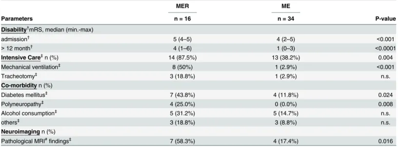
Table 2. Clinical characteristics of severe TBE.
MER ME
Parameters n = 16 n = 34 P-value
Disability†
mRS, median (min.-max) admission†
5 (4–5) 4 (2–5) <0.001
>12 month† 4 (1–6) 1 (0–3) <0.0001
Intensive Care‡
n (%) 14 (87.5%) 13 (38.2%) 0.004
Mechanical ventilation‡
8 (50%) 1 (2.9%) <0.001
Tracheotomy‡ 3 (18.8%) 1 (2.9%) n.s.
Co-morbidityn (%) Diabetes mellitus‡
7 (43.8%) 4 (11.8%) 0.024
Polyneuropathy‡ 4 (25.0%) 0 (0.0%) 0.008
Alcohol consumption‡
5 (31.2%) 5 (14.7%) n.s.
others‡
3 (18.8%) 3 (8.8%) n.s.
Neuroimagingn (%) Pathological MRI#
findings‡
7 (58.3%) 4 (17.4%) 0.016
MER, meningoencephaloradiculitis; ME, meningoendephalitis; mRS, modified RANKIN scale [12]. Statistic was calculated with Mann-Whitney U-test†and with Fisher exact test‡, respectively. #MRI was not available in all patients.
every additional year (ten years) of age results in a 5% (50%) higher risk to switch from cate-gory meningitis to ME and from ME to MER, respectively. Furthermore, male gender is found more frequently in MER (81.2%, p = 0.01) and ME (71%, p = 0.027) than in meningitis (43.3%). Binary logistic regression of gender predicting outcome showed that men, with an odds ratio of 2.41 (95% CI: 1.03–5.63), were at a significantly increased risk (p = 0.043) of unfa-vourable outcome of MER (Table 3). In searching for further clinical risk factors and possible predictors for MER, we identified pre-existing DM (p = 0.024) and PNP (p = 0.008) in the MER group. Using binary logistic regression analysis, DM remained an independent risk factor for MER (p<0.024, odds ratio 5.25, 95% CI: 1.24–22.19) (Table 3). Regression analysis was not feasible for polyneuropathy (PNP) since PNP most likely was a dependent variable of DM as a secondary, long-term complication. MER tended to take a monophasic course.
Neuroimaging in TBE
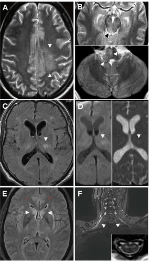
Among patients with severe infections, significantly more MRI abnormalities of any kind were seen in MER (n = 7 of 12, 58.3%, p = 0.0047) than in ME (4 of 23, 17.4%) (Table 2). Differenti-ated further into central brain lesions and peripheral lesions, respectively, brain lesions were present in both MER (n = 3 of 12, 25%) and ME (4 of 23, 17.4%) and did not differ significantly (p = 0.67). The thalamus was involved in all patients presenting with cerebral lesions and in two of them, additional lesions could be detected as depicted inFig 2A–2F. Note that MRI was not available in all patients. HrMRN was not established before 2010.
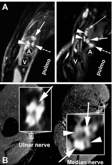
Investigating AMNI in more detail, hrMRN was performed in patients with clinically sus-pected MER. In all investigated cases, lesion contrast was strong, with a pathologically hyperin-tense T2 signal of either nerve roots, plexus trunks, plexus fascicles or peripheral nerves. Furthermore, the calibre of affected neural segments was clearly enlarged. In most cases, the extent of spatial lesions and lesion patterns in hrMRN exceeded the clinical manifestations of disease.Fig 3A and 3Billustrates some examples of hrMRN pathology.
Discussion
Overall, the distribution of the clinical manifestations in our cohort (pure meningitis 57.7%, ME 27.9% and MER 14.4%) is comparable with recent reports [11,16,17], although the pro-portion of MER was higher (14.4% versus 7.6–10%) and that of ME lower (27.9% versus 41– 51.3%). Similarly, mortality was higher in our cohort (3.6% versus 0–1.5%)[11,17,18] and all those who died had presented with MER. The reasons remain unclear: mutations in TBEV causing more virulent strains have been discussed and, indeed, those strains were isolated in
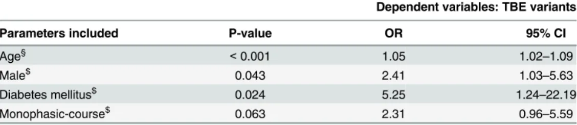
Table 3. Predictors for severe TBE.
Dependent variables: TBE variants
Parameters included P-value OR 95% CI
Age§ <0.001 1.05 1.02–1.09
Male$ 0.043 2.41 1.03–5.63
Diabetes mellitus$ 0.024 5.25 1.24–22.19
Monophasic-course$ 0.063 2.31 0.96–5.59
OR, odds ratio; CI, confidence interval
§
Ordered logistic regression for age: 5% risk per every additional year of age to develop a severe course (meningitis!ME!MER)
$
Binary logistic regression: gender (meningitis/ME!MER), diabetes and infection course (ME!MER).
regions where uncommon, frequently severe cases developed [19]. Indicative of virus strain-dependent virulence and relation to severity of brain infection might be the fact that severe cases in risk areas in Europe range in frequency between 7.6% (e.g. Poland) and 14.1% (e.g. Lat-via)[11,16,17,20,21]. Similarly, mortality in different risk areas in Europe ranges from 0% in Croatia, to 0.6% in Poland, 0.8% in the Czech Republic/Lithuania, and in Germany, respec-tively, 1.5% and 3.6%, as found in our study [11,17,18,21,22].
Recent studies have shown several clinical findings to be associated with severity of symp-toms or late sequelae in TBE [11,17,21]. In our cohort, significantly more MER than ME patients presented with a consciousness disturbance at admission or which developed during the acute course (87.5% versus 45.2%, p = 0.003) (Fig 1A). In contrast, significantly more cere-bellar symptoms such as ataxia (54.8%, p<0.001) and oculomotor disturbances (29%, p = 0.018) were observed in ME. These data are partially consistent with previous clinical find-ings [11,22–24]. However, there are several differences which may in part be due to our strin-gent classification into MER and ME. Although not statistically significant, higher-order brain dysfunction (such as aphasia) and autonomic symptoms were found exclusively in the ME group.
Taking the clinical features of MER and ME, respectively, together with a travel history from an endemic risk area, the evidence of peripheral flaccid paresis or prominent cerebellar signs should prompt the physician to suspect TBE. In cases of pure meningitis, a travel history from an endemic risk area and a history of tick-bite may support the suspected diagnosis but it cannot be clinically distinguished from other viral entities. In all cases, final diagnosis must be confirmed by the presence of TBEV antibodies (for details, see materials and methods, CSF analysis and immunoassays). If more complex symptoms such as delirious state, confusion, sei-zure or aphasia are present, other pathogens, especially members of theherpesviridae, should be taken into account [8,10].
TBE not only has a potentially severe acute course but also produces long-term disability in a considerable number of individuals [21–24]. The mRS is a commonly used instrument to measure the degree of neurological disability affecting activities of daily living (ADL)[12]. Already at admission, the mRS score in MER and ME differed significantly (median 5 in MER versus 4 in ME, p<0.001) (Table 2). At follow-up, the gap between MER and ME had wid-ened, with ongoing severe disability in MER (median mRS 4 in MER versus 1 in ME, p<0.0001). Looking more closely at the changes in mRS at follow-up (Fig 1B), all of the ME patients had mRS scores equal to or lower (“better”) than 3. In contrast, only 37.6% of MER patients scored equal to or lower (“better”) than 3. Another 37.5% were moderately severely disabled (mRS 4). Furthermore, 25% of MER and 3.6% of all patients died during intensive
axial lesions of the left semioval centre (A, arrowhead), right inferior colliculum tecti (B, upper panel) and right medulla oblongata (B, lower panel) Not shown: additional T2-weighted lesions in the thalamus and cerebellar peduncles. (C and D), A 62-year-old male MER patient initially misdiagnosed as lacunar stroke with consciousness disturbance, complete hemiplegia, dysphagia and lethal course due to aspiration pneumonia: axial fluid-attenuated inversion recovery (FLAIR) sequence depicts a rounded hyperintense lesion in the left thalamus (arrowhead) (C) that shows pathological water diffusion on diffusion-weighted imaging (DWI) (D, upper panel). Corresponding ADC map identifies the lesion as vasogenic oedema with enhanced water diffusion (D, lower panel), incompatible with acute or subacute stroke. (E and F), A 49-year-old female MER patient with“man in the barrel syndrome”(unable to move her upper limbs) as long-term outcome. MRI at admission showed extended encephalitis, cervical myelitis and brachial radiculitis: axial FLAIR image depicts bilateral hyperintense lesions of caudate nucleus (arrowheads), cingulate cortex (red arrows), and left external capsule (black arrow) (E). A T1-weighted, fat-saturated coronal image of the brachial plexus shows bilateral contrast enhancement of the displayed nerve roots and fascicles, proving radiculitis (F). Inset: A T2-weighted axial image of the cervical myelon shows bilateral, symmetrical, hyperintense lesions strictly confined to the anterior horn.
care or within the following month due to TBE or related complications (e.g. aspiration pneu-monia). The significantly greater severity of the acute course in MER patients as well as the worse long-term outcome in our cohort are reflected in the significantly more frequent need for intensive care (100%, p<0.0001) and mechanical ventilation (50%, p<0.001) in the MER
Fig 3. High-resolution magnetic resonance neurography in meningoencephaloradiculitis.HrMRN in plexus localisation (A): Pathological signal hyperintensities in high-resolution, T2-weighted coronal images of the brachial plexus. Left: The same patient as inFig 2E and 2F. A representative section with perpendicular orientation to the lateral cord (arrowhead), medial cord (dashed arrow) and posterior cord is shown. The cords display strongly increased T2 signal and are swollen, indicative of a clinically relevant injury (high-grade flaccid tetraparesis, areflexia, no sensory deficit). Right: A 69-year-old female patient with high-(high-grade flaccid paresis of the left arm. A representative section through the supraclavicular plexus at trunk level is shown, with strong T2 signal increase and calibre gain. The thin solid white arrow indicates the dorsal scapular nerve. The two arrowheads show the anterior and posterior divisions of the superior trunk; the thick solid white arrow, the middle trunk; the thick dashed white arrow, the inferior trunk. In both images, the letters A and V denote the subclavian artery and vein, respectively. "Pulmo" denotes the lung apex. hrMRN for peripheral nerve lesion localisation (B): Pathological signal hyperintensities in high-resolution, T2-weighted, axial images at upper arm level in a 62-year-old male patient suffering from flaccid paresis of the right arm. Left: A representative magnified section of the ulnar nerve, with a strong T2 lesion of the ulnar nerve fascicles (solid white arrow on left). Right: A zoomed, representative section of the median nerve of the same patient. The median nerve, in contrast to the ulnar nerve, shows a T2 lesion of some, but not all fascicles of its cross section. The arrowheads on the right indicate fascicles with normal/hypointense T2 signal, whereas the solid white arrows on the right designate lesion fascicles with clearly increased T2 signal. This imaging finding is in close agreement with the clinical symptoms of predominant ulnar neuropathy. Spatial resolution was 0.3– 0.6 × 0.3–0.6mm in-plane at a slice thickness of 3–3.5mm.
group during the acute infection phase (Table 2). Previous publications described rates of 36.5–43.3% moderate TBE and 8.2–12.7% severe TBE[18,21–23]. In contrast to prior studies, we specifically addressed the clinical presentation of TBE with a stringent classification into MER and ME and the use of clinical outcome scales. Furthermore, as already discussed above for mortality, differences in virulence may account for the regional variability in severe courses.
We analysed putative risk factors and co-morbidity that might be associated with MER and a worse outcome. Higher age and male gender were identified as risk factors associated with both MER and ME. Thus, significantly more males were affected by MER (81.2%, p = 0.01); more males also presented with ME (71%, p = 0.027) than with meningitis (54.7%). Higher age was significantly associated with MER (median = 62 years, p<0.001) but the median age was also significantly higher in the ME group (57 years, p = 0.009) than for meningitis (46 years). Gender and age were both confirmed by logistic regression analyses as independent risk fac-tors, especially for development of MER (Table 3). This is consistent with previous findings showing an increase in the relative frequency of severe TBE with increasing age [11,18]. More-over, using ordered logistic regression, we could show that every additional year (ten years) of age results in a 5% (50%) higher risk to switch from category meningitis to ME and from ME to MER, respectively (Table 3).
We collected further data to address the question of whether co-morbidity might be associ-ated with severe course and alpha-motor neuron injury. We found that DM (43.8%, p = 0.024) and PNP (25%, p = 0.008) were observed significantly more frequently in the MER group than in the ME patients (Table 2). Logistic regression analysis showed that DM is an independent risk factor. Diabetic patients are regarded as immunocompromised and have a higher risk for infections in general [25,26]. Interestingly, TBEV does not seem to be the only member of the Flaviviridaethat is associated with diabetes. In addition to hepatitis C virus, most interestingly, DM has also been identified as a risk factor for West Nile virus (WNV) infections [27–29]. The latter is also a vector-borne infection with a clinical presentation quite similar to that of TBEV [4]. Furthermore, the WNV genome shares a high sequence homology with TBEV (approx. 54% sequence identity; sequence comparison with ALIGN, Optimal Global Sequence Align-ment, e.g. Biology Workbench,http://workbenchsdsc.edu). One clue may be the fact that the receptors for virus cell entry, among others, are glycosaminoglycans that might be target pro-teins of non-enzymatic glycosylation on peripheral motor neurons in diabetics and thus may increase susceptibility to peripheral motor neuron infection [30].
MRI brain abnormalities in TBE have been reported in several previous studies. Corre-spondingly, we identified MRI abnormalities in 13% of the ME group and in 58% of the MER group (p = 0.005). In ME, all MRI-positive patients had thalamus lesions, and one patient also had evidence of pan encephalitis (Fig 2A and 2B). This is in accordance with previous findings that the thalamus seems to be a site of predilection [11,31–34]. In some reports, mostly single cases or small case series, the extent of brain injury has been associated with disease severity [11,31,34,35]. However, in these reports severity was defined by clinical presentation at admission, but the duration of follow-up was short or no follow-up was available. Only one comprehensive study conducted by Kaiser et al. showed a positive correlation of MRI abnor-malities with severity of TBE [11], but they examined sequelae in general rather than differenti-ating between ME and MER. In contrast, we found a significant association of MRI findings with MER phenotype and worse long-term outcome (Table 2).
electrophysiological examinations (Fig 3A and 3B)[14,15]. In this way hrMRN contributes substantially to estimating the prognosis of an individual patient.
Conclusions
We have presented a comprehensive analysis of TBE from a high-risk area in south-western Germany. Our study does have a certain weakness in terms of power to compare MER cases. While we had a large initial sample size of 111 cases, breaking them down into clinical presen-tation categories and then examining differences between them weakened our ability to per-form robust statistical analyses. This problem can only be solved by continuing to increase the sample size of cases prospectively and include participants from several European countries endemic for TBEV or by establishing a Europe-wide registry, both with consistent protocols. Nevertheless, our data should support neurologists and treating physicians from other special-ties alike not only in endemic areas but, in particular, for treating travellers returning home from endemic areas to classify TBE and to estimate infection-associated complications and individual prognosis. In this context, hrMRN is a powerful tool to precisely describe peripheral motor neuron involvement in TBE-MER and thus has clinically relevant diagnostic implica-tions for lesion localisation. Furthermore, our results document the increasing risk of severe, disabling infections in the elderly—especially in those individuals with a common and fre-quently observed comorbidity (such as diabetes)–and should prompt general practitioners in endemic countries to recommend vaccination for prevention of disease. Vaccine should also be recommended for those travelling to endemic areas, particularly those individuals who are older and expect to engage in activities that can increase their risk of exposure to ticks.
Supporting Information
S1 Appendix. Late sequelae in pure meningitis.
(DOC)
S2 Appendix. Epidemiological and clinical raw data.
(XLSX)
Author Contributions
Conceived and designed the experiments: TL UML. Performed the experiments: TL DO NJJ PB FMT. Analyzed the data: TL MP. Contributed reagents/materials/analysis tools: TL MP. Wrote the paper: TL DO NJJ MP FMT UML.
References
1. Kovalev SY, Chernykh DN, Kokorev VS, Snitkovskaya TE, Romanenko VV. Origin and distribution of tick-borne encephalitis virus strains of the Siberian subtype in the Middle Urals, the north-west of Rus-sia and the Baltic countries. J Gen Virol. 2009; 90(Pt 12):2884–92. doi:10.1099/vir.0.012419–0PMID:
19656959.
2. Suss J. Tick-borne encephalitis 2010: epidemiology, risk areas, and virus strains in Europe and Asia-an overview. Ticks Tick Borne Dis. 2011; 2(1):2–15. doi:10.1016/j.ttbdis.2010.10.007PMID:
21771531.
3. Donoso Mantke O, Escadafal C, Niedrig M, Pfeffer M, Working Group For Tick-Borne Encephalitis Virus C. Tick-borne encephalitis in Europe, 2007 to 2009. Euro Surveill. 2011; 16(39). PMID:
21968423.
4. Sips GJ, Wilschut J, Smit JM. Neuroinvasive flavivirus infections. Rev Med Virol. 2012; 22(2):69–87. doi:10.1002/rmv.712PMID:22086854.
5. Turtle L, Griffiths MJ, Solomon T. Encephalitis caused by flaviviruses. QJM. 2012; 105(3):219–23. doi:
6. Dantas-Torres F, Chomel BB, Otranto D. Ticks and tick-borne diseases: a One Health perspective. Trends Parasitol. 2012; 28(10):437–46. doi:10.1016/j.pt.2012.07.003PMID:22902521.
7. Hellenbrand W. Fünsfjahresinzidenzen der Frühsommer-Meningoenzephalitis in Kreisen und Kreisre-gionen in Deutschland für die Jahre 1998 bis 2012. Berlin, Germany: Robert-Koch Institut; Epidemiolo-gisches Bulletin 18, 2013. 18:. p. 151–66.
8. Schmutzhard E. Viral infections of the CNS with special emphasis on herpes simplex infections. J Neu-rol. 2001; 248(6):469–77. PMID:11499636.
9. Stahl JP, Mailles A, Dacheux L, Morand P. Epidemiology of viral encephalitis in 2011. Med Mal Infect. 2011; 41(9):453–64. doi:10.1016/j.medmal.2011.05.015PMID:21802875.
10. Solomon T, Michael BD, Smith PE, Sanderson F, Davies NW, Hart IJ, et al. Management of suspected viral encephalitis in adults—Association of British Neurologists and British Infection Association National Guidelines. J Infect. 2012; 64(4):347–73. doi:10.1016/j.jinf.2011.11.014PMID:22120595. 11. Kaiser R. The clinical and epidemiological profile of tick-borne encephalitis in southern Germany 1994–
98: a prospective study of 656 patients. Brain. 1999; 122 (Pt 11):2067–78. PMID:10545392. 12. Bruno A, Shah N, Lin C, Close B, Hess DC, Davis K, et al. Improving modified Rankin Scale
assess-ment with a simplified questionnaire. Stroke. 2010; 41(5):1048–50. doi:10.1161/STROKEAHA.109. 571562PMID:20224060.
13. Youssem DM, Grossman RI. Techniques in neuroimaging. Neuroradiology. The requisites. 3 ed. Phil-adelphia, PA: Mosby, Elsevier; 2010. p. 1–22.
14. Pham M, Oikonomou D, Baumer P, Bierhaus A, Heiland S, Humpert PM, et al. Proximal neuropathic lesions in distal symmetric diabetic polyneuropathy: findings of high-resolution magnetic resonance neurography. Diabetes Care. 2011; 34(3):721–3. doi:10.2337/dc10-1491PMID:21266652; PubMed Central PMCID: PMC3041214.
15. Pham M, Baumer P, Meinck HM, Schiefer J, Weiler M, Bendszus M, et al. Anterior interosseous nerve syndrome: Fascicular motor lesions of median nerve trunk. Neurology. 2014. doi:10.1212/WNL. 0000000000000128PMID:24415574.
16. Lindquist L, Vapalahti O. Tick-borne encephalitis. Lancet. 2008; 371(9627):1861–71. doi:10.1016/ S0140-6736(08)60800-4PMID:18514730.
17. Karelis G, Bormane A, Logina I, Lucenko I, Suna N, Krumina A, et al. Tick-borne encephalitis in Latvia 1973–2009: epidemiology, clinical features and sequelae. Eur J Neurol. 2012; 19(1):62–8. doi:10. 1111/j.1468-1331.2011.03434.xPMID:21615626.
18. Czupryna P, Moniuszko A, Pancewicz SA, Grygorczuk S, Kondrusik M, Zajkowska J. Tick-borne encephalitis in Poland in years 1993-2008—epidemiology and clinical presentation. A retrospective study of 687 patients. Eur J Neurol. 2011; 18(5):673–9. doi:10.1111/j.1468-1331.2010.03278.xPMID:
21143706.
19. Kupca AM, Essbauer S, Zoeller G, de Mendonca PG, Brey R, Rinder M, et al. Isolation and molecular characterization of a tick-borne encephalitis virus strain from a new tick-borne encephalitis focus with severe cases in Bavaria, Germany. Ticks Tick Borne Dis. 2010; 1(1):44–51. doi:10.1016/j.ttbdis.2009. 11.002PMID:21771510.
20. Haglund M, Vene S, Forsgren M, Gunther G, Johansson B, Niedrig M, et al. Characterisation of human tick-borne encephalitis virus from Sweden. J Med Virol. 2003; 71(4):610–21. doi:10.1002/jmv.10497
PMID:14556277.
21. Haglund M, Gunther G. Tick-borne encephalitis—pathogenesis, clinical course and long-term follow-up. Vaccine. 2003; 21 Suppl 1:S11–8. PMID:12628810.
22. Mickiene A, Laiskonis A, Gunther G, Vene S, Lundkvist A, Lindquist L. Tickborne encephalitis in an area of high endemicity in lithuania: disease severity and long-term prognosis. Clin Infect Dis. 2002; 35 (6):650–8. doi:10.1086/342059PMID:12203160.
23. Gunther G, Haglund M, Lindquist L, Forsgren M, Skoldenberg B. Tick-bone encephalitis in Sweden in relation to aseptic meningo-encephalitis of other etiology: a prospective study of clinical course and out-come. J Neurol. 1997; 244(4):230–8. PMID:9112591.
24. Kaiser R. Tick-borne encephalitis: Clinical findings and prognosis in adults. Wien Med Wochenschr. 2012; 162(11–12):239–43. doi:10.1007/s10354-012-0105-0PMID:22695809.
25. Muller LM, Gorter KJ, Hak E, Goudzwaard WL, Schellevis FG, Hoepelman AI, et al. Increased risk of common infections in patients with type 1 and type 2 diabetes mellitus. Clin Infect Dis. 2005; 41(3):281– 8. doi:10.1086/431587PMID:16007521.
27. Antonelli A, Ferri C, Fallahi P, Pampana A, Ferrari SM, Goglia F, et al. Hepatitis C virus infection: evi-dence for an association with type 2 diabetes. Diabetes Care. 2005; 28(10):2548–50. PMID:16186298. 28. Jadoon NA, Shahzad MA, Yaqoob R, Hussain M, Ali N. Seroprevalence of hepatitis C in type 2
diabe-tes: evidence for a positive association. Virol J. 2010; 7:304. doi:10.1186/1743-422X-7-304PMID:
21054842; PubMed Central PMCID: PMC2992065.
29. Montgomery RR, Murray KO. Risk factors for West Nile virus infection and disease in populations and individuals. Expert review of anti-infective therapy. 2015; 13(3):317–25. doi:10.1586/14787210.2015. 1007043PMID:25637260.
30. Perera-Lecoin M, Meertens L, Carnec X, Amara A. Flavivirus entry receptors: an update. Viruses. 2014; 6(1):69–88. doi:10.3390/v6010069PMID:24381034.
31. Alkadhi H, Kollias SS. MRI in tick-borne encephalitis. Neuroradiology. 2000; 42(10):753–5. PMID:
11110080.
32. Jellinger K. The histopathology of tick-borne encephalitis. In: Kunz TH, ed. Tick-borne encephalitis. Vienna: Facultas Verlag,: Vienna: Facultas Verlag; 1981 1981. 59–75. p.
33. Kaiser R. Tick-borne encephalitis in southwestern Germany. Infection. 1996; 24(5):398–9. PMID:
8923056.
34. Marjelund S, Tikkakoski T, Tuisku S, Raisanen S. Magnetic resonance imaging findings and outcome in severe tick-borne encephalitis. Report of four cases and review of the literature. Acta Radiol. 2004; 45(1):88–94. PMID:15164786.