Factors associated with thrombocytopenia in severe
leptospirosis (Weil’s disease)
Elizabeth F. Daher,I,IIGeraldo B. Silva Junior,I,IIICharles O. Silveira,I Felipe S. Falca˜o,IMarı´lia P. Alves,I Jo´rio A. A. A. Mota,IJoyce B. Lima,IRosa M. S. Mota,IVAna Patrı´cia F. Vieira,IRoberto da Justa Pires Neto,V,VIAlexandre B. Libo´rioI
IFederal University of Ceara´, School of Medicine, Department of Internal Medicine, Division of Nephrology, Fortaleza/CE, Brazil.IIFederal University of
Ceara´ School of Medicine, Post-graduate Program in Medical Sciences, Fortaleza/CE, Brazil.IIIUniversity of Fortaleza, School of Medicine, Health Sciences Center, Fortaleza/CE, Brazil.IVFederal University of Ceara´, Department of Statistics, Fortaleza/CE, Brazil.VFederal University of Ceara´, Department of Community Health, Fortaleza/CE, Brazil.VIHospital Sa˜o Jose´ de Doenc¸as Infecciosas, Fortaleza/CE, Brazil.
OBJECTIVE:This study was conducted to investigate factors associated with thrombocytopenia in a large cohort of patients with leptospirosis in an endemic area.
METHODS:This retrospective study included 374 consecutive patients with leptospirosis who were admitted to tertiary hospitals in Fortaleza, Brazil. All patients had a diagnosis of severe leptospirosis (Weil’s disease). Acute kidney injury was defined according to the RIFLE criteria. Thrombocytopenia was defined as a platelet count ,100,000/mm3.
RESULTS:A total of 374 patients were included, with a mean age of 36.1¡15.5 years, and 83.4% were male. Thrombocytopenia was present at the time of hospital admission in 200 cases (53.5%), and it developed during the hospital stay in 150 cases (40.3%). The patients with thrombocytopenia had higher frequencies of dehydration (53% vs. 35.3%, p= 0.001), epistaxis (5.7% vs. 0.8%, p= 0.033), hematemesis (13% vs. 4.6%, p= 0.006), myalgia (91.5%vs. 84.5%,p= 0.038), hematuria (54.8%vs. 37.6%,p= 0.011), metabolic acidosis (18% vs. 9.2%,p= 0.016) and hypoalbuminemia (17.8%vs. 7.5%,p= 0.005). The independent risk factors associated with thrombocytopenia during the hospital stay were lengthy disease (OR: 1.2, p= 0.001) and acute kidney injury (OR: 6.6,p= 0.004). Mortality was not associated with thrombocytopenia at admission (12.5%vs. 12.6%, p= 1.000) or during the hospital stay (12.6%vs. 11.3%,p= 0.748).
CONCLUSIONS:Thrombocytopenia is a frequent complication in leptospirosis, and this condition was present in more than half of patients at the time of hospital admission. Lengthy disease and acute kidney injury are risk factors for thrombocytopenia. There was no significant association between thrombocytopenia and mortality.
KEYWORDS: Leptospirosis; Thrombocytopenia; Platelets; Acute Kidney Injury; Mortality.
Daher EF, Silva Junior GB, Silveira CO, Falca˜o FS, Alves MP, Mota JA, et al. Factors associated with thrombocytopenia in severe leptospirosis (Weil’s disease). Clinics. 2014;69(2):106-110.
Received for publication onJune 4, 2013;First review publication onJuly 6, 2013;Accepted for publication onJuly 27, 2013
E-mail: [email protected] / [email protected]
Tel.: 55 85 3224-9725
& INTRODUCTION
Leptospirosis is an infectious disease caused by the pathogenic spirochete Leptospira interrogans, which has a worldwide distribution (1-3). There is a large range of clinical manifestations in leptospirosis, and infected people can present with asymptomatic illness, self-limited systemic infection or severe and potentially fatal disease (1-5).
Symptomatic disease begins suddenly, with headache, fever, malaise, myalgia, conjunctival suffusion and transient rash (1). The severe form is characterized by jaundice, acute kidney injury (AKI) and hemorrhage, known as Weil’s disease, and is mainly caused by the serovars Icterohaemorrhagiae,
CopenhageniandLai. There are also severe forms of the disease
that occur without jaundice or renal failure, such as hemor-rhagic pneumonitis (1,2).
Hematological manifestations are common in leptospiro-sis and are usually manifested as thrombocytopenia (6-8). Thrombocytopenia is often observed in connection with hemorrhagic pneumopathy and is a significant predictor of the development of acute respiratory failure, which is currently the main cause of death in this disease (6,7,9).
The aim of this study was to investigate the factors associated with thrombocytopenia in a large cohort of patients with severe leptospirosis in an endemic area.
Copyrightß2014CLINICS– This is an Open Access article distributed under the terms of the Creative Commons Attribution Non-Commercial License (http:// creativecommons.org/licenses/by-nc/3.0/) which permits unrestricted non-commercial use, distribution, and reproduction in any medium, provided the original work is properly cited.
No potential conflict of interest was reported.
& PATIENTS AND METHODS
We examined a retrospective cohort of 374 consecutive patients admitted to tertiary hospitals in Fortaleza, north-eastern Brazil. All patients had a diagnosis of leptospirosis confirmed by a microscopic agglutination test (MAT), with titers equal to or higher than 15800. Serological testing was
performed at least 7 days after admission, and patients with titers lower than 15800 were excluded. Patients with
negative serologies or other comorbidities, such as hyper-tension, diabetes and autoimmune diseases, were not included. The protocol of this study was approved by the ethics committees of both institutions.
Demographic characteristics, such as age, gender, the time between the onset of the initial symptoms and hospital admission and the length of hospital stay, were recorded. The clinical investigation included a record of all clinical signs and symptoms presented by each patient at hospital admission and during the hospital stay. Hemorrhagic phenomena, such as gastrointestinal hemorrhage, hemop-tysis or blood-tinged sputum, hematuria and epistaxis, were recorded at admission and during hospitalization. Laboratory data collected during the hospital stay included an assessment of serum urea, creatinine, sodium, potassium, bilirubin, aspartate aminotransaminase (AST), alanine ami-notransaminase (ALT), creatine kinase and lactate dehy-drogenase levels; the total blood count; and the prothrombin time (PT).
Respiratory failure was defined as a need for mechanical ventilation. AKI was defined according to the RIFLE criteria (10). Thrombocytopenia was defined as a platelet count
,100,000/mm3, and severe thrombocytopenia was defined as a count,50,000/mm3. The occurrence of metabolic acido-sis was diagnosed at a pH ,7.35 and HCO3 ,20 mEq/L. Oliguria was defined as a urine volume,400 mL/day after 24 h of effective hydration. Hypotension was defined as a mean arterial blood pressure (MAP) ,60 mmHg, and therapy with vasoactive drugs was initiated when the MAP remained,60 mmHg. The systolic blood pressure (SBP) and diastolic blood pressure (DBP) at admission were also analyzed. Dialysis was indicated in those patients who remained oliguric after effective hydration, in those cases in which uremia was associated with hemorrhagic or severe respiratory failure and in those patients with hyperkalemia or metabolic acidosis that was refractory to clinical treatment. The use of antibiotic therapy in the later phase of leptos-pirosis is still controversial, but penicillin G was adminis-tered to several patients, at the discretion of the assistant, at a dosage of 8 million units/day in the first 7-10 days after the patients were admitted.
& RESULTS
The patients were divided into two groups according to their platelet levels (with thrombocytopenia vs. without thrombocytopenia). A comparison of clinical and labora-tory characteristics was performed to investigate the differences between the two groups. All data were analyzed with the program SPSS ver. 10.0 (Chicago, IL, USA). The comparison of parameters was performed with a Student’s t-test and Fisher’s exact test. The Mann-WhitneyU-test was used for the parameters with a non-normal distribution. A logistic regression model was used for the quantitative variables. Adjusted odds ratios (ORs) and 95% confidence intervals (CIs) were calculated. A
multivariate logistic regression was performed to analyze the possible risk factors associated with thrombocytopenia and death. The factors included in the multivariate model were those that showed significance (p,0.05) in the univariate analysis.
The patients’ mean age was 36.1¡15.5 years, and 312
(83.4%) were male. The time between the onset of symptoms and hospital admission was 4.4¡3.8 days for patients with thrombocytopenia and 5.9¡4.8 days for patients without
thrombocytopenia (p= 0.001). The average length of hospital stay was 10.3¡6.9 days for patients with thrombocytopenia and 9.3¡6.2 days for patients without thrombocytopenia (p= 0.190).
The main signs and symptoms presented at admission were fever (96%), myalgia (88.2%), jaundice (78.9%), calf pain (74.2%), headache (72.2%), vomiting (68.2%), asthenia (55.8%), anorexia (50.7%), chills (49.2%), coluria (47.8%), hematuria (47.1%), abdominal pain (46.6%), dehydration (44.8%), diarrhea (41.2%), hepatomegaly (28.9%) and oli-guria (21.2%).
Thrombocytopenia was present at hospital admission in 200 cases (53.5%), and 150 (40.3%) patients developed this condition during their hospital stay. Severe thrombocyto-penia (,50,000/mm3) was found in 107 patients (29.3%). Patients with thrombocytopenia at admission had higher frequencies of dehydration (53% vs. 35.3%, p= 0.001), epistaxis (5.7% vs. 0.8%, p= 0.033), hematemesis (13% vs. 4.6%, p= 0.006), myalgia (91.5% vs. 84.5%, p= 0.038), hematuria (54.8% vs. 37.6%, p= 0.011), metabolic acidosis
(18% vs. 9.2%, p= 0.016) and hypoalbuminemia (17.8% vs.
7.5%,p= 0.005). The frequency of oliguria was not higher in patients with thrombocytopenia (23.1%vs. 19.0%,p= 0.374). Penicillin use had a tendency to be more frequent in patients without thrombocytopenia at admission (42.6% vs. 32%, p= 0.07). These data are summarized in Table 1.
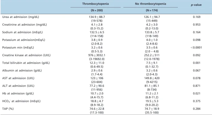
Regarding the laboratory data, patients with thrombocy-topenia at admission presented with hypokalemia (3.2¡0.6
vs. 3.5¡0.6 mEq/L,p,0.0001), hypoalbuminemia (2.9¡0.6
vs. 3.2¡0.6 g/dL, p= 0.067), low hemoglobin levels (10.7¡2.0 vs. 11.2¡2.1 g/dL, p= 0.021) and high total
bilirubin levels (12.3¡11.0 vs. 7.5¡9.1 mg/dL, p= 0.001)
significantly more often than did patients with normal platelet levels (Table 2).
The multivariate analysis showed that hypokalemia (OR: 0.7,p= 0.02), dehydration (OR: 2.1,p= 0.006) and metabolic acidosis (OR: 2.3,p= 0.03) were independent risk factors for thrombocytopenia at admission (Table 3). Lengthy disease (OR: 1.2, p= 0.001) and the presence of AKI (OR: 6.6, p= 0.004) were independent risk factors for thrombocytope-nia during the hospital stay (Table 4).
Death occurred in 22 patients (12.5%) without thrombo-cytopenia and 25 patients (12.6%) with thrombothrombo-cytopenia at admission. Mortality was not associated with the presence of thrombocytopenia at hospital admission (12.5%vs. 12.6%, p= 1.000) or during the hospital stay (12.6% vs. 11.3%, p= 0.748).
Low DBP (OR: 0.9, p= 0.02), advanced age (OR: 1.0, p= 0.001) and oliguria (OR: 5.4,p= 0.006) were independent risk factors for death (Table 5).
& DISCUSSION
between thrombocytopenia and the occurrence of bleeding manifestations (7). In the present study, we evaluated a large number of patients with leptospirosis-associated thrombocy-topenia.
Most studies on leptospirosis have focused on AKI and pulmonary complications. After several outbreaks of pul-monary hemorrhage in association with leptospirosis, attention has shifted to understanding the mechanism of bleeding diathesis in these patients. Initially, thrombocyto-penia in leptospirosis was thought to be mild and rare. However, certain reviews have reported a higher prevalence (12,13). There are several hypotheses about the possible mechanism of this complication: 1) the presence of disseminated intravascular coagulation (12); 2) the partici-pation of cytotoxins (13); and 3) the direct complication of vasculitis, triggered by the Leptospira (14). Further studies are needed to establish the actual pathophysiology of this complication.
The independent risk factors for thrombocytopenia were dehydration, metabolic acidosis and low potassium levels at admission. Low serum potassium was a protective factor against thrombocytopenia, i.e., hyperkalemia is associated with thrombocytopenia. This phenomenon may be due to oliguric AKI and metabolic acidosis because there is no cause-effect relationship between serum potassium and the
occurrence of thrombocytopenia. Dehydration and meta-bolic acidosis are systemic manifestations of leptospirosis that are associated with thrombocytopenia. Hemorrhagic manifestations were more frequent in patients in the thrombocytopenia group. Thrombocytopenia was also associated with poor laboratory findings, such as hypoal-buminemia, lower hemoglobin levels and higher AST and total bilirubin levels. However, this condition did not translate into a worse prognosis. In contrast to our study, Spichler et al. (14) reported that elevated creatinine (.3 mg/dL; OR: 4.2) and total bilirubin (.6 mg/dL; OR: 2.2) levels were associated with a lethal outcome. Turgut et al. (15) showed that there was an inverse correlation between ALT/AST levels and thrombocyte counts (r = –0.360; p= 0.016) and that, consistent with our data, there was no statistically significant correlation between bilirubin levels and thrombocytopenia. Certain researchers have reported that a high serum potassium level at hospital admission was an independent risk factor for mortality (16,17).
In our study, the overall mortality rate was 12.5%, which is comparable with the rate in other studies (17-23). This large range may be due to the variable severity of the clinical picture, which is partly due to differences between
Leptospira strains and the absence of standards for the
Table 1 -Comparison of the demographics and clinical manifestations of leptospirosis patients with and without thrombocytopenia at admission.
Thrombocytopenia No thrombocytopenia p-value
(N = 200) (N = 174)
Age 36.4¡16.3 36.7¡15.0 0.624
(9-82 years) (9-84 years)
Gender
Male 168 (84.0%) 144 (82.8%) 0.781
Female 32 (16.0%) 30 (17.2%)
Length of hospital stay 10.3¡6.9 9.3¡6.2 0.190
Signs and symptoms
Crackles 33 (16.5%) 28 (16.1%) 1.000
Coluria 56 (45.5%) 64 (50%) 0.528
Dehydration 106 (53.0%) 61 (35.3%) 0.001
Diarrhea 79 (39.5%) 75 (43.1%) 0.528
Dyspnea 23 (11.7%) 16 (9.2%) 0.499
Epistaxis 7 (5.7%) 1 (0.8%) 0.033
Fever 194 (97.0%) 165 (94.8%) 0.304
Headache 147 (73.5%) 123 (70.7%) 0.565
Hematemesis 26 (13.0%) 8 (4.6%) 0.006
Hematuria 69 (54.8%) 38 (37.6%) 0.011
Hepatomegaly 65 (32.5%) 43 (24.7%) 0.110
Hypotension 13 (10.6%) 14 (10.9%) 1.000
Jaundice 164 (82.0%) 131 (75.3%) 0.128
Myalgia 183 (91.5%) 147 (84.5%) 0.038
Tachypnea 49 (24.5%) 32 (18.4%) 0.167
Vomiting 144 (72.0%) 111 (63.8%) 0.096
Anorexia 80 (53.7%) 65 (47.4%) 0.344
Asthenia 66 (53.7%) 74 (57.8%) 0.527
Oliguria 33 (19.0%) 46 (23.1%) 0.374
Calf pain 106 (78.5%) 78 (69.0%) 0.109
Abdominal pain 57 (46.3%) 60 (46.9%) 1.000
Metabolic acidosis 36 (18.0%) 16 (9.2%) 0.016
Hypoalbuminemia 35 (17.8%) 13 (7.5%) 0.005
AKI 167(83.9%) 134 (77.5%) 0.145
Need for dialysis 73 (36.5%) 51 (29.3%) 0.153
Penicillin G use 49 (32.0%) 52 (42.6%) 0.079
Death 25 (12.5%) 22 (12.6%) 1.000
diagnostic criteria. In the present study, mortality was not associated with the presence of thrombocytopenia. Other studies also showed that mortality was not associated with the presence of thrombocytopenia (16,17), but Spichler et al. (14) showed that it was associated with lethal outcomes, being an independent risk factor for mortality in leptos-pirosis.
Hemorrhagic manifestations are characteristic of Weil’s disease and are potentially fatal. Patients can develop important hemodynamic abnormalities secondary to hypo-volemia, which is caused by dehydration and the direct effects of Leptospira toxins that damage the vascular endothelium and increase permeability (6). Hemorrhage is recognized as the most important manifestation of human leptospirosis and is being increasingly reported around the world (24). The main hemorrhagic manifestations reported in this study were epistaxis, hemoptysis, hematemesis and hematuria, all of which were more frequent in the patients in the thrombocytopenia group.
AKI in leptospirosis is reported in 40-60% of severe cases (25) and is usually non-oliguric (26,27). However, the
present study found a higher prevalence (80.5%) according to the RIFLE criteria. There were no differences between the two groups regarding renal function or the need for dialysis. This analysis found that lengthy disease and the presence of AKI were associated with thrombocytopenia during the hospital stay. Regarding antibiotic therapy in leptospirosis, in the present study, penicillin G was administered to 152 (41.6%) of the patients, and there was a tendency toward more frequent use of penicillin among those patients with thrombocytopenia.
Thrombocytopenia in leptospirosis is known to be associated with a worse prognosis (9). Tantitanawat and Tanjatham (28) found that platelet counts ,100,000/mm3 were an independent risk factor for death in leptospirosis. However, the present study found similar mortality rates in patients with and without thrombocytopenia. A low DBP, advanced age and oliguria were independent risk factors for death.
In summary, leptospirosis is a globally relevant disease with a potentially fatal outcome. Hemorrhagic complications are common and are reported as main causes of morbidity and mortality in this disease. Although thrombocytopenia
Table 2 -Comparison of laboratory data between leptospirosis patients with and without thrombocytopenia at admission.
Thrombocytopenia No thrombocytopenia p-value
(N = 200) (N = 174)
Urea at admission (mg/dL) 134.9¡88.7 126.1¡94.7 0.169
(19-578) (15-449)
Creatinine at admission (mg/dL) 4.1¡2.8 4.2¡3.0 0.953
(0.3-15.2) (0.2-13.0)
Sodium at admission (mEq/L) 132.5¡6.5 133.8¡5.7 0.164
(114-158) (118-149)
Potassium at admission(mEq/L) 3.8¡0.9 4.0¡1.0 0.098
(2.0-8.2) (2.4-8.6)
Potassium min (mEq/L) 3.2¡0.6 3.5¡0.6 ,0.0001
(0.5-5.3) (2.0 – 4.8)
Creatine kinase at admission (UI/L) 976¡3032.1 252.2¡511 0.092
(3-19692.0) (12.0-1978)
Total bilirubin at admission (g/dL) 12.3¡11.0 7.5¡9.1 0.001
(0.6-49.5) (0.1-32.7)
Albumin at admission (g/dL) 2.9¡0.6 3.2¡0.6 0.067
(1.7-4.4) (2.0-4.3)
AST at admission (UI/L) 123¡106 149.8¡420 0.078
(23-604) (9-4215)
ALT at admission (UI/L) 77.2¡90.6 81.1¡85.1 0.871
(11-956) (8-734)
Hb at admission (g/dL) 10.7¡2.0 11.2¡2.1 0.021
(4.4-15.7) (6.8-11.2)
HCO3-at admission (mEq/L) 18.8¡4.7 19.5¡5.3 0.375
(8.9-18.2) (9.0-20.2)
TAP (%) 74.6¡22.8 74.7¡18.9 0.284
(17.3-100) (35.5-100)
The data are shown as the mean¡SD and range (minimum-maximum) or as numbers with percentages in parentheses. Significance,p,0.05. Student’s t-test and Fisher’s exact test.
Table 3 -Independent risk factors for thrombocytopenia in patients with leptospirosis at admission.
OR 95% CI p-value
Low potassium level 0.7 0.5-0.9 0.02
Dehydration 2.1 1.2-3.5 0.006
Metabolic acidosis 2.3 1.0-5.4 0.03
OR: odds ratio; 95% CI: 95% confidence interval. Multivariate analysis; chi-square test. Significance,p,0.05.
Table 4 -Independent risk factors for thrombocytopenia in patients with leptospirosis during their hospital stay.
OR 95% CI p-value
Lengthy disease 1.2 1.0-1.4 0.001
AKI 6.6 1.8-23 0.004
was associated with mortality in previous studies, in the present study, this complication was not a risk factor for death. Advanced age and oliguria were independent risk factors for death.
& ACKNOWLEDGMENTS
We are very grateful to the team of physicians, residents, medical students and nurses at the Walter Cantı´dio University Hospital and the Sa˜o Jose´ Infectious Diseases Hospital for the assistance that they provided to the patients and for the technical support that they provided for the development of this research. This research was supported by Conselho Nacional de Desenvolvimento Cientı´fico e Tecnolo´gico, CNPq (Brazilian Research Council).
& AUTHOR CONTRIBUTIONS
All authors designed and performed the study. Daher EF, Mota JA and Silva Junior GB wrote the article. All authors have read and approved the final version of the manuscript.
& REFERENCES
1. Adler B, de la Pen˜a Moctezuma A. Leptospira and leptospirosis. Vet Microbiol. 2010;140(3-4):287-96, http://dx.doi.org/10.1016/j.vetmic.2009. 03.012.
2. Bharti AR, Nally JE, Ricaldi JN, Matthias MA, Diaz MM, Lovett MA, et al. Leptospirosis: a zoonotic disease of global importance. Lancet Infect Dis. 2003;3(12):757-71, http://dx.doi.org/10.1016/S1473-3099(03)00830-2. 3. Pappas G, Papadimitriou P, Siozopoulou V, Christou L, Akritidis. The
globalization of leptospirosis: worldwide incidence trends. Int J Infect Dis. 2008;12(4):351-7.
4. McBride AJ, Athanazio DA, Reis MG, Ko AI. Leptospirosis. Curr Opin Infect Dis. 2005;18(5):376-86, http://dx.doi.org/10.1097/01.qco.0000178824. 05715.2c.
5. Vinetz JM. Leptospirosis. Curr Opin Infect Dis. 2001;14(5):527-38, http:// dx.doi.org/10.1097/00001432-200110000-00005.
6. Nicodemo AC, Duarte MIS, Alves VAE, Takakura CEH, Santos RTM, Nicodemo EL. Lung lesions in human leptospirosis, Microscopic, immmunohistochemical and ultrastructural features related to thrombo-cytopenia. Am J Trop Med Hyg. 1997;56(2):181-7.
7. Casiple LC. Thrombocytopenia and Bleeding In Leptospirosis. Phil J Microbiol Infect Dis. 1998;27(1):18-22.
8. Daher EF, Lima RSA, Silva Junior GB, Silva EC, Karbage NN, Kataoka RS, et al. Clinical presentation of leptospirosis: a retrospective study of
201 patients in a metropolitan city of Brazil. Braz J Infect Dis. 2010;14(1):3-10.
9. Edwards CN, Nicholson GD, Everard CO. Thrombocytopenia in leptospirosis. Am J Trop Med Hyg. 1982;31(4):827-9.
10. Kellum JA, Bellomo R, Ronco C. Definition and classification of acute kidney injury. Nephron Clin Pract. 2008;109(4):c182-7, http://dx.doi. org/10.1159/000142926.
11. Vijayachari P, Sugunan AP, Shriram. Leptospirosis: an emerging global public health problem. J Biosci. 2008;33(4):557-69.
12. Sitprija V, Pipatanagul V, Mertowidjojo K, Boonpucknavig V, Boonpucknavig S. Pathogenesis of renal disease: clinical and experi-mental studies. Kidney Int. 1980;17(6):827-36, http://dx.doi.org/10. 1038/ki.1980.95.
13. O9Neil K, Richman LS, Lazarus AA. Pulmonary manifestations of leptospirosis. Rev Infect Dis. 1991;13(4):705-9, http://dx.doi.org/10.1093/ clinids/13.4.705.
14. Spichler AS, Vilaca PJ, Athanazio DA, Albuquerque JO, Buzzar M, Castro B, et al. Predictors of lethality in severe leptospirosis in urban Brazil. Am J Trop Med Hyg. 2008;79(6):911-4.
15. Turgut M, Sunbul M, Bayirli D, Bilge A, Leblebicioglu H, Haznedaroglu I. Thrombocytopenia complicating the clinical course of leptospiral infection. J Int Med Res. 2002;30(5):535-40, http://dx.doi.org/10.1177/ 147323000203000511.
16. Esen S, Sunbul M, Leblebicioglu H, Eroglu C, Turan D. Impact of clinical and laboratory findings on prognosis in leptospirosis. Swiss Med Wkly. 2004;134(23-24):347-52.
17. Dupont H, Dupont-Perdrizet D, Perie JL, Zehner-Hansen S, Jarrige B, Daijardin JB. Leptospirosis: prognostic factors associated with mortality. Clin Infect Dis. 1997;25(3):720-4, http://dx.doi.org/10.1086/513767. 18. Ko AI, Galvao Reis M, Ribeiro Dourado CM, Johnson WD Jr, Riley LW.
Urban epidemic of severe leptospirosis in Brazil. Salvador Leptospirosis Study Group. Lancet. 1999;354(9181):820-5.
19. Daher EF, Zanetta DM, Cavalcante MB, Abdulkader RC. Risk factors for death and changing patterns in leptospirosis acute renal failure. Am J Trop Med Hyg. 1999;61(4):630-4.
20. Pappachan MJ, Mathew S, Aravindan KP, Khader A, Bharghavan PV, Kareem MM, et al. Risk factors for mortality in patients with leptospirosis during an epidemic in northern Kerala. Natl Med J India. 2004;17(5):240-2.
21. Kuriakose M, Eapen CK, Paul R. Leptospirosis in Kolenchery, Kerala, India: epidemiology, prevalent local serogroups and serovars and a new serovar. Eur J Epidemiol 1997;13(6):691-697.
22. Pinn TG. Leptospirosis in the Seychelles. Med J Aust. 1992;156(3):163–7. 23. Park SK, Lee SH, Rhee YK, Kang SK, Kim KJ, Kim MC, et al. Leptospirosis in Chonbuk Province of Korea in 1987: a study of 93 patients. Am J Trop Med Hyg. 1989;41(3):345-51.
24. Wagenaar JFP, Goris MGA, Partiningrum DL, Isbandrio B, Hartskeerl RA, Brandjes DP, et al. Coagulation disorders in patients with severe leptospirosis are associated with severe bleeding and mortality. Trop Med Int Health. 2010;15(2):152-9, http://dx.doi.org/10.1111/j.1365-3156. 2009.02434.x.
25. Andrade L, Daher EF, Seguro AC. Leptospiral nephropathy. Semin Nephrol. 2008;28(4):383-94, http://dx.doi.org/10.1016/j.semnephrol.2008. 04.008.
26. Daher EF, Zanetta DM, Abdulkader RC. Pattern of renal function recovery after leptospirosis acute renal failure. Nephron Clin Pract. 2004;98(1):8-14, http://dx.doi.org/10.1159/000079922.
27. Seguro AC, Lomar AV, Rocha AS. Acute renal failure of leptospirosis: nonoliguric and hypokalemic forms. Nephron. 1990;55(2):146-51, http:// dx.doi.org/10.1159/000185943.
28. Tantitanawat S, Tanjatham S. Prognostic factors associated with severe leptospirosis. J Med Assoc Thai. 2003;86(10):925-31.
Table 5 -Independent risk factors for death in patients with leptospirosis.
OR 95% CI p-value
Diastolic blood pressure 0.9 0.8-0.9 0.02
Advanced age 1.0 1.0-1.1 0.001
Oliguria 5.4 1.6-18 0.006