Generates Non Amyloidal Structures
Sudeshna Ghosh, Nitin Kumar Pandey, Atanu Singha Roy, Debi Ranjan Tripathy, Amit Kumar Dinda,
Swagata Dasgupta
*Department of Chemistry, Indian Institute of Technology Kharagpur, Kharagpur, India
Abstract
Glycation causes severe damage to protein structure that could lead to amyloid formation in special cases. Here in this report, we have shown for the first time that hen egg white lysozyme (HEWL) does not undergo amyloid formation even after prolonged glycation in the presence of D-glucose, D-fructose and D-ribose. Cross-linked oligomers were formed in all the cases and ribose was found to be the most potent among the three sugars. Ribose mediated oligomers, however, exhibit Thioflavin T binding properties although microscopic images clearly show amorphous and globular morphology of the aggregates. Our study demonstrates that the structural damage of hen egg white lysozyme due to glycation generates unstructured aggregates.
Citation: Ghosh S, Pandey NK, Singha Roy A, Tripathy DR, Dinda AK, et al. (β01γ) Prolonged Glycation of Hen Egg White Lysozyme Generates Non Amyloidal Structures. PLoS ONE 8(9): e74γγ6. doi:10.1γ71/journal.pone.0074γγ6
Editor: Rizwan H. Khan, Aligarh Muslim University, India
Received April β5, β01γ; Accepted August β, β01γ; Published September 16, β01γ
Copyright: © β01γ Ghosh et al. This is an open-access article distributed under the terms of the Creative Commons Attribution License, which permits unrestricted use, distribution, and reproduction in any medium, provided the original author and source are credited.
Funding: SD is grateful to the Department of Science and Technology, Government of India for financial assistance for the chemicals and reagents. SG, ASR, DRT and AKD thank Council of Scientific and Industrial Research, New Delhi and NKP thank IIT Kharagpur for their senior research fellowship respectively. The funders had no role in study design, data collection and analysis, decision to publish, or preparation of the manuscript.
Competing interests: The authors have declared that no competing interests exist. * E-mail: [email protected]
Introduction
Non-enzymatic glycation of proteins is a post-translational modification process which involves covalent bond formation between free amino groups of proteins and reducing sugars [1]. It results in the modification of proteins which leads to the formation of groups of heterogeneous compounds commonly referred to as advanced glycation end products (AGEs) [β,γ]. AGEs play a crucial role in aging processes and debilitating diseases such as Alzheimer’s disease [4]. Conformational alterations in the protein structure result due to glycation, which affects the physical and functional properties of proteins [5]. Glycation can also lead to protein aggregation [4]. Formation of toxic protein aggregates commonly known as amyloid fibrils, is associated with neurological disorders such as Alzheimer’s, transmissible spongiform encephalopathies etc [6,7]. AGE modified amyloid fibrils have been found in the brain tissues of patients suffering from Alzheimer’s, transmissible spongiform encephalopathy and the islets of Langerhans of diabetic patients [8-10]. Fibrils possess a common core crossed -sheet structural motif [11] and glycation facilitates the formation of the cross-linked beta structure [4]. Moreover, a recent report has shown that the glycated A 1-4β peptide with an altered
secondary structure is more toxic with respect to the pathogenesis associated with Alzheimer’s disease [1β]. These
findings have encouraged researchers to investigate the structural changes of proteins that evolve due to glycation and to explore the possible link between glycation of proteins and amyloid formation.
Hen egg white lysozyme (HEWL) is known to possess a well-defined three dimensional structure, folding mechanism and thermodynamic parameters [1γ-16]. Human lysozyme is a structural homologue of HEWL, which is known to be involved in a systemic amyloidosis disease due to a point mutation in the lysozyme gene [17]. Fibrils obtained from human lysozyme
in vitro were found to be similar with those obtained from patients and also fibrils obtained from human lysozyme bear a notable resemblance with HEWL fibrils [18,19]. A recent report has shown that human lysozyme fibrils can induce the secretion of innate immune receptors where the cross -sheet structure plays a crucial role [β0]. Membrane activity and the ability of inducing apoptosis in neuroblastoma cells by HEWL fibrils support the use of HEWL as a suitable model to study the mechanistic aspects of amyloid formation in vitro [β1,ββ].
Several studies have revealed the involvement of different proteins in the formation of AGEs and their relation with protein aggregation and amyloid formation [4,γ1-γ8]. Based on the information above and considering the role of lysozyme in our natural immune system [γ9, 40], we have chosen HEWL to
examine the effect of glycation and its consequences on HEWL aggregation in vitro. In the present article, we have treated HEWL over a prolonged period (~180 days) in the presence of D-glucose (glucose), D-fructose (fructose) and D-ribose (ribose) at pH 7.4 at γ7 °C. Glycation of HEWL was characterized using different spectroscopic and microscopic techniques. HEWL was found to form cross-linked oligomeric species in the presence of all the three sugars. Ribose was found to exert the most proficient role in comparison to fructose and glucose. Our study demonstrates that glycation of HEWL promotes aggregation but no fibrillar species was observed. Recent studies have shown that methylglyoxal (glycating agent) favors formation of native like aggregates of insulin [41] and cytochrome c [4β]. This study will be beneficial in terms of understanding the effects of glycation on the structural aspects of proteins and its consequences in protein aggregation.
Materials and Methods
Materials
Hen Egg White Lysozyme (HEWL), Thioflavin T (ThT) were purchased from Sigma Chemical Co. (St. Louis, USA) and used as received. D-glucose, D-fructose and D-ribose and all other chemicals were obtained from SRL (India). Protein molecular weight marker (14.γ-97.4 kDa) was purchased from Bangalore, Genei.
Preparation of glycated HEWL: prolonged incubation to facilitate aggregation
HEWL (10 mg/ml) [εβ80=γ7646 M-1cm-1] [4γ] was incubated in
0.1 M sodium phosphate buffer of pH 7.4 at γ7 °C for γ0 days in the absence and presence of three reducing sugars such as D-glucose (glucose), D-fructose (fructose) and D-ribose (ribose) of 0.5 M concentration in each case. This incubation was extended up to 180 days to promote aggregation. Sodium azide (1 mM) was used to prevent bacterial growth. Incubation process was carried out under sterile conditions. Aliquots were withdrawn at definite time intervals and sodium phosphate buffer (10 mM) of pH 7.4 was used for dilution to achieve the final desired concentration of protein in each study. In each study, the control is native HEWL incubated in the absence of sugars at pH 7.4 at γ7 °C keeping the other conditions similar to that of sets (HEWL-glucose/ HEWL-fructose/ HEWL-ribose).
Fluorescence measurements: steady state fluorescence
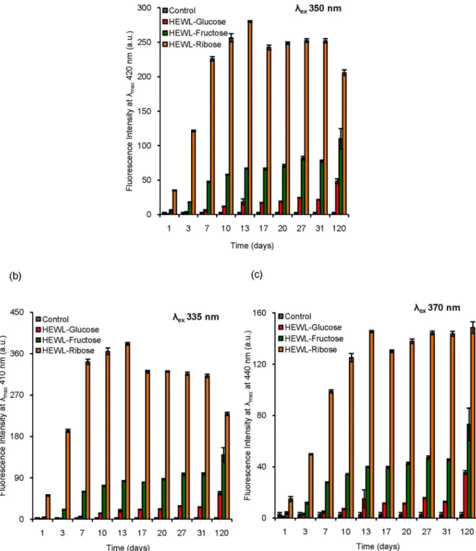
The formation of AGEs was monitored using different fluorescence spectroscopic techniques. At definite intervals (in days), aliquots were withdrawn and used for fluorescence measurements using a final protein concentration of 5 µM. Fluorescence of the samples was monitored using excitation wavelengths of β95, γγ5, γ50 and γ70 nm respectively in a
Horiba Jobin Yvon Fluoromax-4 spectrofluorimeter. Slit width and integration time were kept at 5 nm and 0.β sec respectively. Tryptophan (Trp) fluorescence was monitored using λex of β95 nm ensuring no contribution from Tyrosine
(Tyr) residues. Measurement of fluorescence intensity using excitation and emission at γ50 nm and 450 nm respectively indicates formation of AGEs [44]. Excitation at γγ5 nm and γ70 nm correspond to the formation of two AGE products such as pentosidine and malondialdehyde (MDA) [45,46]. The fluorescence intensity values corresponding to different sets (HEWL-glucose/HEWL-fructose/HEWL-ribose) have been corrected with respect to the blank (glucose/fructose/ribose) and thus represent the difference fluorescence intensity in each case.
Synchronous fluorescence study
Aliquots were withdrawn from HEWL solutions incubated in the presence of glucose, fructose and ribose respectively after an incubation of γ1 days at pH 7.4 at γ7 °C to examine the synchronous fluorescence. Samples were scanned using a protein concentration of 10 µM between β00 to 600 nm keeping the offset value (Δλ) at 40 nm in Horiba Jobin Yvon Fluoromax 4 spectrofluorimeter [47]. Slit width and integration time were kept at 5 nm and 0.β sec respectively. The number of band components and respective peak positions were determined from the second derivative spectra.
Sodium dodecyl sulfate-polyacrylamide gel electrophoresis (SDS-PAGE)
SDS-PAGE was carried out under reducing conditions. Aliquots (100 µM) withdrawn at different times were mixed with sample buffer (βX) containing sodium dodecyl sulfate (SDS) (4 g), bromophenol blue (1%, 4 ml), -mercaptoethanol (10 ml), glycerol (β0 ml) and Tris–HCl [pH 6.8, 1 M, 1β.5 ml] (the volume was adjusted to 100 ml with water for the sample buffer). Prior to loading, samples were boiled for β min and then applied to a 15% resolving gel and electrophoresis conducted in a Mini Dual vertical electrophoresis unit (Tarson). Gels were stained with Coomassie brilliant blue (SRL, India) and destained using a mixture of CH γCOOH/MeOH/H βO (γ7.5
ml, β5 ml, 4γ0 ml) with gentle agitation.
Circular dichroism (CD) measurements
Aliquots withdrawn at definite time intervals were scanned in a Jasco-810 spectrophotometer. Far UV–CD spectra were accumulated between 190 to β40 nm at a scan rate of 50 nm/min keeping the protein concentration at β0 µM using a quartz cuvette having a path length of 0.1 cm. Protein secondary structure contents were estimated using an online server DICHROWEB [48]. Near UV–CD spectra were acquired between β50 to γ50 nm at a scan rate of 100 nm/min keeping protein concentration of 100 µM. Sodium phosphate buffer (10 mM) of pH 7.4 was used for dilution in each case.
SDS-PAGE: detection of glycoprotein using fuchsin staining
SDS-PAGE was performed as mentioned earlier in the previous section. After the completion of the gel run, it was removed and placed in 8%/6% (v/v) AcOH/MeOH solution for gel fixing. The gel was then washed with an oxidizing agent (Sodium periodate in the presence of β drops of concentrated HβSO4) for 15 min. The gel was washed with γ% (v/v) AcOH
solution twice for 5 min followed by Fuchsin staining. Destaining was achieved using sodium bisulfite (NaHSOγ) for
10 min and the gel finally washed with γ% (v/v) AcOH solution to acquire the required staining. Horseradish peroxidase was used as a marker as it is a glycoprotein and develops magenta color in the presence of Fuchsin staining [49, 50].
Matrix assisted laser desorption ionization-time of flight (MALDI-TOF)
Incubated HEWL solutions were diluted up to a concentration of 100 µM and then mixed with matrix (saturated solution of sinapic acid in 50% (v/v) CHγCN/HβO solution) in a 1:1 ratio.
Samples were then placed on the spot plate, air dried and spectra obtained in a VOYAGER-DE PRO instrument.
Thioflavin T (ThT) fluorescence
ThT is an amyloid marker dye which intensely fluoresces at ~485 nm upon binding with amyloid fibrils [51,5β]. Aliquots from each set were withdrawn at definite interval of days and mixed with ThT to accomplish final protein and dye concentrations of β µM and 5 µM respectively. Samples were incubated for β min and scanned in a Horiba Jobin Yvon Fluoromax 4 spectrofluorimeter. Excitation and emission maxima were kept at 450 nm and 485 nm respectively. Slit width and integration time were kept at 5 nm and 0.γ sec respectively. Sodium phosphate buffer (10 mM) of pH 7.4 was used for dilution and spectra were corrected with respect to the corresponding blank.
Fluorescence microscopy
ThT (10 µl of 1 mM) was mixed well with the glycated protein solutions (5 µl) after γ0 days and 180 days of incubation to achieve the required staining and placed on a glass slide after covering with a cover slip. Images were obtained using a Leica DM β500M microscope equipped with a fluorescence attachment. Filter cube no β (Leica Iγ 1151γ878, BZ: 01) was used for ThT excitation and emission. The images were acquired with a Leica DFC γ10 FX camera attached with the microscope. All observations were performed at 10X/0.β5 (N PLAN EPI).
Field emission scanning electron microscopy (FESEM)
Aliquots were withdrawn (β µl) from each set after an incubation of 60 days at γ7 °C at pH 7.4 in the absence and presence of sugars and placed on glass pieces (thoroughly cleaned), air dried and gold coated. Samples were then scanned using a Carl Zeiss field emission electron microscope operating at 5 kV.
Transmission electron microscopy (TEM)
HEWL solutions incubated in the absence and presence of sugars at γ7 °C at pH 7.4 after 180 days of incubation were diluted to a final concentration of 50 µM and placed on TEM grids. Uranyl acetate [1% (w/v)] was used to accomplish the required staining, air dried and scanned in a TECNAI Gβ
β0S-TWIN transmission electron microscope operating at an accelerating voltage of 80 kV.
Results and Discussion
Glycation results in the structural alteration of proteins. Formation of crossed beta structures in proteins is often associated with glycation and even amyloidal aggregates have been observed as a result of glycation [4,γ1,γβ]. Studies have revealed a relationship between the effect of glycation on protein structure, protein aggregation, and fibrillation [γ1-γ8]. Recent studies have shown the presence of high levels of AGEs in the brains of patients suffering from neurological disorders and their involvement in amyloid deposition [8,5γ]. In addition, glycation has been shown to enhance the severity linked with the neurotoxicity of A 1-4β peptide [1β]. Sugars such
as glucose, fructose and ribose are common reducing sugars which are well known glycating agents among which the effect of ribose is less studied. In the present article, we have investigated whether prolonged glycation of HEWL can lead to amyloid formation as no such report is available till date, to the best of our knowledge, in case of HEWL. We have used different spectroscopic and microscopic techniques to examine glycation of HEWL and its consequences in aggregation of HEWL in the presence of three reducing sugars such as glucose, fructose and ribose over a prolonged period.
Fluorescence study: steady state measurements
HEWL was subjected to prolonged glycation in the presence of glucose, fructose and ribose to observe whether glycation could lead to amyloid formation of HEWL. Steady state fluorescence methods were employed to investigate structural changes of HEWL during the incubation period. Formation of AGEs is monitored using excitation and emission at γ50 nm and 450 nm respectively [44]. Two different AGEs, pentosidine and malondialdehyde (MDA) formation can be monitored using λex values of γγ5 nm and γ70 nm respectively [45,46]. We have
measured the fluorescence properties of incubated HEWL solutions using λex values of β95 nm, γγ5 nm, γ50 nm and γ70
nm respectively [Figure 1 and Figure β]. We have found a considerable reduction in Trp (λex=β95 nm) fluorescence with
increasing incubation period (~1β0 days) in the presence of each sugar [Figure 1 (a)]. The relative reduction in Trp fluorescence intensity with respect to the control is maximum for ribose (~8γ% loss) followed by fructose (~60% loss) and glucose (~51% loss). The Trp fluorescence spectra of HEWL solutions after an incubation of γ1 days are given in Figure 1 (b). We have noticed that λmax of Trp fluorescence of the control
remains the same as would have been expected [Figure 1 (b)]. In the presence of glucose, no prominent shift in λmax was found
indicative of an exposure of Trp residues of HEWL to a more polar medium in presence of sugars which is most pronounced in case of ribose [Figure 1 (b)]. Earlier studies have also shown a loss in Trp fluorescence of human serum albumin (HSA) and -crystallin due to glycation [54,55]. The red shift in λmax of Trp
fluorescence spectrum of -crystallin occurs on glycation which has been attributed to exposure of Trp residues to the solvent medium that is similar to the results obtained in this study [55]. We have found that on excitation at γ50 nm (indicative of other types of AGEs), incubated HEWL solutions show strong fluorescence in each case (λem=4β0 nm) [Figure β (a)]. HEWL
solutions, upon excitation at γγ5 nm (λem=410 nm) and γ70 nm
(λem=440 nm) [Figure β (b) and (c)] exhibit intense fluorescence
which also increases with increasing incubation period. This clearly indicates the formation of two specific AGE products, pentosidine and malondialdehyde (MDA) respectively. In each case, we have noticed that the formation of AGEs reaches a maximum in the presence of ribose implying that ribose plays the most effective role among the sugars used. This is in good agreement with the previously observed fact that the glycating ability of these sugars lies in the order: D-glucose < D-fructose < D-ribose [56]. The higher glycation ability of ribose is
attributed to its puckered aldopentose ring structure, which makes it more reactive towards the amino groups in comparison to the other sugars [57,58]. Therefore, it is evident that treatment with sugars such as glucose, fructose and ribose cause prominent structural changes of HEWL along with the formation of AGE products where the effect of ribose is predominant.
Synchronous fluorescence
Synchronous fluorescence studies have been performed to get more detail about the fluorescence property of treated HEWL solutions after γ1 days of incubation at γ7 °C at pH 7.4. We have noticed that the control does not show any peak in the range from γ50 to 550 nm; whereas in the presence of sugars two major peaks are observed [Figure γ (a)]. The peak near γ80 nm is more prominent compared to the other peak near ~415-4β0 nm in all the cases [Figure γ (a)]. The maximum intensity of the peak at γ80 nm was obtained in the presence of ribose followed by fructose and glucose [Figure γ (a)]. Our observation is similar to that found earlier in AGE formation of bovine serum albumin (BSA) where ribose was found to be the most potent in generating the γ80 nm band [59]. This signifies
Figure 1. Determination of Trp fluorescence on glycation of HEWL in the presence of different sugars using fluorescence spectroscopy. (a) Histogram represents Trp fluorescence intensity of different HEWL solutions incubated in the presence of glucose, fructose and ribose respectively over a period of 1β0 days. (b) Representative Trp fluorescence spectra of different HEWL solutions incubated in the presence of glucose, fructose and ribose respectively obtained after an incubation of γ1 days. Control represents native HEWL incubated in the absence of sugars at pH 7.4 at γ7 °C keeping other conditions similar as that of sets in each case.
doi: 10.1γ71/journal.pone.0074γγ6.g001
Figure 2. Characterization of different AGE products formed during glycation of HEWL in the presence of different sugars using fluorescence spectroscopy. Histograms represent fluorescence intensity of different HEWL solutions incubated in the presence of glucose, fructose and ribose respectively over a period of 1β0 days. Formation of different AGE products such as (a) other AGE products (λex=γ50 nm), (b) pentosidine (λex=γγ5 nm) and (c) malondialdehyde (MDA) (λex=γ70 nm). [HEWL]=5 µM in
each case. Control represents native HEWL incubated in the absence of sugars at pH 7.4 at γ7 °C keeping other conditions similar as that of sets in each case.
that the structural changes of proteins are most prominent in the presence of ribose followed by fructose and glucose. To further illustrate this result, we have plotted the second derivative spectra of synchronous fluorescence in each case. We have found two peaks, one at ~γ75-γ80 nm and another one at ~415-4β0 nm respectively [Figure γ (b)]. Synchronous fluorescence reveals that the effect of ribose is most dominant among the three sugars used which are in accordance with the steady state fluorescence studies.
Formation of higher oligomers: SDS PAGE
To monitor the formation of HEWL aggregates in presence of sugars, SDS-PAGE has been performed under reducing conditions. We have found that after an incubation of 1 day, in the presence of glucose, only monomeric HEWL exists (lanes β-4) [Figure 4 (a)]. On the other hand, in the presence of fructose, dimeric species are also formed (lane 5-7) [Figure 4 (a)]. Ribose imparts the most noteworthy effect which is clear from SDS-PAGE as trimeric species are formed within a day (lanes β-4) [Figure 4 (b)]. After β0 days of incubation we have found that even in the presence of glucose, both dimer and trimers were generated (lanes β-4) [Figure 4 (a)]. Trimer and tetramer formation was also observed in the presence of fructose (lanes 5-7) [Figure 4 (a)] which is most prominent in case of ribose as expected (lanes β-4) [Figure 4 (b)]. After
incubation of γ1 days, this becomes more evident as ribose shows formation of oligomeric species in HEWL most effectively followed by fructose and glucose [Figure 4]. This scenario becomes distinctly clear upon quantitation of oligomer formation via densitometric analysis of SDS-PAGE [Figure 5]. We have found that the presence of ribose results in the formation of dimeric species to a higher extent as compared to fructose after 1 day of incubation [Figure 5 (a)]. After β0 days and γ1 days of incubation, the formation of higher oligomeric species was found to be maximum in the presence of ribose and minimum in presence of glucose [Figure 5 (b) and (c)]. In each case the relative concentration of oligomers varies in the order: dimer > trimer > tetramer. Therefore, it appears that aggregated cross-linked products of HEWL are indeed formed in the presence of all the three sugars with ribose playing the most potent role. Our observations are similar to those found earlier in the fructosylation reaction of hemoglobin, where the presence of a cross-linked aggregated population was noticed [60].
Circular dichroism study: conformational aspects of glycation
Circular dichroism is a useful technique to explore conformational changes in protein structure, especially α-helix to -sheet transformations [61]. The far UV-CD spectrum of
Figure 3. Synchronous fluorescence characteristics of HEWL during glycation in the presence of different sugars. (a) Synchronous fluorescence spectra of control and HEWL solutions treated in the presence of glucose, fructose and ribose respectively after γ1 days of incubation at γ7 °C at pH 7.4; (b) Second derivative plot of synchronous fluorescence spectra of HEWL solutions incubated in the presence of glucose, fructose and ribose respectively obtained after γ1 days of incubation at γ7 °C at pH 7.4. Protein concentration=10 µM and Δλ=40 nm in each case.
doi: 10.1γ71/journal.pone.0074γγ6.g00γ
control shows a minimum at β08 nm [Figure 6], which is indicative of α-helical structure [6β]. We have noticed that in the presence of glucose, fructose and ribose, the CD mdeg value at β08 nm decreases with increasing incubation period which points toward a loss in helicity [Figure 6]. Lowering in the mdeg
value at β08 nm is relatively less in the presence of glucose which is more pronounced in the presence of fructose and ribose. Thus presence of sugars causes prominent secondary structural changes in HEWL (loss in helicity) that is shown to be more effective in the presence of ribose. Quantitative
Figure 4. Detection of oligomerization of HEWL during glycation in the presence of glucose, fructose and ribose. Representative SDS polyacrylamide gel electrophoresis of different HEWL solutions obtained after incubation at pH 7.4 at γ7 °C in the presence of different sugars at definite intervals of time (1 day, β0 days and γ1 days respectively). (a) In each case lane 1: molwt marker; lane β-4: HEWL-glucose; lane: 5-7: HEWL-fructose respectively; (b) In each case lane 1: molwt marker; lane β-4: HEWL-ribose; lane: 5: Control (native HEWL incubated at pH 7.4 at γ7 °C in the absence of sugars) respectively.
analysis of CD spectra reveals that with increasing incubation period, there is an increase in % -sheet content of HEWL solutions in presence of all the sugars [Figure 7]. Though increasing trend is more or less maintained throughout the
incubation period, we have found slight fluctuation in the % -sheet content of HEWL solutions in presence of three sugars. Earlier reports have revealed that glycation over a longer period has led to cross-linked beta sheet structure which might
Figure 5. Densitometric analysis of SDS-PAGE. Histograms represent relative mean band intensity of different oligomeric species (dimer, trimer and tetramer) with respect to their corresponding monomer at definite time intervals (a) 1 day, (b) β0 days and (c) γ1 days respectively.
doi: 10.1γ71/journal.pone.0074γγ6.g005
be true in this case as well [4]. Near UV-CD spectra of control shows maxima around ~β8β, ~β85 and ~β95 nm respectively [Figure 8]. Near UV-CD spectra of HEWL solutions is disrupted to the greatest extent in the presence of ribose followed by fructose and glucose. Thus it becomes evident that maximum perturbation of the tertiary structure of HEWL occurs in the presence of ribose followed by fructose and glucose [Figure 8]. However, in presence of glucose, minimum changes in shape of the spectrum were observed. Thus ribose exhibits most potent role in affecting protein structural changes in comparison to fructose and glucose. This is in accordance with the findings obtained from steady state fluorescence measurements where we have found the maximum red shift in λmax of Trp fluorescence spectrum of HEWL in presence of
ribose.
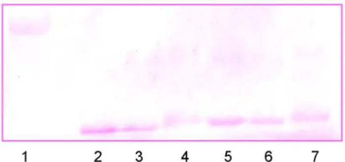
Detection of glycated proteins: Fuchsin based SDS-PAGE
SDS-PAGE has been performed (under reducing conditions) using Fuchsin staining to determine the attachment of the sugar moiety to HEWL. Periodic acid-Schiff base (PAS) reaction is the governing protocol, where periodic acid oxidizes vicinal diol groups to form aldehyde followed by reaction with Schiff reagent to generate a magenta color. This staining is achieved with fuchsin [50]. We have found that horseradish peroxidase, the marker protein is stained in the presence of fuchsin producing a magenta color as would have been expected [Figure 9]. The controls in this experiment are native HEWL and the individual sugars. It was found that they do not develop any color in the Fuchsin based SDS-PAGE assay [Figure S1]. After an incubation of γ1 days, HEWL solutions in the presence of glucose (lane β and 5), fructose (lane γ and 6) and ribose (lane 4 and 7) subjected to SDS-PAGE (fuchsin based) develop a magenta color similar to that of horseradish peroxidase [Figure 9]. This clearly indicates the attachment of sugar moiety to HEWL. This further confirms the formation of AGEs in HEWL in the presence of all the sugars which is strongly supported by other studies as well. To further ensure addition of sugar moiety to HEWL, we have performed MALDI-TOF experiments and found that in each case the molecular mass of native HEWL [Figure 10 (a)] increases which corresponds to the attachment of ~6 glucose [Figure 10 (b)], ~9 fructose [Figure 10 (c)] and ~1γ ribose [Figure 10 (d)] sugar moieties respectively.
ThT fluorescence study: nature of the cross-linked aggregates
We have found that cross-linked oligomers of HEWL formed during treatment with sugars. We have also noticed that the % -sheet content of HEWL solutions increases during the incubation period. This has encouraged us to investigate the ThT binding property of these aggregates. ThT is an amyloid specific dye which fluoresces strongly upon binding with amyloid fibrils [51,5β]. We have found that up to ~180 days of incubation, there is no significant change in ThT fluorescence for HEWL solutions in the presence of glucose [Figure 11]. HEWL solutions in the presence of fructose show slightly more ThT fluorescence which is ~β.5 times compared to the control
solution (native HEWL incubated at pH 7.4 at γ7 °C keeping the other conditions similar to that of sets incubated with the individual sugars) [Figure 11]. Ribose treated HEWL solutions
Figure 9. Identification of attachment of sugar moieties to
HEWL: Fuchsin based SDS PAGE. Representative SDS
polyacrylamide gel electrophoresis of different HEWL solutions obtained after incubation at pH 7.4 at γ7 °C for γ1 days in the presence of different sugars using Fuchsin staining. lane 1: Horseradish peroxidase; lane β and 5: HEWL-glucose; lane γ and 6: HEWL-fructose; lane 4 and 7: HEWL-ribose.
doi: 10.1γ71/journal.pone.0074γγ6.g009
Figure 10. Determination of the mass of glycated HEWL. MALDI TOF spectra of different HEWL solutions obtained after an incubation of γ1 days at pH 7.4 at γ7 °C (a) Native HEWL (14γ45.γ0 Da) (b) HEWL-glucose (15401.ββ Da) (c) HEWL-fructose (15955.81 Da) (d) HEWL-ribose (16βγγ.48 Da).
show a prominent ThT fluorescence (~5 times compared to the control) for up to ~γ1 days. After γ1 days of incubation we observe a decreasing trend in the ThT intensity (HEWL-ribose solutions) up to ~180 days which then becomes similar to that in the presence of fructose. Thus it appears that HEWL oligomers formed in the presence of ribose possess ThT
binding property, other oligomeric species formed in the presence of fructose and glucose do not bind prominently with ThT. We speculate that oligomeric aggregates formed in the presence of glucose and fructose might be amorphous whereas HEWL solutions treated in the presence of ribose might be fibrillar in nature.
Figure 6. Variation of secondary structural components during glycation of HEWL in the presence of different sugars. Representative far UV-CD spectra of (a) HEWL-glucose, (b) HEWL-fructose and (c) HEWL-ribose solutions respectively obtained after incubation at pH 7.4 at γ7 °C at different time intervals. [HEWL]=β0 µM in each case.
doi: 10.1γ71/journal.pone.0074γγ6.g006
Fluorescence microscopy, FESEM and TEM
Microscopic studies were undertaken to analyze the nature of the HEWL aggregates formed. We have found that after γ0 days of incubation none of the treated HEWL solutions (in the presence of three sugars) exhibited any prominent fluorescence, not even in the presence of ribose [Figure Sβ]. Thus HEWL aggregates formed, during the course of incubation in presence of sugars are amorphous in nature. However, after an incubation of 180 days also these treated HEWL solutions do not show any notable change [Figure Sβ]. Thus it is evident that HEWL does not undergo fibrillation during treatment with sugars. To get a further insight about the morphology of the species formed, we have obtained FESEM and TEM images. We have found that none of them display fibrillar morphology, only globular and amorphous structures are visible in the FESEM images after 60 days of incubation where the control does not show any aggregated morphology [Figure 1β (a-d)]. Our findings are similar to the results obtained earlier where glycation (in presence of D-ribose) results in formation of globular aggregates of α-synuclein which show ThT binding affinity [γ5]. Furthermore, TEM images clearly show that globular structures are visible in all the cases after 180 days of incubation. Here again the control does not exhibit any aggregated morphology [Figure 1β (e-h)]. Therefore it is clear that extended incubation of HEWL with sugars generates amorphous and globular aggregates. A previous study has shown the formation of nanofibrillar structures of HSA in the presence of glucose, fructose and ribose after prolonged glycation [γ1]. Apart from fibrillar morphology, globular and amorphous types of aggregates were also visible
in this case [γ1]. However, we have found that glycation of HEWL in the presence of three different sugars does not induce amyloid formation even after ~180 days of incubation. Oligomeric species of amorphous and globular morphology were formed with enhanced -sheet content. Earlier reports have shown that amorphous aggregates could also be cytotoxic in comparison to the fibrillar morphology [6γ,64]. Therefore, our study adds fruitful information regarding the morphological diversity of protein aggregates. This will be beneficial in terms of a further understanding of the nature and type of protein aggregates generated during glycation of globular proteins.
Conclusions
In the present article, we have investigated the effect of glycation on HEWL from the structural point of view and its outcome after a prolonged period. We have found that glycation promotes generation of cross-linked oligomers in HEWL instead of amyloidal aggregates. Ribose was found to be the most effective sugar in facilitating HEWL aggregation. Conformational alteration that is helix to -sheet transition occurs during glycation of HEWL. Though oligomeric species formed in presence of fructose and mainly ribose, possess ThT binding affinity, they did not show any fibrillar morphology in the microscopic studies. Therefore it appears that prolonged glycation of HEWL results in the formation of crosslinked -sheet rich oligomers which are amorphous and globular in nature. Our study reflects that glycation causes structural changes of HEWL, generates cross-linked oligomers, but failed
Figure 7. Estimation of β-sheet content of HEWL solutions incubated in the presence of different sugars. Percentage -sheet content of different HEWL solutions (HEWL-glucose, HEWL-fructose and HEWL-ribose) obtained after incubation at pH 7.4 at γ7 °C estimated using online server DICHROWEB at different time intervals.
to generate fibrillar species. An earlier study has shown glycation induced fibrillation of HSA after an incubation of ~β0 weeks [γ1]. Here we have found that even after ~180 days (~β4 weeks) of incubation in the presence of sugars, HEWL does not undergo fibrillation. Ribose has been used as a
bioactive component over a longer period. In vivo
administration of ribose is associated with several risk factors such as hypoglycemia, enhanced insulin levels as well as formation of cross-linked protein aggregates (glycation reaction) which results in cellular dysfunction [65]. A recent
Figure 8. Tertiary structural alterations of HEWL solutions during glycation in the presence of three different sugars. Representative near UV-CD spectra of (a) HEWL-glucose, (b) HEWL-fructose and (c) HEWL-ribose solutions respectively obtained after incubation at pH 7.4 at γ7 °C at different time intervals.
doi: 10.1γ71/journal.pone.0074γγ6.g008
Figure 11. Variation in the ThT intensity during glycation of HEWL in the presence of three different sugars. Histogram represents ThT fluorescence of different HEWL solutions after incubation at pH 7.4 at γ7 °C at different time intervals in the presence of different sugars such as glucose, fructose and ribose respectively.
doi: 10.1γ71/journal.pone.0074γγ6.g011
Figure 12. Identification of the nature of the aggregates formed during glycation of HEWL. FESEM (a-d) and TEM images (e-h) of different HEWL solutions obtained after an incubation at pH 7.4 at γ7 °C at different time intervals in the presence of different sugars. For FESEM images scale bars represent β µm for HEWL-glucose and HEWL-fructose; β0 µm for Control and HEWL-ribose respectively. For TEM images scale bars represent β00 nm.
study has revealed that ribose is an effective glycating agent compared to glucose both in vitro and in vivo [58]. Our observations will help in structural analysis of sugar induced damages of HEWL which is a structural homologue of human lysozyme (responsible for systemic amyloidosis disease). Glycation has been found to exert notable effects in relation to neurological disorders. Therefore, our study adds meaningful information regarding glycation related structural alterations of globular proteins and their contribution to amyloid formation. Our study also gives an insight into sugar mediated aggregation of HEWL through the formation of cross-linked oligomers (non amyloidal) and specifically emphasizes the importance of ribose in HEWL glycation.
Supporting Information
Figure S1. Fuchsin based SDS-PAGE of Controls. Lane 1: Horseradish peroxidase (marker); lane β: Control (native HEWL in the absence of sugars incubated at pH 7.4 at γ7 °C keeping the other conditions similar as that of sets); lane γ: glucose; lane 4: fructose; lane 5: ribose.
(TIF)
Figure S2. Characterization of oligomeric species formed during glycation of HEWL in the presence of different sugars. Fluorescence microscopic images of different HEWL solutions after incubation at pH 7.4 at γ7 °C in the presence of different sugars such as glucose, fructose and ribose respectively at different time intervals (a-d) γ0 days, scale bars represent 100 µm; (e-h) 180 days, scale bars represent β0 µm. (TIF)
Acknowledgements
The authors would like to acknowledge the Central Research Facility, IIT Kharagpur for providing experimental facilities.
Author Contributions
Conceived and designed the experiments: SG NKP SD. Performed the experiments: SG NKP ASR DRT AKD. Analyzed the data: SG NKP. Contributed reagents/materials/analysis tools: SD. Wrote the manuscript: SG NKP SD.
References
1. Cloos PAC, Christgau S (β00β) Non-enzymatic covalent modifications of proteins: mechanisms, physiological consequences and clinical applications. Matrix Biol β1: γ9–5β. doi:10.1016/ S0945-05γX(01)00188-γ. PubMed: 118β7791.
β. Lapolla A, Traldi P, Fedele D (β005) Importance of measuring products of non-enzymatic glycation of proteins. Clin Biochem γ8: 10γ–115. doi: 10.1016/j.clinbiochem.β004.09.007. PubMed: 1564ββ71.
γ. Brownlee M (1995) Advanced protein glycosylation in diabetes and aging. Annu Rev Med 46: ββγ–βγ4. doi:10.1146/annurev.med. 46.1.ββγ. PubMed: 7598459.
4. Bouma B, Kroon-Batenburg LMJ, Wu YP, Brünjes B, Posthuma G et al. (β00γ) Glycation induces formation of amyloid cross- structure in albumin. J Biol Chem β78: 41810–41819. doi:10.1074/ jbc.Mγ0γ9β5β00. PubMed: 1β9096γ7.
5. Chevalier F, Chobert JM, Dalgalarrondo M, Choiset Y, Haertlé T (β00β) Maillard glycation of beta-lactoglobulin induces conformation changes. Nahrung 46: 58–6γ. doi:10.100β/15β1-γ80γ(β00β0γ01)46:β. PubMed: 1β017991.
6. Jackson GS, Clarke AR (β000) Mammalian prion proteins. Curr Opin Struct Biol 10: 69–74. doi:10.1016/S0959-440X(99)00051-β. PubMed: 10679460.
7. Tan SY, Pepys MB (1994) Amyloidosis. Histopathology β5: 40γ–414. doi:10.1111/j.1γ65-β559.1994.tb00001.x. PubMed: 7868080. 8. Vitek MP, Bhattacharya K, Glendening JM, Stopa E, Vlassara H et al.
(1994) Advanced glycation end products contribute to amyloidosis in Alzheimer disease. Proc Natl Acad Sci U S A 91: 4766–4770. doi: 10.107γ/pnas.91.11.4766. PubMed: 81971γγ.
9. Sasaki N, Takeuchi M, Chowei H, Kikuchi S, Hayashi Y et al. (β00β) Advanced glycation end products (AGE) and their receptor (RAGE) in the brain of patients with Creutzfeldt-Jakob disease with prion plaques. Neurosci Lett γβ6: 117–1β0. doi:10.1016/S0γ04-γ940(0β)00γ10-5. PubMed: 1β05784β.
10. Kapurniotu A, Bernhagen J, Greenfield N, Al-Abed Y, Teichberg S et al. (1998) Contribution of advanced glycosylation to the amyloidogenicity of islet amyloid polypeptide. Eur J Biochem β51: β08–β16. doi: 10.1046/j.14γβ-1γβ7.1998.β510β08.x. PubMed: 949ββ86.
11. Sunde M, Serpell LC, Bartlam M, Fraser PE, Pepys MB et al. (1997) Common core structure of amyloid fibrils by synchrotron X-ray diffraction. J Mol Biol β7γ: 7β9–7γ9. doi:10.1006/jmbi.1997.1γ48. PubMed: 9γ56β60.
1β. Li XH, Du LL, Cheng XS, Jiang X, Zhang Y et al. (β01γ) Glycation exacerbates the neuronal toxicity of -amyloid. Cell Death Dis 4: e67γ. 1γ. Blake CCF, Koenig DF, Mair GA, North ACT, Phillips DC et al. (1965)
Structure of hen egg-white lysozyme. A three-dimensional Fourier
synthesis at β Angstrom resolution. Nature β06: 757–761. doi: 10.10γ8/β06757a0. PubMed: 5891407.
14. Dobson CM, Evans PA, Radford SE (1994) Understanding how proteins fold: the lysozyme story so far. Trends Biochem Sci 19: γ1–γ7. doi:10.1016/0968-0004(94)90171-6. PubMed: 8140619.
15. Arnaudov LN, de Vries R (β005) Thermally induced fibrillar aggregation of hen egg white lysozyme. Biophys J 88: 515–5β6. doi:10.15β9/ biophysj.104.048819. PubMed: 15489β99.
16. Mishra R, Sörgjerd K, Nyström S, Nordigården A, Yu YC et al. (β007) Lysozyme amyloidogenesis is accelerated by specific nicking and fragmentation but decelerated by intact protein binding and conversion. J Mol Biol γ66: 10β9–1044. doi:10.1016/j.jmb.β006.11.084. PubMed: 17196616.
17. Pepys MB, Hawkins PN, Booth DR, Vigushin DM, Tennent GA et al. (199γ) Human lysozyme gene mutations cause hereditary systemic amyloidosis. Nature γ6β: 55γ–557. doi:10.10γ8/γ6β55γa0. PubMed: 8464497.
18. Morozova-Roche LA, Zurdo J, Spencer A, Noppe W, Receveur V et al. (β000) Amyloid fibril formation and seeding by wild-type human lysozyme and its disease-related mutational variants. J Struct Biol 1γ0: γγ9-γ51. doi:10.1006/jsbi.β000.4β64. PubMed: 10940βγ7.
19. Arnaudov LN, de Vries R (β005) Thermally induced fibrillar aggregation of hen egg white lysozyme. Biophys J 88: 515-5β6. doi:10.15β9/ biophysj.104.048819. PubMed: 15489β99.
β0. Gustot A, Raussens V, Dehousse M, Dumoulin M, Bryant CE et al. (β01γ) Activation of innate immunity by lysozyme fibrils is critically dependent on cross- sheet structure. Cell Mol Life Sci 70: β999-γ01β. doi:10.1007/s00018-01β-1β45-5. PubMed: βγγγ4185.
β1. Chaudhary N, Nagaraj R (β009) Hen lysozyme amyloid fibrils induce aggregation of erythrocytes and lipid vesicles. Mol Cell Biochem γβ8: β09-β15. doi:10.1007/s11010-009-0091-8. PubMed: 19γββ6γ9. ββ. Gharibyan AL, Zamotin V, Yanamandra K, Moskaleva OS, Margulis BA
et al. (β007) Lysozyme amyloid oligomers and fibrils induce cellular death via different apoptotic/necrotic pathways. J Mol Biol γ65: 1γγ7-1γ49. doi:10.1016/j.jmb.β006.10.101. PubMed: 171γ4716. βγ. Tagami U, Akashi S, Mizukoshi T, Suzuki E, Hirayama K (β000)
Structural studies of the Maillard reaction products of a protein using ion trap mass spectrometry. J Mass Spectrom γ5: 1γ1-1γ8. doi: 10.100β/(SICI)1096-9888(β0000β)γ5:β. PubMed: 1067997β.
β4. Lapolla A, Fedele D, Aronica R, Baldo L, D’Alpaos M et al. (1996) The in vitro glycation of lysozyme and the influence of buffer concentration investigated by mass spectrometry. Rapid Commun Mass Spectrom 10: 151β-1518. doi:10.100β/(SICI)1097-0βγ1(199609)10:1β. PubMed: 88854β1.
β5. Bathaie SZ, Nobakht BB, Mirmiranpour H, Jafarnejad A, Moosavi-Nejad SZ (β011) Effect of chemical chaperones on glucose-induced lysozyme modifications. Proteins J γ0: 480–489. doi:10.1007/s109γ0-011-9γ5γ-x. PubMed: β188β049.
β6. López-Jaramillo FJ, Pérez-Banderas F, Hernández-Mateo F, Santoyo-González F (β005) Production, crystallization and X-ray characterization of chemically glycosylated hen egg-white lysozyme. Acta Crystallogr Sect Struct Biol Cryst Commun 61: 4γ5-4γ8. doi: 10.1107/S0108768105015004. PubMed: 1651106β.
β7. Yeboah FK, Alli I, Yaylayan VA, Konishi Y, Stefanowicz P (β000) Monitoring glycation of lysozyme by electrospray ionization mass spectrometry. J Agric Food Chem 48: β766-β774. doi:10.10β1/ jf990978j. PubMed: 108986β0.
β8. Seo S, Karboune S, L’Hocine L, Yaylayan V (β01γ) Characterization of glycated lysozyme with galactose, galactooligosaccharides and galactan: effect of glycation on structural and functional properties of conjugates. Food Sci Technol 5γ: 44-5γ.
β9. Muthenna P, Akileshwari C, Reddy GB (β01β) Ellagic acid, a new antiglycating agent: its inhibition of Nϵ-(carboxymethyl)lysine. Biochem J 44β: ββ1-βγ0..
γ0. Zheng F, Cai W, Mitsuhashi T, Vlassara H (β001) Lysozyme enhances renal excretion of advanced glycation endproducts in vivo and suppresses adverse age-mediated cellular effects in vitro: a potential AGE sequestration therapy for diabetic nephropathy? Mol Med 7: 7γ7– 747. PubMed: 11788787.
γ1. Sattarahmady N, Moosavi-Movahedi AA, Habibi-Rezaei M, Ahmadian S, Saboury AA et al. (β008) Detergency effects of nanofibrillar amyloid formation on glycation of human serum albumin. Carbohydr Res γ4γ: βββ9–ββγ4. doi:10.1016/j.carres.β008.04.0γ6. PubMed: 1851γ709. γβ. Wei Y, Chen L, Chen J, Ge L, He RQ (β009) Rapid glycation with
D-ribose induces globular amyloid-like aggregations of BSA with high cytotoxicity to SH-SY5Y cells. BMC Cell Biol 10: 10. doi: 10.1186/1471-β1β1-10-10. PubMed: 19β16769.
γγ. Wei Y, Han CS, Zhou J, Liu Y, Chen L et al. (β01β) D-ribose in glycation and protein aggregation. Biochim Biophys Acta 18β0: 488-494. doi:10.1016/j.bbagen.β01β.01.005. PubMed: βββ741γβ. γ4. Chen L, Wei Y, Wang X, He R (β009) D-Ribosylated tau forms globular
aggregates with high cytotoxicity. Cell Mol Life Sci 66: β559–β571. doi: 10.1007/s00018-009-0058-7. PubMed: 1951706β.
γ5. Chen L, Wei Y, Wang X, He R (β010) Ribosylation rapidly induces alpha-synuclein to form highly cytotoxic molten globules of advanced glycation end products. PLOS ONE 5: e905β. doi:10.1γ71/journal.pone. 000905β. PubMed: β0140ββγ.
γ6. Kong FL, Cheng W, Chen J, Liang Y (β011) D-Ribose glycates (β)-microglobulin to form aggregates with high cytotoxicity through a ROS-mediated pathway. Chem Biol Interact 194: 69-78. doi:10.1016/j.cbi. β011.08.00γ. PubMed: β1864514.
γ7. Seneff S, Wainwright G, Mascitelli L (β011) Nutrition and Alzheimer’s disease: the detrimental role of a high carbohydrate diet. Eur J Intern Med ββ: 1γ4–140. doi:10.1016/j.ejim.β010.1β.017. PubMed: β140ββ4β. γ8. Khan TA, Saleemuddin M, Naeem A (β011) Partially folded glycated state of human serum albumin tends to aggregate. Int J Pept Res Ther 17: β71–β79. doi:10.1007/s10989-011-9β67-7.
γ9. Saurabh S, Sahoo PK (β008) Lysozyme: an important defence molecule of fish innate immune system. Aquacult Res γ9: ββγ-βγ9. doi: 10.1111/j.1γ65-β109.β007.0188γ.x.
40. Ganz T (β004) Antimicrobial polypeptides. J Leukoc Biol 75: γ4-γ8. PubMed: 1β960β78.
41. Oliveira LM, Lages A, Gomes RA, Neves H, Família C et al. (β011) Insulin glycation by methylglyoxal results in native-like aggregation and inhibition of fibril formation. BMC Biochem 1β: 41. doi: 10.1186/1471-β091-1β-41. PubMed: β1819598.
4β. Oliveira LM, Gomes RA, Yang D, Dennison SR, Família C et al. (β01γ) Insights into the molecular mechanism of protein native-like aggregation upon glycation. Biochim Biophys Acta 18γ4: 1010-10ββ. doi:10.1016/j.bbapap.β01β.1β.001. PubMed: βγββ89β9.
4γ. Pace CN, Vajdos F, Fee L, Grimsley G, Gray T (1995) How to measure and predict the molar absorption coefficient of a protein. Protein Sci 4: β411–β4βγ. doi:10.100β/pro.55600411β0. PubMed: 856γ6γ9. 44. Ahmed N, Furth AJ (199β) Failure of common glycation assays to
detect glycation by fructose. Clin Chem γ8: 1γ01–1γ0γ. PubMed: 16βγ595.
45. Handl M, Filová E, Kubala M, Lánský Z, Kolácná L et al. (β007) Fluorescent advanced glycation end products in the detection of factual
stages of cartilage degeneration. Physiol Res 56: βγ5-β4β. PubMed: 16555949.
46. Yamamoto Y, Sakata N, Meng J, Sakamoto M, Noma A et al. (β00β) Possible involvement of increased glycoxidation and lipid peroxidation of elastin in atherogenesis in haemodialysis patients. Nephrol Dial Transplant 17: 6γ0–6γ6. doi:10.109γ/ndt/17.4.6γ0. PubMed: 11917057.
47. Vo-Dinh T (198β) Synchronous luminescence spectroscopy: methodology and applicability. Appl Spectrosc γ6: 576-581. doi: 10.1γ66/000γ70β8β46γ9510.
48. Whitmore L, Wallace BA (β004) DICHROWEB, an online server for protein secondary structure analyses from circular dichroism spectroscopic data. Nucleic Acids Res γβ: W668–W67γ. doi: 10.109γ/nar/gkhγ71. PubMed: 15β1547γ.
49. Pio R, Martinez A, Unsworth EJ, Kowalak JA, Bengoechea JA et al. (β001) Complement factor H is a serum-binding protein for adrenomedullin, and the resulting complex modulates the bioactivities of both partners. J Biol Chem β76: 1ββ9β-1βγ00. doi:10.1074/ jbc.M0078βββ00. PubMed: 11116141.
50. Roth Z, Yehezkel G, Khalaila I (β01β) Identification and quantification of protein glycosylation. Int J Carbohyd Chem β01β: 1-10.
51. Naiki H, Higuchi K, Hosokawa M, Takeda T (1989) Fluorometric determination of amyloid fibrils in vitro using the fluorescent dye, thioflavin T1. Anal Biochem 177: β44–β49. doi: 10.1016/000γ-β697(89)90046-8. PubMed: β7β954β.
5β. Biancalana M, Makabe K, Koide A, Koide S (β009) Molecular mechanism of thioflavin-T binding to the surface of beta-rich peptide self-assemblies. J Mol Biol γ85: 105β–106γ. doi:10.1016/j.jmb. β008.11.006. PubMed: 190γ8β67.
5γ. Kikuchi S, Shinpo K, Takeuchi M, Yamagishi S, Makita Z et al. (β00γ) Glycation: a sweet tempter for neuronal death. Brain Res Brain. Res Rev 41: γ06–γβγ. doi:10.1016/S0165-017γ(0β)00β7γ-4.
54. Coussons PJ, Jacoby J, McKay A, Kelly SM, Price NC et al. (1997) Glucose modification of human serum albumin: a structural study. Free Radic Biol Med ββ: 1β17-1ββ7. doi:10.1016/S0891-5849(96)00557-6. PubMed: 9098096.
55. Luthra M, Balasubramanian D (199γ) Nonenzymatic glycation alters protein structure and stability. A study of two eye lens crystallins. J Biol Chem β68: 18119-181β7. PubMed: 8γ49689.
56. Monnier VM (1990) Nonenzymatic glycosylation, the Maillard reaction and the aging process. J Gerontol 45: B105–B111. doi:10.109γ/geronj/ 45.4.B105. PubMed: β195101.
57. Harvey SC, Prabhakaran M (1986) Ribose puckering: structure, dynamics, energetics, and the pseudorotation cycle. J Am Chem Soc 108: 61β8–61γ6. doi:10.10β1/ja00β80a004.
58. Han C, Lu Y, Wei Y, Liu Y, He R (β011) D-Ribose induces cellular protein glycation and impairs mouse spatial cognition. PLOS ONE 6: eβ46βγ. doi:10.1γ71/journal.pone.00β46βγ. PubMed: β1966γ6γ. 59. Das BK, Sun TX, Akhtar NJ, Chylack LT Jr, Liang JJ (1998)
Fluorescence and immunochemical studies of advanced glycation-related lens pigments. Invest Ophthalmol Vis Sci γ9: β058-β066. PubMed: 9761β84.
60. GhoshMoulick R, Bhattacharya J, Roy S, Basak S, Dasgupta AK (β007) Compensatory secondary structure alterations in protein glycation. Biochim Biophys Acta 1774: βγγ–β4β. doi:10.1016/j.bbapap. β006.11.018. PubMed: 17βγ446γ.
61. Kirkitadze MD, Condron MM, Teplow DB (β001) Identification and characterization of key kinetic intermediates in amyloid beta–protein fibrillogenesis. J Mol Biol γ1β: 110γ-1119. doi:10.1006/jmbi.β001.4970. PubMed: 11580β5γ.
6β. Vaney MC, Maignan S, Riès-Kautt M, Ducriux A (1996) High-resolution structure (1.γγ A) of a HEW lysozyme tetragonal crystal grown in the APCF apparatus. Data and structural comparison with a crystal grown under microgravity from SpaceHab-01 mission. Acta Crystallogr D Biol Crystallogr 5β: 505–517. doi:10.1107/S090744499501674X. PubMed: 15β9967β.
6γ. Bucciantini M, Giannoni E, Chiti F, Baroni F, Formigli L et al. (β00β) Inherent toxicity of aggregates implies a common mechanism for protein misfolding diseases. Nature 416: 507-511. doi: 10.10γ8/416507a. PubMed: 119γβ7γ7.
64. Ishimaru D, Andrade LR, Teixeira LSP, Quesado PA, Maiolino LM et al. (β00γ) Fibrillar aggregates of the tumor suppressor p5γ core domain. Biochemistry 4β: 90ββ-90β7. doi:10.10β1/bi0γ4β18k. PubMed: 1β885βγ5.