Strains Associated with Chronic Intestinal Diseases
Nadeem O. Kaakoush1, Nandan P. Deshpande2, Marc R. Wilkins1,2, Chew Gee Tan1, Jose A. Burgos-Portugal1, Mark J. Raftery3, Andrew S. Day4,5, Daniel A. Lemberg5, Hazel Mitchell1*
1School of Biotechnology and Biomolecular Sciences, The University of New South Wales, Sydney, New South Wales, Australia,2Systems Biology Initiative, The University of New South Wales, Sydney, New South Wales, Australia,3Biological Mass Spectrometry Facility, The University of New South Wales, Sydney, New South Wales, Australia, 4Department of Paediatrics, University of Otago (Christchurch), Christchurch, New Zealand,5Department of Gastroenterology, Sydney Children’s Hospital, Sydney, New South Wales, Australia
Abstract
Campylobacter concisushas garnered increasing attention due to its association with intestinal disease, thus, the pathogenic potential of strains isolated from different intestinal diseases was investigated. A method to isolate C. concisus was developed and the ability of eight strains from chronic and acute intestinal diseases to adhere to and invade intestinal epithelial cells was determined. Features associated with bacterial invasion were investigated using comparative genomic
analyses and the effect ofC. concisuson host protein expression was examined using proteomics. Our isolation method
from intestinal biopsies resulted in the isolation of threeC. concisusstrains from children with Crohn’s disease or chronic gastroenteritis. FourC. concisusstrains from patients with chronic intestinal diseases can attach to and invade host cells using mechanisms such as chemoattraction to mucin, aggregation, flagellum-mediated attachment, ‘‘membrane ruffling’’, cell penetration and damage.C. concisusstrains isolated from patients with chronic intestinal diseases have significantly higher invasive potential than those from acute intestinal diseases. Investigation of the cause of this increased pathogenic potential revealed a plasmid to be responsible. 78 and 47 proteins were upregulated and downregulated in cells infected withC. concisus, respectively. Functional analysis of these proteins showed thatC. concisusinfection regulated processes related to interleukin-12 production, proteasome activation and NF-kB activation. Infection with all eightC. concisusstrains resulted in host cells producing high levels of interleukin-12, however, only strains capable of invading host cells resulted in interferon-cproduction as confirmed by ELISA. These findings considerably support the emergence of C. concisusas an intestinal pathogen, but more significantly, provide novel insights into the host immune response and an explanation for the heterogeneity observed in the outcome ofC. concisusinfection. Moreover, response to infection with invasive strains has substantial similarities to that observed in the inflamed mucosa of Crohn’s disease patients.
Citation:Kaakoush NO, Deshpande NP, Wilkins MR, Tan CG, Burgos-Portugal JA, et al. (2011) The Pathogenic Potential ofCampylobacter concisusStrains Associated with Chronic Intestinal Diseases. PLoS ONE 6(12): e29045. doi:10.1371/journal.pone.0029045
Editor:Dipshikha Chakravortty, Indian Institute of Science, India
ReceivedSeptember 16, 2011;AcceptedNovember 18, 2011;PublishedDecember 14, 2011
Copyright:ß2011 Kaakoush et al. This is an open-access article distributed under the terms of the Creative Commons Attribution License, which permits unrestricted use, distribution, and reproduction in any medium, provided the original author and source are credited.
Funding:This work was supported by the National Health and Medical Research Council, Australia (grant number APP510113). NPD and MRW wish to acknowledge funding support from the Education Investment Fund Super Science Scheme, the New South Wales State Government Science Leveraging Fund and The University of New South Wales. The funders had no role in study design, data collection and analysis, decision to publish, or preparation of the manuscript.
Competing Interests:The authors have declared that no competing interests exist.
* E-mail: [email protected]
Introduction
The human host first comes in contact with a rich array of intestinal bacteria, both non-pathogenic and potentially patho-genic, at the surface of the thick mucus layer that covers the mucosal surface of the intestine. Under specific conditions, some of these bacteria can penetrate the mucus layer, adhere to and invade the mucosa, and subsequently cause chronic intestinal diseases.
Crohn’s disease (CD) is one of two major types of inflammatory bowel diseases. It is a chronic, relapsing active inflammatory disease affecting any part of the human gastrointestinal tract. Currently, the major differential diagnosis of CD from acute and self-limited gastroenteritis relies upon the presence of particular pathological findings including acute and chronic inflammatory cell infiltrates, the branching of intestinal crypts, granulomata and remodelling of the epithelial layer as well as the presence of symptoms for several weeks and recurrent symptomatic bouts of
disease [1,2]. Despite much research over many decades, no consensus has been reached regarding its etiology, however, there is strong evidence to support the role of bacteria in this disease [3]. It has been postulated that mucosa-associated bacteria (MAB) due to their morphological and motility features may penetrate and break the mucus barrier, thus allowing them to adhere to, invade, and subsequently colonize the intestinal mucosa layer [4]. These MAB include the spiral-shapedCampylobacterspecies, many of which are equipped with corkscrew-like motion that allows them by means of their flagella to move through the mucus layer to the epithelial surface [5].
In 2009, Zhang et al [6] reported the molecular detection of
Campylobacter species in biopsy samples of children with newly diagnosed CD and controls. Interestingly, C. concisus DNA was found to be significantly more prevalent in children with CD (51%) than in controls (2%). Importantly in this study,C. concisus
that in the early stages of CD, viableC. concisusspecies are present in the intestinal tracts of CD children. In 2010, further support for the possible role ofC. concisusin CD was provided in a study by Man et al who reported the prevalence of C. concisus to be significantly higher in fecal samples of CD children as compared with that in non-CD inflammatory and healthy control groups [7]. Studies on C. concisusUNSWCD showed that this strain had an increased ability to invade the intestinal cell line Caco-2 as compared with strains isolated from patients with acute gastroen-teritis and healthy controls [8]. In addition, a range of virulence factors have been identified to be secreted by C. concisus
UNSWCD, including a RTX toxin and an outer membrane fibronectin binding protein [9].
Given that the current literature suggests that C. concisus is genetically and taxonomically diverse [4,10], further studies investigating whether other isolates from chronic intestinal diseases have similar invasive abilities as the UNSWCD strain were required. In this study, a method to isolate MAB from intestinal biopsies of patients with chronic intestinal diseases and healthy controls was developed and, the ability of eightC. concisusstrains isolated from patients with chronic intestinal diseases (three of which were isolated in this study) to adhere to and invade intestinal epithelial cells was investigated. The feature that likely confers the invasive phenotype ofC. concisuswas elucidated. Furthermore, we examined the effect of C. concisus UNSWCD on the protein expression in the human intestinal epithelial cell-line, Caco-2, using two dimensional (2D) gel electrophoresis coupled with tandem mass spectrometry. The regulation of inflammatory pathways identified through proteomics were confirmed with ELISA.
Materials and Methods
Isolation of mucosa-associated bacteria from intestinal biopsies
A method to isolate MAB from intestinal biopsies based on a two-step enrichment-filtration procedure was developed. For the enrichment step, Ham’s F-12 was employed as an enrichment broth as this medium had been reported to have the unique property of providing stable growth of Helicobacter pylori even without the addition of serum [11]. The second step involved filtration of the complex growth mixtures from the enrichment broth through size-specific porous membranes that allowed for the separation of highly motile MAB from other non-motile or less motile bacteria [12]. Mucosal biopsy specimens from symptomatic children undergoing colonoscopy at the Sydney Children’s Hospital (Randwick, Australia) were collected from an area adjacent to areas of inflammation within the ileo-colonic region of the intestine. Biopsy specimens were enriched in 3 ml Ham’s F-12 media (Invitrogen) containing 5% fetal bovine serum (FBS) and vancomycin (10mg ml21) for 48 h at 37uC after which 200ml of the growth mixture was filtered through a 0.6mm Whatman filter (Interpath Services) onto Horse Blood Agar containing vancomy-cin (10mg ml21) and incubated at 37uC under microaerobic conditions generated by a Campylobacter gas generating system (Oxoid) for a further 48 h. Colonies were visualized under phase contrast microscopy and near complete 16S rRNA gene sequencing using the primers F27 and R1494 [13] was performed on all colonies of interest (spiral morphology).
Ethics approval
This study was approved by the Research Ethics Committees of the University of New South Wales and the South East Sydney Area Health Service-Eastern Section, Sydney (Ethics No.: 06/
164). Written consent was obtained from all subjects, or their guardians, participating in this study.
Bacterial species and strains, and growth conditions Campylobacter concisus strains UNSWCD, UNSW1, UNSW2, UNSW3, ATCC 51561, ATCC 51562, UNSWCS and BAA-1457 were used in this study. All strains were grown on Horse Blood Agar (HBA) plates [Blood Agar Base No. 2 supplemented with 6% defibrinated horse blood (Oxoid)], and incubated at 37uC under microaerobic conditions for 48 h. Salmonella Typhimurium LT2 and Escherichia coli K-12 were grown on Nutrient agar (Oxoid) under atmospheric conditions at 37uC for 24 h.
Cell culture
Three cell lines were used in this study, the human intestinal epithelial cell line Caco-2 (American Type Culture Collection; HTB-37), the human mucin producing intestinal cell line LS174T (American Type Culture Collection; CL-188) and the human monocytic leukemia THP-1 cell line (ATCC No.: TIB-202).
Caco-2 cells
Cells were grown in 10 ml cell culture media comprised of Minimum Essential Medium (MEM), (Invitrogen) supplemented with 10% FBS, 1 mM sodium pyruvate, 0.1 mM non-essential amino acids, 2.25 mg 121sodium bicarbonate and 100mg ml21 penicillin and streptomycin (Invitrogen) in 25 cm2 tissue culture flasks (In Vitro Technologies; Noble Park, VIC, Australia) at 37uC with 5% CO2. After 1 week of culture, cells were harvested by
trypsinization. Cells were either passaged at a concentration of 16105cells ml21into 25 cm2tissue culture flasks and maintained for a week or seeded at a concentration of 56105cells ml21into 24-well plates and kept for 2 days at 37uC with 5% CO2in order
to form a confluent monolayer for the adherence and invasion assays. Prior to seeding, the wells were coated with 1 ml collagen (0.338 mg ml21) and incubated for 20 min at 37uC with 5% CO2.
Intestinal cell line LS174T
Cells were grown in 10 ml cell culture media comprising Roswell Park Memorial Institute (RPMI)-1640 medium (Invitro-gen) supplemented with 10% FBS and 100mg ml21penicillin and streptomycin in 25 cm2tissue culture flasks at 37uC with 5% CO2.
After 2 days of culture, cells were harvested by trypsinization. Cells were then either passaged at a concentration of 56105cells ml21 into 25 cm2tissue culture flasks and kept for 2 days or seeded at a concentration of 56105cells ml21
into 24-well plates and kept for 2 days to form a confluent monolayer. The confluent monolayer was incubated at 37uC with 5% CO2for an extra 3 days to allow
the development of a mucin layer for the adherence and invasion assays. The medium was changed daily until the development of a mucin layer.
THP-1 cells
Cells were cultured in RPMI 1640 medium containing 2 mM L-glutamine (Invitrogen) supplemented with 10% FBS, 1 mM sodium pyruvate, 2.25 mg l21
sodium bicarbonate and 100 U ml21
was replaced and the cells were used for ELISA assays the following day.
Gentamicin protection (invasion) and adherence assays
Monolayers were infected with the bacteria at a Multiplicity of Infection (MOI) of 200. Following the addition of the bacteria, the 24-well plates were centrifuged at 2326gfor 5 min to promote bacterial-human cell contact. Infected monolayers were then co-incubated with the bacteria for 6 h at 37uC with 5% CO2to allow
adherence and invasion to occur.
Invasion assays were performed as previously described by Man
et al[8]. AsC. concisusUNSW1 exhibited decreased sensitivity to gentamicin a modification was made where monolayers were treated with cell culture media containing 100mg ml21penicillin and streptomycin plus 200mg ml21 gentamicin during the 1 h incubation to kill any extracellular bacteria.
For the adherence assays, the monolayers were washed four times with antibiotic-free cell culture media to remove extracel-lular bacteria, and were lysed with 0.5 ml 1% Triton X-100 for 5 min to release internalized bacteria. The lysate solutions from each monolayer were plated in quadruplicate on suitable media. All adherence assays were performed in duplicate and all experiments were repeated three times. Bacterial adherence was calculated by subtracting the internalized bacteria determined using the gentamicin protection assay from the bacterial counts obtained using the adherence assay, and expressed as a relative percentage of inoculated bacteria.
The statistical significance of the differences between the levels of adherence and invasion (mean6standard deviation) achieved by the different strains of C. concisus was determined using the unpaired t-test using Prism GraphPad version 5.0 (GraphPad Software; San Diego, CA, USA).
Antibiotic susceptibility testing
As gentamicin failed to kill all extracellularC. concisusUNSW1, the susceptibility of C. concisus UNSW1 to gentamicin was examined using the Epsilometer (E)-test system according to the manufacturer’s instructions (AB Biodisk; Solna, Sweden). Based on the E-test, the minimum inhibitory concentration (MIC) of gentamicin required to inhibit C. concisus UNSW1 was 1.5mg ml21. Unfortunately, no adequate standard for gentamicin susceptibility testing forCampylobacterstrains are available [14], and thus, we were unable to determine ifC. concisusUNSW1 fell into the susceptible, intermediate or resistant category. Despite this, the MIC value for UNSW1 is considerably higher than that previously reported for otherC. concisusstrains (,0.03mg ml21) examined by Vandenberg et al [15]. This reduced susceptibility is likely to explain the failure of gentamicin to successfully kill the extracellularC. concisusUNSW1.
Scanning Electron Microscopy
Caco-2 or LS174T cells were grown at 37uC with 5% CO2on
poly-L-lysine coated glass cover slips in 24-well plates at a concentration of 56105 cells per well for 2 and 5 days, respectively. Cells were then infected with bacteria at a MOI of 200 and samples were visualized on a Hitachi S3400-X Scanning Electron Microscope (Hitachi High-Technologies Corporation; Tokyo, Japan) as previously described [8].
Plasmid purification and PCR
Plasmid DNA was extracted and purified using the low copy number protocol from the HiYield Plasmid mini kit (Real Biotech Corporation; Banqiao City, Taipei County, Taiwan). Circular
plasmid visualization was performed using the CGView web-server. The exotoxin 9 PCR was performed using the primer pair exotox-F (GAGACAAAGCTGCTTTAT) and exotox-R (CTAT-CAAGATTAAAGCCG), which amplifies a 291 bp region. The thermal cycling conditions for this reaction was: 94uC for 5 min, 30 cycles of 94uC for 20 s, 53uC for 20 s, and 72uC for 30 s, followed by 72uC for 5 min.
Preparation of cell-free protein extracts for two-dimensional electrophoresis
To study the effects of C. concisus UNSWCD on the human proteome, Caco-2 cells were grown with and without bacteria (MOI 200) at a density of 26105cfu ml21. Cyclohexamide was added to human cell cultures after 48 h of co-incubation withC. concisus. Cultures were detached and centrifuged at 3006g for 10 min at 4uC, and the pellet was washed three times with 0.2 M ice cold sucrose. After the final wash, the cell pellet was disrupted by twice freeze-thawing, sonication with a Branson sonifier for five cycles of 30 s at an amplitude of 30% keeping the cell suspension in ice, and resuspended in 1 ml TSU buffer (50 mM Tris pH 8.0, 0.1% SDS, 2.5 M urea). Estimation of the protein content of the samples was performed using the bicinchoninic acid method employing a microtitre protocol (Pierce; Rockford, ILL, USA). Absorbances were measured using a Beckman Du 7500 spectrophotometer.
Two-dimensional polyacrylamide gel electrophoresis and mass spectrometry
Strip rehydration, isoelectric focusing and SDS-PAGE were carried out according to the protocol supplied with the ReadyStrip IPG strips (Bio-Rad). For each strip, protein aliquots (200mg) were suspended in 245ml of a rehydration buffer consisting of 8 M urea, 100 mM DTT, 65 mM CHAPS, 40 mM Tris-HCl pH 8.0, 10ml pH 4–7 and IPG buffer. Nuclease buffer (5ml) was added, and the mixture was incubated at 4uC for 20 min. The sample was then centrifuged at 72306 g for 15 min at 4uC, and the supernatant loaded for the first dimension chromatography onto an 11 cm ReadyStrip IPG (Bio-Rad) of the appropriate pI range, and left to incubate sealed for 24 h at room temperature. Isoelectric focusing was performed using an IsoeletrIQTM Focusing System (Proteome Systems; Sydney, NSW, Australia). The machine was programmed to run at 300 V for 4 h, 10,000 V for 8 h, and 10,000 V for 22 h or until 80,000 Vh was reached. After focusing, strips were equilibrated sequentially in two buffers of 6 M urea, 20% (w/w) glycerol, 2% (w/v) SDS, 375 mM Tris-HCl, the first one contained 130 mM DTT, and the second one contained 135 mM IA. Strips were rinsed briefly with 25% 1.5 M pH 8.0 Tris before SDS-PAGE was performed using Criterion 12.5% Tris-HCl Precast gels (Bio-Rad), run at 200 V for approximately 45 min. Gels were fixed individually in 0.1 l fixing solution (50% (v/v) methanol, 10% (v/v) acetic acid) for a minimum of 1 h, and were subsequently stained using a sensitive ammoniacal silver method based on silver nitrate.
trypsin-digested with 10 ng/ml of trypsin. After digestion for 14 h at 37uC, peptides were extracted by washing the gel slice for 15 min with 25ml 1% formic acid, followed by dehydration in acetonitrile. Digests were then driedin vacuo, resuspended in 10ml 1% formic acid. Proteins were separated by nano-LC using an Ultimate/Famos/Switchos system (LC Packings, Dionex). Sam-ples (5ml) were loaded on to a C18 precolumn (Micron; 500mm62 mm) with buffer A (98% H2O, 2% CH3CN, 0.1%
formic acid) and eluted at 25ml/min. After a 4 min wash, the flow was switched into line with a C18 RP analytical column (PEPMAP; 75mm615 cm) and eluted for 30 min using buffer A at 200 nl/min. Liquid chromatography–tandem mass spectrom-etry (LC-MS/MS) analysis was performed using a Quadrupole-TOF (Q-Quadrupole-TOF) Ultima mass spectrometer. The Q-Quadrupole-TOF instru-ment was operated in data-dependent acquisition mode. A time-of-flight mass spectrometry survey scan was acquired (1 s), and the most intense ions present in the spectrum were selected sequentially by Q1 for tandem MS analysis. Database searches with the Mascot search engine (Matrix Science Ltd.; Boston, MA, USA) were performed and proteins were identified with high confidence according to the matching scores and p-values. Pathway analysis on the regulated proteins was performed using IPAH(Ingenuity Systems; Redwood City, CA, USA).
ELISA
To study the effects ofC. concisusstrains isolated from subjects with CD, acute gastroenteritis and a healthy control andE. colion the secretion of cytokines, THP-1 cells were grown with and without bacteria (MOI 200) at a density of 26105cfu ml21. The supernatants were collected, and the levels of interleukin-12 (IL-12)+p40 and interferon-c(IFN-c) secreted into the supernatant by differentiated THP-1 cells (these monocyte-derived macrophages were employed as IL-12 is produced by macrophages) were measured using the human IL-12 ELISA kit (Invitrogen) and the human IFN-cELISA kit (Invitrogen) according to the manufac-turer’s instructions.
Results and Discussion
Previous epidemiological studies have shown a significant association between C. concisus and newly diagnosed CD [6,7]. Preliminary investigations of aC. concisusstrain isolated from an intestinal biopsy of a child with CD have shown this strain to have the ability to invade Caco-2 cells [8]. While this preliminary study
would suggest that C. concisus from CD patients can invade epithelial cells, further studies on additional clinical isolates were essential to confirm this finding.
Isolation of Campylobacter concisus from intestinal biopsies of patients
Our novel two-step enrichment-filtration procedure was used in an attempt to isolate MAB from 11 intestinal biopsies collected from children undergoing colonoscopy (Table 1). This resulted in the isolation of three C. concisus strains from three individual children (Table 1). Upon further examination, only 6 of the 11 patients were found to beCampylobacter-positive using a previously validatedCampylobacter-specific PCR [6], thus the isolation rate for
C. concisusin this study was 50%. This isolation rate is higher than that reported by Zhanget alwho isolatedC. concisusfrom only 1 of 18 biopsies (5.5%), all of which wereC. concisus-PCR positive [6]. In addition to C. concisus, a further MAB was isolated, namely
Desulfovibrio fairfieldensis (Table 1), which has been implicated in bacteremia and gastrointestinal diseases [16,17]. This latter isolate was not investigated in the current study.
Investigation of the invasive and adherence potential of Campylobacter concisus
It has been recognized that host cell invasion represents a major virulence factor of C. jejuni, a clear correlation between the invasiveness and the pathogenic potential of specific strains having been reported [18]. Adherence ofC. jejuni to host cells has also been shown to be a critical step for host cell invasion [19,20]. Given this, we evaluated the ability of eight strains ofC. concisus
isolated from children with chronic intestinal diseases (UNSWCD, UNSW2, UNSW3 and UNSW1), acute intestinal diseases (BAA-1457, UNSWCS and ATCC 51562) and a health control (ATCC 51562) to adhere to and invade the intestinal epithelial cell line Caco-2.
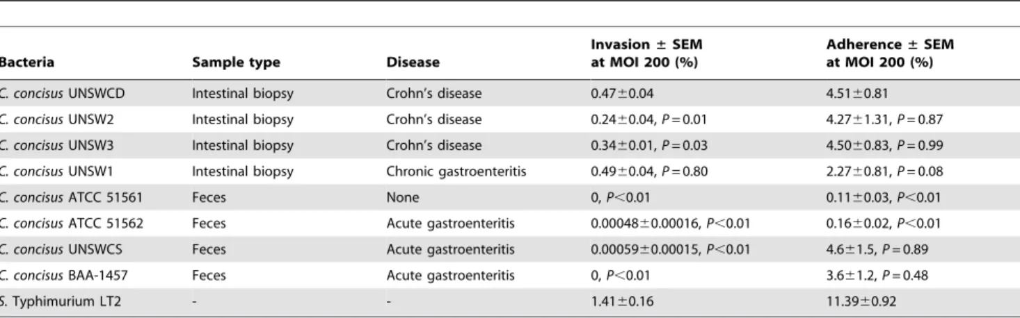
At a MOI of 200C. concisusUNSWCD was observed to be the most efficient among the 3 CD strains, followed by C. concisus
UNSW3, and thenC. concisusUNSW2 (Table 2). Interestingly, the level of invasion observed forC. concisusUNSW1 at a MOI of 200 was similar to that ofC. concisusUNSWCD (Table 2). The levels of invasion quantified for the ATCC 51562 and UNSWCS isolated from a patients with acute gastroenteritis were negligible as compared with the chronic strains, whereas no invasion was observed for BAA-1457 and ATCC 51561 (Table 2).
Table 1.Mucosa-associated bacteria isolated from child intestinal biopsies using the Ham’s F-12 enrichment-filtration method.
Child Diagnosis Age Gender Campylobacterdetection Bacterial species Strain
1 Normal 8 M 2 2 2
2 Normal 16 F + 2 2
3 Chronic gastroenteritis 13 M + C. concisus UNSW1
4 H. pyloriinfection 15 F 2 2 2
5 Crohn’s disease 3 M + C. concisus UNSW2
6 Crohn’s disease 5 M 2 2 2
7 Crohn’s disease 12 F + D. fairfieldensis UNSW1
8 Crohn’s disease 8 M 2 2 2
9 Crohn’s disease 12 F + 2 2
10 Crohn’s disease 12 M + C. concisus UNSW3
11 Ulcerative colitis 15 M 2 2 2
The results of the adherence assays at a MOI of 200 showed that the percentage adherence for six of theC. concisus strains was very similar (Table 2). The level of adherence observed in
C. concisusUNSW2,C. concisusUNSW3,C. concisusUNSW1,C. concisus BAA-1457 and C. concisus UNSWCS were not significantly different to that in C. concisus UNSWCD. Interestingly, the levels of adherence for ATCC 51562 and ATCC 51561 were significantly different to the other six strains (Table 2).
These results would suggest that although all fourC. concisus
strains isolated from chronic intestinal diseases have similar abilities to adhere to Caco-2 cells, the percentage invasion into the Caco-2 cell line remained strain-dependent. While significant differences were observed in the percentage invasion of the four chronic strains examined, they all showed significantly increased adherence and invasion as compared with the percentages observed for the acute gastroenteritis strain (ATCC 51562) and a non-invasive healthy control strain (ATCC 51561). Indeed, the percentages of invasion observed for C. concisus UNSW2, C. concisusUNSW3,C. concisusUNSWCD, andC. concisusUNSW1 were 500, 708, 979 and 1021 times higher than that found forC. concisusATCC 51562, respectively. Interestingly,C. concisus BAA-1457 had similar adherence levels to the chronic strains yet did not invade host cells, suggesting this strain could have a unique mechanism of pathogenesis. These results indicate that the pathogenic potential of C. concisus strains isolated from patients with chronic intestinal diseases is higher than those of strains isolated from patients with acute intestinal diseases and healthy controls. Based on these findings it could be postulated thatC. concisus strains associated with chronic intestinal diseases may belong to the same genomospecies, while those strains associated with acute gastroenteritis and healthy controls may belong to different genomospecies.
Visualization of Campylobacter concisus adherence and invasion to host cells
Scanning electron microscopy (ScEM) was used to further investigate the mechanisms employed by the four highly invasive
C. concisus strains (UNSWCD, UNSW1, UNSW2, UNSW3) to adhere to and invade the human intestinal cell lines Caco-2 and LS174T. The typical morphologies ofC. concisusstrains UNSW2, UNSW3, UNSW1 and UNSWCD are shown in Figures 1A, B, C and D, respectively.
ScEM clearly illustrated that the four C. concisus strains investigated had similar host epithelial cell-bacterial interactions. Given this, representative ScEM images have been used to portray the interactions betweenC. concisusand Caco-2 cells (Figure 2). An overview of uninfected Caco-2 cells (Figure 2A) showed the expression of intact differentiating and differentiated microvilli on the surface of Caco-2 monolayers (Figures 2A1, 2A2).C. concisus
tended to aggregate upon interaction with the host cells (Figures 2B, 2B1, 2B2).C. concisus mediated initial contact with host cells via flagellum-microvilli interactions, their polar flagellum binding to the tips of different host cell microvilli (indicated by arrows in Figure 2C). Unlike areas where noC. concisusinfection was found (indicated by an asterisk in Figure 2D), abnormalities in the microvilli and host cell structures were observed in areas where bacterial infection was present (indicated by a ring in Figure 2D and arrows in Figure 2E). Following adherence, C. concisus
appeared to induce a ‘‘membrane ruffling’’-like effect on the host cell membrane (indicated by an asterisk in Figure 2F) with penetration of the host cell membrane occurring from the non-flagellated end (indicated by arrows in Figures 2G, 2H, 2I).
Table 2.Comparison of the percentage invasion and adherence into Caco-2 cells of eightCampylobacter concisusstrains.
Bacteria Sample type Disease
Invasion±SEM at MOI 200 (%)
Adherence±SEM at MOI 200 (%)
C. concisusUNSWCD Intestinal biopsy Crohn’s disease 0.4760.04 4.5160.81
C. concisusUNSW2 Intestinal biopsy Crohn’s disease 0.2460.04,P= 0.01 4.2761.31,P= 0.87
C. concisusUNSW3 Intestinal biopsy Crohn’s disease 0.3460.01,P= 0.03 4.5060.83,P= 0.99
C. concisusUNSW1 Intestinal biopsy Chronic gastroenteritis 0.4960.04,P= 0.80 2.2760.81,P= 0.08
C. concisusATCC 51561 Feces None 0,P,0.01 0.1160.03,P,0.01
C. concisusATCC 51562 Feces Acute gastroenteritis 0.0004860.00016,P,0.01 0.1660.02,P,0.01
C. concisusUNSWCS Feces Acute gastroenteritis 0.0005960.00015,P,0.01 4.661.5,P= 0.89
C. concisusBAA-1457 Feces Acute gastroenteritis 0,P,0.01 3.661.2,P= 0.48
S.Typhimurium LT2 - - 1.4160.16 11.3960.92
The data shown are representative of viable invading or adhering bacteria relative to the viable initial inoculum of three independent experiments6Standard Error of the Mean (SEM), with each experiments being performed in duplicate.P,0.05 was considered significant.
doi:10.1371/journal.pone.0029045.t002
Figure 1. Scanning electron microscopy of fourCampylobacter concisusstrains.C. concisusUNSW2 was observed as spiral curved-shaped bacteria with rounded ends and a single polar flagellum as shown in Panel A (bar = 3mm). In Panel B (bar = 1.5mm) C. concisus
UNSW3 was observed to be curved-shaped bacteria with rounded ends and a single polar flagellum, while in Panels C (bar = 2mm) and D
(bar = 2.5mm)C. concisusstrains UNSW1 and UNSWCD were shown to
be spiral curved-shaped bacterium with rounded ends and a single flagellum.
Invasion of bacteria into the host cell was associated with irregular shaped membrane protrusions (indicated by asterisks in Figures 2G, 2H, 2I) with the uptake of C. concisus, resulting in bacteria inducing host cell damage (indicated by ‘‘#’’ in Figures 2G, 2H, 2I).
The cell line LS174T exhibits characteristics of enterocyte-morphology and is able to produce a mucin layer inin vitroculture, thus, more closely mimicking the human gastrointestinal tract [21]. Further investigation of theseC. concisusstrains using the cell line LS174T was considered to be important as this novel model provides the opportunity to studyin vitrothe role of mucus on the pathogenic behavior ofC. concisusstrains. ScEM clearly revealed that all fourC. concisusstrains had very similar host epithelial cell-bacterium interactions on LS174T cells (Figure 3). An overview of uninfected LS174T cells (Figure 3A) showed the expression of differentiated goblet cells (indicated by an arrow in Figure 3B) with sparse microvilli (indicated by a ring in Figure 3B) being observed on the apical surface of LS174T monolayers. The mucus layer (indicated by an ‘‘#’’ in Figure 3C) was observed on the monolayer surface of LS174T cells. C. concisus appeared to be attracted to the intestinal mucus layer (indicated by arrows in Figure 3D) using their single polar flagellum (indicated by arrows
in Figure 3E). The bacteria aggregated upon interaction with the mucus layer of the LS174T cells (more bacterial aggregation was observed for LS174T cells than Caco-2 cells) (Figure 3F). C. concisusused its flagellum (indicated by an arrow in Figure 3G) to adhere to the microvilli (indicated by a ring in Figure 3G) and goblet cells (indicated by an arrow in Figure 3H) of the LS174T monolayers which appeared to mediate initial contact with host cells. Following adherence, C. concisus induced a ‘‘membrane ruffling’’-like effect (indicated by an asterisk in Figure 3I) on the host cell membrane and appeared to penetrate host cell membrane from the non-flagellated end (indicated by an arrow in Figure 3I), leading to host cell damage (indicated by ‘‘#’’ in Figure 3I).
Previous studies have shown that C. jejuni expresses the fibronectin-binding outer membrane protein (CadF) that mediates adherence by binding to the cell matrix protein fibronectin located on epithelial cells [22,23]. CadF is involved in the ‘‘membrane ruffling’’ observed prior toC. jejuniinvasion [24]. Moreover, ScEM studies have shown thatC. jejunienters intestinal cells with its tip followed by the flagellar end [24]. Our findings thatC. concisus
secretes the outer membrane fibronectin binding protein [9], is associated with a ‘‘membrane ruffling’’-like effect on the intestinal cell membrane prior to invasion, and that invasion occurred from
Figure 2. Scanning electron microscopy of human intestinal cell line Caco-2 infected withCampylobacter concisusstrains for six hours.Panel A shows an overview of uninfected Caco-2 monolayer. The Caco-2 cells expressed differentiating microvilli (Panel A1) and differentiated microvilli (Panel A2).C. concisuswas shown to aggregate upon interaction with host cells as shown in Panel B (Panel B1, bar = 1.5mm and Panel B2,
bar = 2mm). In Panel C, the polar flagellum ofC. concisusis shown binding to the tips of host cell microvilli which mediated initial contact with host
cells (as indicated by the arrows). Abnormalities in the epithelial host cell structure and microvilli were observed following infection withC. concisus (indicated by a ring in Panel D and arrows in Panel E). Panel F shows the flagellum ofC. concisusappeared to wrap itself around the microvilli (as indicated by arrows). Following adherence,C. concisusinduced a ‘‘membrane ruffling’’-like effect on the host cell membrane (indicated by an asterisk in Panel F), and penetrated the host cell membrane from the non-flagellated end (indicated by an arrow in Panel G).C. concisuswas observed invading the host cell (indicated by arrows in Panels G, H and I) resulting in irregular shaped membrane protrusions (indicated by asterisks in Panels G, H and I), leading to host cell damage (indicated by ‘‘#’’ in Panels G, H and I).
the non-flagellated end would suggest thatC. concisus has a very similar mechanism of invasion to C. jejuni. Interestingly, as aggregation of C. concisus strains upon interaction with the intestinal mucus layer was observed, it is possible that this aggregation of C. concisus may involve biofilm formation. Such aggregation is similar to that previously reported in C. concisus
ATCC 33237, which was shown to form biofilms on glass [25].
Investigation of the invasive phenotype of
Campylobacter concisus
Investigation of the invasive phenotype of C. concisus was undertaken due to the observed differences in invasive potential among strains isolated from chronic and acute intestinal diseases. One feature of interest was a 30 kb plasmid that we had recently detected in UNSWCD and that was different to the two plasmids found in BAA-1457 [26]. Assembly of theC. concisusUNSWCD plasmid sequence was performed in this study using sequencing data generated in a previously published study [26]. The plasmid contained several virulence determinants from various organisms not closely related to C. concisus (Figure 4). Genes within this plasmid encoded the toxin-antitoxin (TA) replicon stabilization system StbD and StbE, mobilization protein MobA, exotoxin 9, restriction endonuclease R.Ecl19kI, DNA-cytosine methyltrans-ferase, two site-specific recombinases, TonB-dependent receptor, mature parasite-infected erythrocyte surface antigen (MESA), a sodium/solute symporter, choline kinase, glycosyl transferase, a membrane spanning protein, 3 stress-related proteins and 10
hypothetical proteins. Importantly, analysis of C. concisus
UNSWCD whole lysate expression data generated in a previously published study [26] revealed several of the proteins encoded by these genes were expressed under normal growth conditions.
The plasmid contains a TA system that is composed of two components, a stable toxin and an unstable antitoxin that interferes with the lethal action of the toxin. StbD and StbE homologues are commonly found in other pathogenic bacteria such as Vibrio cholerae and Haemophilus influenzae, a finding that suggests that they may have a function in virulence [27]. Of particular interest, were two genes encoding a Gram-positive exotoxin 9 and MESA. Exotoxin 9 has very high homology with exotoxins in Gram-positive bacteria, and contains a CYCLIN domain within its sequence. Cyclin homologues have been found in various viruses, where these viral homologues differ from their cellular counterparts in that the viral proteins are modified to harness the cell and benefit the virus [28]. MESA has been found to play a major role in intra-erythrocytic malarial viability [29]. It competes with P55 for the erythrocyte skeletal muscle protein, and hence regulates stability and mechanical properties of the erythrocyte plasma membrane [30]. Although the ubiquitously expressed P55 has been identified as a scaffolding protein in erythrocytes that stabilizes the actin cytoskeleton to the plasma membrane, its function in non-erythroid cells remains poorly understood [30]. Recently, P55 has been found to regulate neutrophil polarity, and function as a positive upstream effector of Akt phosphorylation [31]. Thus, the competition of MESA with
Figure 3. Scanning electron microscopy of human mucin producing intestinal cell line LS174T infected withCampylobacter concisus
strains for six hours.Panel A shows uninfected LS174T monolayers. LS174T cells expressing microvilli (indicated by a ring) and goblet cells (indicated by a arrow) are shown in Panel B. The mucus layer was found on the monolayer surface of LS174T cells as indicated by an ‘‘#’’ in Panel C.C. concisusappeared to be attracted to the mucus layer of host cells (indicated by ‘‘#’’ in Panel D) using their single polar flagellum (indicated by arrows in Panel E) and upon the interaction with host cells tended to aggregate (Panel F). Panel G shows the polar flagellum (as indicated by an arrow) ofC. concisusbinding to the tips of host cell microvilli (as indicated by a ring) and goblet cells (as indicated by an arrow in Panel H) which appeared to mediate initial contact with host cells. Following adherence,C. concisusinduced a ‘‘membrane ruffling’’-like effect on the host cell membrane (indicated by an asterisk in Panel I) and penetrated the host cell membrane from the non-flagellated end (indicated by an arrow in Panel I) resulting in cell damage (indicated by ‘‘#’’ in Panel I).
P55, and the functional role of P55 in the host may imply thatC. concisus UNSWCD employs this protein to modulate the host innate immune response.
The presence of this plasmid was investigated in the other seven strains through a PCR targeting the gene encoding the exotoxin 9, and significantly, the four highly invasive strains from chronic intestinal diseases (UNSWCD, UNSW1, UNSW2 and UNSW3) were the only strains to contain this gene (Figure 5). Four further plasmid genes (encoding: DNA-cytosine methyltransferase, mobi-lization protein MobA, site-specific recombinase and restriction
endonuclease R.Ecl18kl) were confirmed to be present in the chronic strains and absent in the other four strains (data not shown). This provides further evidence that this plasmid, with the possibility of some minor modifications, may be responsible for the heterogeneity in the invasive potential ofC. concisus.
Effect ofCampylobacter concisus on host cell protein expression
The effect ofC. concisusUNSWCD on host cells was examined by determining the change in protein expression upon infection
Figure 4. Graphical representation of the genes encoded by the plasmid purified fromCampylobacter concisusUNSWCD.Outer circle (blue) represents the coding sequences within the plasmid; inner circle (black) represents the GC content; inner circle (purple/green) represents the GC skew.
with the bacterium. The response of Caco-2 cells to C. concisus
UNSWCD infection was analyzed using 2D gel electrophoresis to determine the changes in the proteome of the human cells (Figure 6). 2D gel electrophoresis was performed on proteins extracted from pairs of human cultures grown with and withoutC. concisus; they included four independent biological repeats. The
four pairs of gels obtained from cultures under both conditions were analyzed to identify, using tandem mass spectrometry, spots corresponding to proteins whose expression was regulated upon infection with bacteria. One hundred and twenty five proteins were differentially expressed (6.71% of the total spots detected on the gels), of which 78 were upregulated and 47 were
downreg-Figure 5. PCR analysis of the exotoxin 9 gene in the eightCampylobacter concisusstrains.Lane 1: FN-1 marker, lane 2: UNSWCD, lane 3: UNSW2, lane 4: UNSW3, lane 5: UNSW1, lane 6: BAA-1457, lane 7: UNSWCS, lane 8: ATCC 51562, lane 9: ATCC 51651 and lane 10: negative control. doi:10.1371/journal.pone.0029045.g005
Table 3.Caco-2 cell proteins whose expression is upregulated in the presence ofCampylobacter concisusUNSWCD.
ID Symbol Gene Name Location Type*
28614 ALDOA Fructose-bisphosphate aldolase Cytoplasm Enzyme
521205 APOC3 Apolipoprotein C-III Extracellular Space Transporter
5031593 ARPC5 Actin related protein (16 kDa) Cytoplasm Other
32189394 ATP5B ATP synthase Cytoplasm Transporter
4757880 BUB3 Budding-related yeast homolog Nucleus Other
3355455 C19ORF10 Chromosome 19 ORF 10 Extracellular Space Cytokine
4757900 CALR Calreticulin Cytoplasm Transcription reg
2809324 CALU Calumenin Cytoplasm Other
119617636 CCT2 Chaperonin containing TCP1 Cytoplasm Kinase
180570 CKB Creatine kinase Cytoplasm Kinase
38201710 DDX17 DEAD box polypeptide 17 Nucleus Enzyme
4758138 DDX5 DEAD box polypeptide 5 Nucleus Enzyme
499719 DLST Dihydrolipoamide succinyltransferase Cytoplasm Enzyme
219588 DNAJA1 DnaJ (Hsp40) homolog Nucleus Other
181608 DSP Desmoplakin Plasma Membrane Other
1922287 ECHS1 Enoyl Coenzyme A hydratase Cytoplasm Enzyme
4503481 EEF1G Eukaryotic translation elongation factor Cytoplasm Translation reg
4503545 EIF5A Eukaryotic translation initiation factor Cytoplasm Translation reg
693933 ENO1 Enolase 1 Cytoplasm Transcription reg
52487191 ERP44 Endoplasmic reticulum protein 44 Cytoplasm Enzyme
19743875 FH Fumarate hydratase Cytoplasm Enzyme
17402900 FUBP1 FUSE binding protein 1 Nucleus Transcription reg
7669492 GAPDH GAP dehydrogenase Cytoplasm Enzyme
4504035 GMPS Guanine monophosphate synthetase Nucleus Enzyme
4504327 HADHB Enoyl-Coenzyme A hydratase Cytoplasm Enzyme
1568551 HIST1H2BE Histone cluster 1, H2be Nucleus Other
55956919 HNRNPAB Ribonucleoprotein A/B Nucleus Enzyme
14110414 HNRNPD Ribonucleoprotein D Nucleus Transcription reg
16876910 HNRNPF Ribonucleoprotein F Nucleus Other
14141157 HNRNPH3 Ribonucleoprotein H3 (2H9) Nucleus Other
14110407 HNRPDL Ribonucleoprotein D-like Nucleus Other
4507677 HSP90B1 Heat shock protein 90 kDa beta Cytoplasm Other
16507237 HSPA5 Heat shock 70 kDa protein 5 Cytoplasm Other
5729877 HSPA8 Heat shock 70 kDa protein 8 Cytoplasm Enzyme
12653415 HSPA9 Heat shock 70 kDa protein 9 Cytoplasm Other
3641398 IDH1 Isocitrate dehydrogenase 1 Cytoplasm Enzyme
55957496 LMNA Lamin A/C Nucleus Other
2906146 MDH2 Malate dehydrogenase 2 Cytoplasm Enzyme
4758756 NAP1L1 Nucleosome assembly protein 1-like 1 Nucleus Other
189306 NCL Nucleolin Nucleus Other
5729953 NUDC Nuclear distribution gene C homolog Cytoplasm Other
20070125 P4HB Prolyl 4-hydroxylase Cytoplasm Enzyme
2697005 PA2G4 Proliferation-associated 2G4 (38 kDa) Nucleus Transcription reg
460771 PCBP1 Poly(rC) binding protein 1 Nucleus Translation reg
14141166 PCBP2 Poly(rC) binding protein 2 Nucleus Other
387011 PDHA1 Pyruvate dehydrogenase Cytoplasm Enzyme
21361657 PDIA3 Protein disulfide isomerase family A Cytoplasm peptidase
1710248 PDIA6 Protein disulfide isomerase family A Cytoplasm Enzyme
4505763 PGK1 Phosphoglycerate kinase 1 Cytoplasm kinase
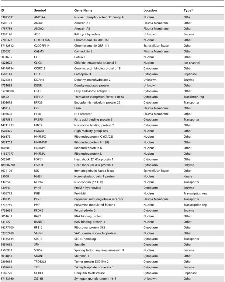
ulated in the presence ofC. concisus(Tables 3 and 4, respectively; Table S1 and S2).
Analysis of the response of Caco-2 cells toC. concisusinfection revealed a significant impact on host cell metabolism, specifically, the upregulation of creatine kinase (CK) and processes involved in energy production, and inhibition of proteases (Tables 3, 4). CK catalyzes the conversion of creatine to phosphocreatine (PCr) through the consumption of adenosine triphosphate (ATP). PCr serves as an energy reservoir for the rapid buffering and regeneration of ATP in situ, as well as for intracellular energy transport by the PCr shuttle [32]. The increase in energy production through enzymes such as ATP synthase, dihydroli-poamide succinyltransferase, enoyl coenzyme A hydratase, enolase, fumarate dehydratase, GAP dehydrogenase, isocitrate dehydrogenase, malate dehydrogenase, pyruvate dehydrogenase and pyruvate kinase indicate that the cells are producing more energy to combat the damage caused by bacterial infection. In addition, it appears that the cells have downregulated the activity of proteases either directly through the downregulation of cathepsin D or indirectly through the upregulation of serpin peptidase inhibitors. This may relate to the fact that during
infection bacteria produce proteases that target the innate immune response and degrade host proteins [33].
One likely avenue for the increase in energy production is for reinforcement of the structural integrity of the cell following cellular damage by the bacteria. This is supported by the upregulation of an actin-related protein and the downregulation of Hsp27, a heat shock protein known to inhibit F-actin polymerization [34]. Interestingly, we observed the downregula-tion of cofilin, a protein that is known to promote rapid actin filament turnover through severing actin filaments [35]. One possible explanation for this downregulation is that followingC. concisus infection, host cells protect themselves against further severing of actin filaments. Moreover,b-tubulin was upregulated and stathmin was downregulated, a finding that further supports the view that the cell was strengthening its structural integrity, given that stathmin is known to promote microtubule disassembly by sequestering b-tubulin into the tight ternary T2S complex thereby rendering it non-polymerizable [36].
Another indication that C. concisus affected the structural integrity of the cell monolayer was the upregulation of desmopla-kin. This protein is an essential component of functional
ID Symbol Gene Name Location Type*
10863927 PPIA Cyclophilin A Cytoplasm Enzyme
6166493 PRDX5 Peroxiredoxin 5 Cytoplasm Enzyme
62896529 PSMC3 Proteasome 26S subunit, ATPase, 3 Nucleus Transcription reg
976227 PSMC5 Proteasome 26S subunit, ATPase, 5 Nucleus Transcription reg
1526426 PSMC6 Proteasome 26S subunit, ATPase, 6 Nucleus Peptidase
13477197 QPRT Quinolinate phosphoribosyltransferase Extracellular Space Enzyme
4506387 RAD23B RAD23 homolog B Nucleus Other
2078529 RBM4 RNA binding motif protein 4 Nucleus Other
3256007 RBMX RNA binding motif protein, X-linked Nucleus Other
4502801 RCC1 Reg of chromosome condensation 1 Nucleus Other
33150766 RPL22 Ribosomal protein L22 Nucleus Other
4506605 RPL23 Ribosomal protein L23 Cytoplasm Other
5032051 RPS14 Ribosomal protein S14 Cytoplasm Other
4506695 RPS19 Ribosomal protein S19 Cytoplasm Other
15080499 SERPINA1 Serpin peptidase inhibitor, clade A Extracellular Space Other
30130 SERPINH1 Serpin peptidase inhibitor, clade H Extracellular Space Other
119608226 SET SET nuclear oncogene Nucleus Phosphatase
25777713 SKP1 S-phase kinase-associated protein 1 Nucleus Transcription reg
5902090 SLC2A3 Solute carrier family 2 (glucose) Plasma Membrane Transporter
19923193 ST13 Suppression of tumorigenicity 13 Cytoplasm Other
7305503 STOML2 Stomatin (EPB72)-like 2 Plasma Membrane Other
3037013 SYNCRIP RNA interacting protein Nucleus Other
37267 TKT Transketolase Cytoplasm Enzyme
35959 TUBB4 b-Tubulin Cytoplasm Other
833999 TUFM Tu translation elongation factor Cytoplasm Translation reg
4507797 UBE2V2 Ubiquitin-conjugating enzyme E2 Cytoplasm Enzyme
46593007 UQCRC1 Ubiquinol-cytochrome c reductase Cytoplasm Enzyme
4507879 VDAC1 Voltage-dependent anion channel 1 Cytoplasm Ion channel
Statistical data were acquired and analyzed using PDQuest 2-D. Proteins with changes in their intensity$2-fold (P,0.05) were identified by tandem mass spectrometry analyses. Spot numbers, mascot scores and number of identified peptides are listed in Table S1.
*Reg = Regulator.
doi:10.1371/journal.pone.0029045.t003
Table 4.Caco-2 cell proteins whose expression is downregulated in the presence ofCampylobacter concisusUNSWCD.
ID Symbol Gene Name Location Type*
33875631 ANP32A Nuclear phosphoprotein 32 family A Nucleus Other
4502101 ANXA1 Annexin A1 Plasma Membrane Other
4757756 ANXA2 Annexin A2 Plasma Membrane Other
1263196 ATIC IMP cyclohydrolase Unknown Enzyme
7706322 C14ORF166 Chromosome 14 ORF 166 Nucleus Other
37182312 C20ORF114 Chromosome 20 ORF 114 Extracellular Space Other
825635 CALM3 Calmodulin 3 Plasma Membrane Other
5031635 CFL1 Cofilin 1 Nucleus Other
4323622 CLIC3 Chloride intracellular channel 3 Nucleus Ion channel
14149734 CORO1B Coronin, actin binding protein, 1B Cytoplasm Other
4503143 CTSD Cathepsin D Cytoplasm Peptidase
7524354 DDAH2 Dimethylaminohydrolase 2 Unknown Enzyme
4755083 DENR Density-regulated protein Unknown Other
55770888 EEA1 Early endosome antigen 1 Cytoplasm Other
38522 EEF1D Translation elongation factor 1 delta Cytoplasm Translation reg
5803013 ERP29 Endoplasmic reticulum protein 29 Cytoplasm Transporter
340217 EZR Ezrin Plasma Membrane Other
8393638 F11R F11 receptor Plasma Membrane Other
4557581 FABP5 Fatty acid binding protein 5 Cytoplasm Transporter
14211923 HINT2 Nucleotide binding protein 2 Cytoplasm Other
4504425 HMGB1 High-mobility group box 1 Nucleus Other
306875 HNRNPC Ribonucleoprotein C (C1/C2) Nucleus Other
5031753 HNRNPH1 Ribonucleoprotein H1 (H) Nucleus Other
460789 HNRNPK Ribonucleoprotein K Nucleus Other
11527777 HNRNPL Ribonucleoprotein L Nucleus Other
662841 HSPB1 Heat shock 27 kDa protein 1 Cytoplasm Other
189502784 HSPD1 Heat shock 60 kDa protein 1 Cytoplasm Enzyme
16741061 IGK Immunoglobulin kappa locus Extracellular Space Other
35068 NME1 Non-metastatic cells 1 protein Nucleus Kinase
432654 NUP62 Nucleoporin (62 kDa) Nucleus Transporter
339647 P4HB Prolyl 4-hydroxylase Cytoplasm Enzyme
4505773 PHB Prohibitin Nucleus Transcription reg
238236 PIGR Polymeric immunoglobulin receptor Plasma Membrane Transporter
5737759 PMF1 Polyamine-modulated factor 1 Nucleus Transcription reg
4758638 PRDX6 Peroxiredoxin 6 Cytoplasm Enzyme
8051631 RALY RNA binding protein Nucleus Other
431422 RANBP1 RAN binding protein 1 Nucleus Other
14277700 RPS12 Ribosomal protein S12 Cytoplasm Other
62202489 SARNP SAP domain ribonucleoprotein Nucleus Other
34335134 SEC13 SEC13 homolog Cytoplasm Transporter
5454052 SFN Stratifin Cytoplasm Other
4506903 SFRS9 Splicing factor, arginine/serine-rich 9 Nucleus Enzyme
5031851 STMN1 Stathmin 1 Cytoplasm Other
2895085 TPD52L2 Tumor protein D52-like 2 Cytoplasm Other
4507645 TPI1 Triosephosphate isomerase 1 Cytoplasm Enzyme
4185720 UCHL1 Ubiquitin thiolesterase Cytoplasm Peptidase
37183160 ZG16B Zymogen granule protein 16 B Unknown Other
Statistical data were acquired and analyzed using PDQuest 2-D. Proteins with changes in their intensity#0.5-fold (P,0.05) were identified by tandem mass spectrometry analyses. Spot numbers, mascot scores and number of identified peptides are listed in Table S2.
*Reg = Regulator.
desmosomes, intercellular junctions that tightly link adjacent cells, and is responsible for anchoring intermediate filaments to desmosomal plaques [37]. This finding is supported by our previous study that showedC. concisusUNSWCD to preferentially attach to intercellular junctional spaces, and that this spatial distribution was concomitantly associated with a loss of mem-brane-associated ZO-1 and occludin [8].
IPA analysis revealed that the pathway involved in the production of IL-12 was upregulated in cells exposed toC. concisus
UNSWCD, 28 proteins directly or indirectly involved in the IL-12 pathway being found to be upregulated (Figure S1). Those proteins directly involved in the production of IL-12 complex, included fuse binding protein 1, nuclear distribution gene C homolog, heat shock protein 90 kDa beta, endoplasmic reticulum protein 44, serpin peptidase inhibitor clade A, apolipoprotein C-III, voltage-dependent anion channel 1 and SET nuclear oncogene (Figure S1). IL-12 is of particular interest due to its induction of intestinal mucosal inflammation through an IFN-c -dependent manner [38]. The significance of this cytokine is discussed further below.
Infection of Caco-2 cells with C. concisus resulted in the upregulation of three proteins involved in the proteasome complex, namely the proteasome 26S subunit ATPases 3, 5 and 6. In conjunction with this, ubiquitin-conjugating enzyme E2 and
ubiquinol-cytochromec reductase, which are involved in protein ubiquitination were upregulated, and ubiquitin thiolesterase, which is involved in protein deubiquitination, was downregulated. Proteasomes are part of the protein degradation machinery of the cell that regulate the concentration of particular proteins and degrade misfolded proteins [39]. In this process, proteins are initially tagged for degradation with a small protein called ubiquitin, which provides a signal to other ubiquitinating enzymes to attach additional ubiquitin molecules, thus forming a poly-ubiquitin chain that is bound by the proteasome, thereby allowing it to degrade the tagged protein [40].
One indication that NF-kB might be activated upon infection withC. concisusUNSWCD is the upregulation of the proteasome and protein ubiquitination pathways that are involved in the degradation of NF-kB inhibitors [41]. Direct evidence of NF-kB activation upon infection comes from the finding that one pathway leading to the ser/thr kinase Akt is upregulated, namely the proteins ATPase, EEF1G, ATP5B, P4HB, PDIA3, CALR and RPS14 (Figure S2). Akt functions through Ikb kinase (IKK) to promote the transactivation potential and phosphorylation of NF-kB [42,43]. More recently, Akt has been found to promote IKK-dependent activation of NF-kB via mTOR and Raptor [44].
Further evidence includes the downregulation of NF-kB inhibitors such as annexin 1 (ANXA1) and prohibitin (PHB).
Figure 7. Levels of interleukin-12 and interferon-cproduced by the human monocytic leukemia cell line THP-1 following infection with Campylobacter concisus strains and Escherichia coli K-12. * represents P,0.05; ** represents P,0.01. Data of three independent experiments6standard error of the mean.
ANXA1 has been found to suppress the transcriptional activity of NF-kB by preventing it from binding to DNA [45]. This inhibitory activity has also been found in the intestinal mucosa of mice treated with an agent that induces ANXA1 [45]. Furthermore, PHB has been found to decrease TNF-a-induced nuclear translocation of the NF-kB protein p65, NF-kB/DNA binding, and NF-kB-mediated transcriptional activationin vitroandin vivo, despite continual IkB-a phosphorylation and degradation and increased cytosolic p65 [46]. The downregulation of polyamine-modulated factor 1 (PMF1), a binding partner of NF-E2 related factor-2 (Nrf2), was a further indication that NF-kB was activated. PMF1 binds to Nrf2 to regulate gene transcription [47]. Nrf2 over-expression has been shown to suppress NF-kB DNA binding activity [48]. Furthermore, it has been suggested that Nrf2 activation induces intracellular events that concur with NF-kB suppression [49].
Cytokine production in response toCampylobacter concisusinfection
Proteomics coupled with tandem mass spectrometry established that the pathway leading to the production of IL-12 was upregulated in cells infected with C. concisus UNSWCD. Thus, we confirmed using ELISA the production of IL-12 in monocyte-derived macrophages upon infection with the eight C. concisus
strains andE. coliK-12. Significantly increased levels of IL-12 were found to be produced in cells exposed toC. concisusUNSWCD as compared with controls (Figure 7), thereby validating the expression results observed through proteomics. However, cells
exposed to any of the eightC. concisusstrains were also found to produce significantly increased levels of IL-12 as compared with controls (Figure 7), indicating that production of IL-12 upon exposure to C. concisus does not correlate with the pathogenic potential of the bacterium. Cells exposed toE. coli K-12 or no bacteria produced negligible amounts of IL-12 (Figure 7), confirming that the production of IL-12 by cells exposed to C. concisuswas due to the bacterium. While it is possible that a small amount of the measured IL-12 may have resulted from IL-23, due to the two cytokines sharing the p40 subunit [50], our proteomics findings and the high levels measured (.600 pg ml21
) both support the importance of IL-12 inC. concisusinfection.
IL-12 is known to stimulate mouse peritoneal macrophages to express and secrete IFN-c[51]. In addition, IFN-cpromotes the accumulation of immunoproteasomes [52], and bothin vitroandin vivo IFN-c is essential for upregulation of immunoproteasome subunits in mice [53]. Together with the upregulation of the proteasome in our study, these findings led us to investigate the production of IFN-cin cells infected withC. concisus. This showed that only C. concisus strains that were capable of invading into human cells stimulated the production of IFN-c, withC. concisus
UNSWCD inducing the highest quantity of this cytokine (Figure 7). This was of particular significance as although all strains of C. concisusproduced high amounts of IL-12, only theC. concisusstrains capable of internalizing into host cells induced a significantly increased quantity of IFN-cwith respect to both controls.
Overall our findings suggest that non-invasiveC. concisusstrains can induce IL-12 upon adherence to human cells, however, this
Figure 8. Proposed immune response toCampylobacter concisusUNSWCD.(A) Non-invasiveC. concisusstrains adhere to the host cell and induce the production of IL-12. (B) InvasiveC. concisusstrains adhere to and invade the host cell inducing both IL-12 and IFN-c, which in turn activate the immunoproteosome. The bacterial insult upregulates ubiquitinating and downregulates de-ubiquitinating enzymes which leads to the ubiquitination of NF-kB inhibitors. The immunoproteosome targets these inhibitors which activates NF-kB.
does not translate to the production of IFN-c (Figure 8). In contrast, in human cells exposed to invasiveC. concisusstrains, the production of IL-12 results in the induction of IFN-c, which in turn activates the immunoproteasome (Figure 8). Concurrently, the human cells exposed to the invasiveC. concisusstrains regulate ubiquitination pathways and these enzymes tag NF-kB inhibitors for degradation by the immunoproteasome, leading to the activation of NF-kB (Figure 8). These findings are of great significance when the association ofC. concisuswith pediatric CD [6,7] is taken into consideration. The tissue damaging inflamma-tory reaction in CD is driven by activated type 1 helper T-cells (Th1), with IL-12 being a major Th1-inducing factor, a view that is supported by the observation that an accumulation of macrophages making IL-12 occurs in CD patients [54]. Further evidence of the importance of IL-12 in CD is the finding that administration of a monoclonal antibody blocking the IL-12/p40 subunit can induce and maintain clinical remission in CD patients [55]. Significantly in relation to our findings, the 26S proteasome has been shown to play an important role in the inflammatory cascade and chronic gut inflammation in particular in CD [56]. Indeed, high expression of immunoproteasome subunits and enhanced processing of the NF-kB precursor p105 and degrada-tion of the NF-kB inhibitor, IkBa, by immunoproteasomes is a characteristic of the inflamed mucosa of CD patients [57]. Enhanced NF-kB activity has also been shown to be involved in the pathology of CD [57]. Furthermore, our finding that PHB, an NF-kB inhibitor, was downregulated upon infection of cells with
C. concisusUNSWCD, is in line with the decreased expression of PHB reported in subjects with CD [58].
Conclusions
This study has not only provided novel information on the mechanisms by whichC. concisusstrains interact with host intestinal cells, but has also provided important evidence that strains ofC. concisusisolated from patients with chronic intestinal diseases have a significantly increased ability to invade intestinal cell lines as compared withC. concisusstrains isolated from patients with acute gastroenteritis and healthy controls. Importantly, we have elucidated the feature that may be responsible for the heteroge-neity in invasive potential ofC. concisus. Moreover, this study has revealed novel information on the host immune response to C. concisusinfection, and has shown that this response has substantial similarities with that observed in the mucosa of CD patients.
Supporting Information
Figure S1 Network associated with the production of IL-12 generated through IPAH(Ingenuity Systems).Proteins colored in grey were upregulated. Proteins highlighted with an asterisk were directly associated with the IL-12 complex. (PPT)
Figure S2 Network associated with the upregulation of Akt generated through IPAH(Ingenuity Systems).Proteins colored in grey were upregulated. Proteins highlighted with an asterisk were associated with one pathway that lead to the expression of Akt.
(PPT)
Table S1 Mass spectrometry results of Caco-2 cell proteins whose expression is upregulated in the pres-ence ofCampylobacter concisus UNSWCD.Proteins with changes in their intensity$2.0-fold (P,0.05) were identified by tandem mass spectrometry analyses. Cut off scores of.58 and$2 peptide matches were employed.
(DOC)
Table S2 Mass spectrometry results of Caco-2 cell proteins whose expression is downregulated in the presence ofCampylobacter concisusUNSWCD. Proteins with changes in their intensity#0.5-fold (P,0.05) were identified by tandem mass spectrometry analyses. Cut off scores of.58 and $2 peptide matches were employed.
(DOC)
Acknowledgments
The authors would like to thank Jennifer Norman from the Electron Microscopy unit (UNSW) for her technical assistance.
Author Contributions
Conceived and designed the experiments: NOK HM. Performed the experiments: NOK NPD CGT JAB-P MJR. Analyzed the data: NOK NPD CGT JAB-P MJR MRW ASD DAL HM. Contributed reagents/ materials/analysis tools: MRW ASD DAL HM. Wrote the paper: NOK HM. Provided human samples: ASD DAL.
References
1. Cho JH, Abraham C (2007) Inflammatory bowel disease genetics: Nod2. Annu Rev Med 58: 401–416.
2. Theodossi A, Spiegelhalter DJ, Jass J, Firth J, Dixon M, et al. (1994) Observer variation and discriminatory value of biopsy features in inflammatory bowel disease. Gut 35: 961–968.
3. Sartor RB (2006) Mechanisms of disease: pathogenesis of Crohn’s disease and ulcerative colitis. Nat Clin Pract Gastroenterol Hepatol 3: 390–407. 4. Engberg J, On SL, Harrington CS, Gerner-Smidt P (2000) Prevalence of
Campylobacter,Arcobacter,Helicobacter, andSutterellaspp. in human fecal samples as estimated by a reevaluation of isolation methods for Campylobacters. J Clin Microbiol 38: 286–291.
5. On SL (1996) Identification methods for campylobacters, helicobacters, and related organisms. Clin Microbiol Rev 9: 405–422.
6. Zhang L, Man SM, Day AS, Leach ST, Lemberg DA, et al. (2009) Detection and isolation ofCampylobacterspecies other thanC. jejunifrom children with Crohn’s disease. J Clin Microbiol 47: 453–455.
7. Man SM, Zhang L, Day AS, Leach ST, Lemberg DA, et al. (2010)Campylobacter concisusand otherCampylobacterspecies in children with newly diagnosed Crohn’s disease. Inflamm Bowel Dis 16: 1008–1016.
8. Man SM, Kaakoush NO, Leach ST, Nahidi L, Lu HK, et al. (2010) Host attachment, invasion, and stimulation of proinflammatory cytokines by Campylobacter concisus and other non-Campylobacter jejuni Campylobacterspecies. J Infect Dis 202: 1855–1865.
9. Kaakoush NO, Man SM, Lamb S, Raftery MJ, Wilkins MR, et al. (2010) The secretome ofCampylobacter concisus. FEBS J 277: 1606–1617.
10. Aabenhus R, Permin H, On SL, Andersen LP (2002) Prevalence ofCampylobacter concisusin diarrhoea of immunocompromised patients. Scand J Infect Dis 34: 248–252.
11. Sainsus N, Cattori V, Lepadatu C, Hofmann-Lehmann R (2008) Liquid culture medium for the rapid cultivation ofHelicobacter pylorifrom biopsy specimens. Eur J Clin Microbiol Infect Dis 27: 1209–1217.
12. Lastovica AJ, le Roux E (2000) Efficient isolation of campylobacteria from stools. J Clin Microbiol 38: 2798–2799.
13. Kaakoush NO, Holmes J, Octavia S, Man SM, Zhang L, et al. (2010) Detection of Helicobacteraceae in intestinal biopsies of children with Crohn’s disease. Helicobacter 15: 549–557.
14. Luber P, Bartelt E, Genschow E, Wagner J, Hahn H (2003) Comparison of broth microdilution, E Test, and agar dilution methods for antibiotic susceptibility testing ofCampylobacter jejuniandCampylobacter coli. J Clin Microbiol 41: 1062–1068.
15. Vandenberg O, Houf K, Douat N, Vlaes L, Retore P, et al. (2006) Antimicrobial susceptibility of clinical isolates of non-jejuni/colicampylobacters and arcobacters from Belgium. J Antimicrob Chemother 57: 908–913.
17. Urata T, Kikuchi M, Hino T, Yoda Y, Tamai K, et al. (2008) Bacteremia caused byDesulfovibrio fairfieldensis. J Infect Chemother 14: 368–370.
18. Russell RG, O’Donnoghue M, Blake DC, Jr., Zulty J, DeTolla LJ (1993) Early colonic damage and invasion ofCampylobacter jejuniin experimentally challenged infantMacaca mulatta. J Infect Dis 168: 210–215.
19. Bereswill S, Kist M (2002) Molecular microbiology and pathogenesis of HelicobacterandCampylobacterupdated: a meeting report of the 11th conference onCampylobacter,Helicobacterand related organisms. Mol Microbiol 45: 255–262. 20. Ketley JM (1997) Pathogenesis of enteric infection byCampylobacter.
Microbiol-ogy 143: 5–21.
21. Tom BH, Rutzky LP, Jakstys MM, Oyasu R, Kaye CI, et al. (1976) Human colonic adenocarcinoma cells. I. Establishment and description of a new line. In Vitro 12: 180–191.
22. Konkel ME, Christensen JE, Keech AM, Monteville MR, Klena JD, et al. (2005) Identification of a fibronectin-binding domain within the Campylobacter jejuni CadF protein. Mol Microbiol 57: 1022–1035.
23. Konkel ME, Garvis SG, Tipton SL, Anderson DE, Jr., Cieplak W, Jr. (1997) Identification and molecular cloning of a gene encoding a fibronectin-binding protein (CadF) fromCampylobacter jejuni. Mol Microbiol 24: 953–963. 24. Krause-Gruszczynska M, Rohde M, Hartig R, Genth H, Schmidt G, et al.
(2007) Role of the small Rho GTPases Rac1 and Cdc42 in host cell invasion of Campylobacter jejuni. Cell Microbiol 9: 2431–2444.
25. Gunther NWt, Chen CY (2009) The biofilm forming potential of bacterial species in the genusCampylobacter. Food Microbiol 26: 44–51.
26. Deshpande N, Kaakoush NO, Mitchell H, Janitz K, Raftery MJ, et al. (2011) Sequencing and validation of the genome of aCampylobacter concisusreveals extensive intra-species diversity. PLoS One 6: e22170.
27. Hayes F (1998) A family of stability determinants in pathogenic bacteria. J Bacteriol 180: 6415–6418.
28. Hardwick JM (2000) Cyclin’ on the viral path to destruction. Nat Cell Biol 2: E203–204.
29. Magowan C, Coppel RL, Lau AO, Moronne MM, Tchernia G, et al. (1995) Role of thePlasmodium falciparummature-parasite-infected erythrocyte surface antigen (MESA/PfEMP-2) in malarial infection of erythrocytes. Blood 86: 3196–3204.
30. Waller KL, Nunomura W, An X, Cooke BM, Mohandas N, et al. (2003) Mature parasite-infected erythrocyte surface antigen (MESA) ofPlasmodium falciparum binds to the 30-kDa domain of protein 4.1 in malaria-infected red blood cells. Blood 102: 1911–1914.
31. Quinn BJ, Welch EJ, Kim AC, Lokuta MA, Huttenlocher A, et al. (2009) Erythrocyte scaffolding protein p55/MPP1 functions as an essential regulator of neutrophil polarity. Proc Natl Acad Sci U S A 106: 19842–19847.
32. Wallimann T, Wyss M, Brdiczka D, Nicolay K, Eppenberger HM (1992) Intracellular compartmentation, structure and function of creatine kinase isoenzymes in tissues with high and fluctuating energy demands: the ‘phosphocreatine circuit’ for cellular energy homeostasis. Biochem J 281(Pt 1): 21–40.
33. Potempa J, Pike RN (2009) Corruption of innate immunity by bacterial proteases. J Innate Immun 1: 70–87.
34. Piotrowicz RS, Hickey E, Levin EG (1998) Heat shock protein 27 kDa expression and phosphorylation regulates endothelial cell migration. FASEB J 12: 1481–1490.
35. Lappalainen P, Drubin DG (1997) Cofilin promotes rapid actin filament turnoverin vivo. Nature 388: 78–82.
36. Jourdain L, Curmi P, Sobel A, Pantaloni D, Carlier MF (1997) Stathmin: a tubulin-sequestering protein which forms a ternary T2S complex with two tubulin molecules. Biochemistry 36: 10817–10821.
37. Bornslaeger EA, Corcoran CM, Stappenbeck TS, Green KJ (1996) Breaking the connection: displacement of the desmosomal plaque protein desmoplakin from cell-cell interfaces disrupts anchorage of intermediate filament bundles and alters intercellular junction assembly. J Cell Biol 134: 985–1001.
38. Chikano S, Sawada K, Shimoyama T, Kashiwamura SI, Sugihara A, et al. (2000) IL-18 and IL-12 induce intestinal inflammation and fatty liver in mice in an IFN-cdependent manner. Gut 47: 779–786.
39. Peters JM, Franke WW, Kleinschmidt JA (1994) Distinct 19 S and 20 S subcomplexes of the 26 S proteasome and their distribution in the nucleus and the cytoplasm. J Biol Chem 269: 7709–7718.
40. van Deventer S, Neefjes J (2010) The Immunoproteasome Cleans up after Inflammation. Cell 142: 517–518.
41. Karin M, Ben-Neriah Y (2000) Phosphorylation meets ubiquitination: the control of NF-kB activity. Annu Rev Immunol 18: 621–663.
42. Madrid LV, Mayo MW, Reuther JY, Baldwin AS, Jr. (2001) Akt stimulates the transactivation potential of the RelA/p65 Subunit of NF-kB through utilization of the IkB kinase and activation of the mitogen-activated protein kinase p38. J Biol Chem 276: 18934–18940.
43. Sizemore N, Lerner N, Dombrowski N, Sakurai H, Stark GR (2002) Distinct roles of the IkB kinase alpha and beta subunits in liberating nuclear factor kappa B (NF-kB) from IkB and in phosphorylating the p65 subunit of NF-kB. J Biol Chem 277: 3863–3869.
44. Dan HC, Cooper MJ, Cogswell PC, Duncan JA, Ting JP, et al. (2008) Akt-dependent regulation of NF-kB is controlled by mTOR and Raptor in association with IKK. Genes Dev 22: 1490–1500.
45. Zhang Z, Huang L, Zhao W, Rigas B (2010) Annexin 1 induced by anti-inflammatory drugs binds to NF-kB and inhibits its activation: anticancer effects in vitroandin vivo. Cancer Res 70: 2379–2388.
46. Theiss AL, Jenkins AK, Okoro NI, Klapproth JM, Merlin D, et al. (2009) Prohibitin inhibits tumor necrosis factor alpha-induced nuclear factor-kappa B nuclear translocation via the novel mechanism of decreasing importin alpha3 expression. Mol Biol Cell 20: 4412–4423.
47. Wang Y, Devereux W, Stewart TM, Casero RA, Jr. (2002) Polyamine-modulated factor 1 binds to the human homologue of the 7a subunit of the ArabidopsisCOP9 signalosome: implications in gene expression. Biochem J 366: 79–86.
48. Song MY, Kim EK, Moon WS, Park JW, Kim HJ, et al. (2009) Sulforaphane protects against cytokine- and streptozotocin-induced beta-cell damage by suppressing the NF-kB pathway. Toxicol Appl Pharmacol 235: 57–67. 49. Bellezza I, Mierla AL, Minelli A (2010) Nrf2 and NF-kB and Their Concerted
Modulation in Cancer Pathogenesis and Progression. Cancers 2: 483–497. 50. Oppmann B, Lesley R, Blom B, Timans JC, Xu Y, et al. (2000) Novel p19
protein engages IL-12p40 to form a cytokine, IL-23, with biological activities similar as well as distinct from IL-12. Immunity 13: 715–725.
51. Puddu P, Fantuzzi L, Borghi P, Varano B, Rainaldi G, et al. (1997) IL-12 induces IFN-gamma expression and secretion in mouse peritoneal macrophages. J Immunol 159: 3490–3497.
52. Fabunmi RP, Wigley WC, Thomas PJ, DeMartino GN (2001) Interferon gamma regulates accumulation of the proteasome activator PA28 and immunoproteasomes at nuclear PML bodies. J Cell Sci 114: 29–36. 53. Barton LF, Cruz M, Rangwala R, Deepe GS, Jr., Monaco JJ (2002) Regulation
of immunoproteasome subunit expressionin vivofollowing pathogenic fungal infection. J Immunol 169: 3046–3052.
54. Peluso I, Pallone F, Monteleone G (2006) Interleukin-12 and Th1 immune response in Crohn’s disease: pathogenetic relevance and therapeutic implication. World J Gastroenterol 12: 5606–5610.
55. Mannon PJ, Fuss IJ, Mayer L, Elson CO, Sandborn WJ, et al. (2004) Anti-interleukin-12 antibody for active Crohn’s disease. N Engl J Med 351: 2069–2079.
56. Conner EM, Brand S, Davis JM, Laroux FS, Palombella VJ, et al. (1997) Proteasome inhibition attenuates nitric oxide synthase expression, VCAM-1 transcription and the development of chronic colitis. J Pharmacol Exp Ther 282: 1615–1622.
57. Visekruna A, Joeris T, Seidel D, Kroesen A, Loddenkemper C, et al. (2006) Proteasome-mediated degradation of IkBaand processing of p105 in Crohn disease and ulcerative colitis. J Clin Invest 116: 3195–3203.