1Laboratório de Câncer de Mama, Canais Iônicos e AMP Cíclico, Faculdade de Ciências Humanas, Sociais e da Saúde,
Universidade FUMEC, Belo Horizonte, MG, Brasil
2Laboratório de Membranas Excitáveis e de Biologia Cardiovascular, Departamento de Bioquímica e Imunologia, Instituto de
Ciências Biológicas, Universidade Federal de Minas Gerais, Belo Horizonte, MG, Brasil
Abstract
Breast cancer is the most common cancer among women and its metastatic potential is responsible for numerous deaths. Thus, the need tofind new targets for improving treatment, and evenfinding the cure, becomes increasingly greater. Ion channels are known to participate in several physiological functions, such as muscle contraction, cell volume regulation, immune response and cell proliferation. In breast cancer, different types of ion channels have been associated with tumorigenesis. Recently, voltage-gated Na+channels (VGSC) have been implicated in the processes that lead to increased tumor aggressiveness. To explain this relationship, different theories, associated with pH changes, gene expression and intracellular Ca2+, have been proposed in an attempt to better understand the role of these ion channels in breast cancer. However, these theories are having difficulty being accepted because most of thefindings are contrary to the present scientific knowledge. Several studies have shown that VGSC are related to different types of cancer, making them a promising pharmacological target against this debilitating disease. Molecular biology and cell electrophysiology have been used to look for new forms of treatment aiming to reduce aggressiveness and the disease progress.
Key words: Breast cancer; Ion channels; Metastasis; Nav1.5 channel
Introduction
Cancer is one of the main causes of death worldwide, and understanding the origins and progression of the disease is essential for better treatments and diagnosis. Death is mainly caused by metastasis, in a stage where tumor cells disassociate from each other, destroy the basal membrane, and enter the blood stream, forming secondary tumors in other parts of the body (Figure 1) (1–4).
Tumors are classified into benign and malignant. A benign tumor is characterized by a localized mass of cells, which multiplies slowly and resembles the original tissue, and normally is not life threatening. A malignant tumor shows a higher degree of mutations and autonomy, can invade adjacent tissue and present migration of tumor cells to new places in the body, forming metastatic tumors. Metastasis is caused by the loss of cell adhesion, which leads to their migration to the extracellular matrix where they can reach the blood or lymphatic vessels. The rate of metastasis increases with the degree of aggressiveness of the tumor cells. This process is affected by different molec-ular and cellmolec-ular factors, and also by the microenvironment in which the tumor is located. All these factors will determine the occurrence or absence of metastasis (2,3).
Ion channels, which are transmembrane proteins that function as gates that control the flux of ions across the cell membrane, have been associated with tumorigene-sis and tumor progression, although their role in those processes is not totally understood. Many studies have demonstrated the participation of voltage-gated Na+
chan-nels (VGSC) in the progression of different tumors, such as prostate cancer, small cell lung cancer, breast cancer and others, linking VGSC to the invasion capacity of tumor cells (4–9). With regard to breast cancer, it has been
found that VGSC are more expressed in cells with a high metastatic capacity, such as MDA-MB-231 cell line, when compared to weakly metastatic breast cancer cells, as MCF-7 cell line (10–13). It has been shown that the
inva-sion capacity of MDA-MB-231 was reduced by approxi-mately 30% when Na+ currents were blocked with
tetrodotoxin (TTX), a potent VGSC blocker (14). Results reported by Fraser and others (15) show that Na+
chan-nels’activity was associated with the increase in cellular motility, endocytosis and invasion of metastatic human breast cancer cells. They also revealed that expression of a ‘‘neonatal’’splicing form of VGSC was increased in
Correspondence: A.L.P. Rodrigues:<[email protected]>
these cells and was directly associated with the metastatic potential.
Taking into account the role of VGSC in the progres-sion of breast cancer, this protein should be considered as a new target for developing anti-cancer therapies. This paper reviews recent information about these ion chan-nels and their participation in the tumorigenesis process. We also discuss the possibilities of VGSC being a poten-tial therapeutic target.
Cancer statistics
Breast cancer is the second most common cancer in the world and the most frequent among women. Breast cancer is the fifth most common cause of overall death from cancer, only behind lung, liver, colorectal and stomach. Predictive statistics indicate that the number of new cases will rise from 14 million in 2012 to 22 million in the next two decades (16,17), and the number of deaths will be increased by 45% until 2030 (18).
It is believed that thefirst event that occurs in the cells during carcinogenesis is the accumulation of DNA genetic alterations, which results in erroneous regulation of expres-sion levels and/or patterns of certain proteins. Among these alterations are mutations in proto-oncogenes and tumor suppressor genes, and hyper- or hypo-methylation of DNA, which are caused by interactions with chemical, biological and physical carcinogenic factors, such as radiation, tobac-co, alcohol, and infections by some viruses and bacteria (1,17,19,20).
Voltage-gated Na
+channels
Ion channels are signaling molecules expressed through-out the human body, being responsible for processes such as cell proliferation, solute transport, maintenance of mem-brane potential, nerve signaling and control of muscle contraction, secretion, invasion and many other activities (15,21–24). Consequently, alterations in the expression and/
or function of these proteins may lead to the development of
different diseases, like cardiac arrhythmias, epilepsy, multiple sclerosis and the progression of different types of cancer to advanced stages (3,15).
Among the existing ion channels, VGSC are mostly expressed in excitable cells, including neurons and mus-cle cells, and are responsible for initiating and propagating electrical signals. Studies have shown that these channels are also present in non-excitable cells, although their phy-siological function in these cells is not yet well understood (8,11,24,25).
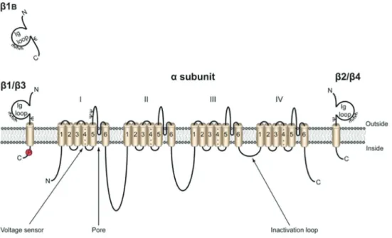
The VGSC are composed of a pore-forming a sub-unit that can be associated with one or more bsubunits (Figure 2). Na+channelasubunits are composed of four
homologous domains, each of which has six transmem-brane segments. The VGSC family has 9 members, Nav1.1 through Nav1.9, encoded by the genes SCN1A-SCN5AandSCN8A-SCN11A. As for thebsubunits,five members have been found, b1 tob4 andb1B (an alter-native splicing of b1), and are encoded by the genes SCN1BthroughSCN4B. These are mainly composed of an extracellular N-terminal segment, a single transmem-brane segment and a short intracellular segment. Pore-forming subunits may be expressed alone, since their operation is independent of the presence of the b sub-units. However, these subunits are essential, as they are responsible for modulating the function of the channel (opening, closing and inactivating it) and allow the asso-ciation with cell adhesion molecules, extracellular matrix and cytoskeleton (11,26,27).
VGSC and intracellular acidification
In recent years, many studies with cell cultures and analysis of biopsy tissues have provided evidence that VGSC are responsible for increasing the invasive potential of tumor cells, participating in the processes of galvano-taxis, cellular motility, migration, and others. Inhibition of VGSC has been linked with reduction of metastatic be-havior (4–8,28).The main question is how the influx of Na+
through these channels increases the invasive and meta-static potential of tumors. Different groups have presented
Figure 1.Tumorigenesis process. The interaction between DNA and different carcinogenic factors can cause mutations in genes that
distinct theories (associated with pH, gene expression and intracellular Ca2+) in an attempt to better understand the
role of these channels and allow the development of new antineoplastic drugs (9,27). Influx of Na+ through the
Nav1.5 channels resulted in intracellular alkalinization and consequent acidification of the extracellular space close to the cell membrane (6,29,30). The mechanism that explains pH variation is associated with co-expression of Na+/H+ (NHE)-1 exchanger, an important protein that
transports hydrogen ions. The influx of Na+through VGSC
increases the efflux of H+, resulting in a high intracellular
and a low extracellular pH. Low pH favors the activation of cathepsins B and S, which are proteolytic enzymes respon-sible for the extracellular matrix degradation (31,32), thus enhancing pH-dependent extracellular matrix degradation and invasion (Figure 3). In another study, Brisson et al. (33) demonstrated that expression of Nav1.5 also promotes modification of the F-actin network and enhances NHE-1 activity in breast cancer cells, resulting in increased invasiveness of malignant cells. This proposition of how VGSC and NHE-1 affect extracellular pH is opposite to the traditional view of how these two proteins interact. It is important to note, however, that cancer cells might have a completely different organization and functioning than normal cells.
Another important point that is not taken into account is the relative contribution of the endo/lysosome H+
extru-sion to the formation of an acidic tumor environment and
to increase cathepsins activity. Endosomes are spherical structures formed from the cellular membrane, which contain approximately 40 hydrolytic enzymes capable of digest cellular components, like mitochondria, intracellular vesicles and even the whole cell. After its constitution, the endosome can be transformed in lysosome or recycled back to cell membrane (34). Lysosome formation is one of the main roles of endosomes. According to Carrithers et al. (35) and Black et al. (36), the lysosomal system is com-plex and highly dynamic. It begins as an early endosome and through a maturation process turns into a late endo-some and then to a lysoendo-some. Different changes occur in this process, such as pH reduction, reception of vesicles coming from the Golgi apparatus, and the activation of lysosomal enzymes (35,37).
The acidic environment of endolysosomes is attribut-ed to V-ATPase, proteins capable of using the energy of ATP hydrolysis to transport H+through intracellular
mem-brane compartments. Recently, it was demonstrated that Nav1.5, classically a cardiac isoform of VGSC, is also pres-ent in late endosomes being responsible for an extra-acidification of this vesicle. The authors suggested a model to explain how these ion channels work and the general idea is that they provide a passage for positive charges (Na+) from inside of the endosome to cytoplasm. This
trans-port enables the entry of more protons into the endosome through voltage-dependent channels CIC or H+-ATPase
pump (35).
Figure 2.Voltage-gated Na+channel. The pore-formingasubunit is composed by four homologous domains with 6 transmembrane
Lysosomal trafficking and cathepsins activation
It is well known that lysosome trafficking is altered in tumor cells (38). Therefore, the actual contribution of this process to the acidification of extracellular environment and, consequently, the cathepsins role during cancer progression must be considered and further investigated. Cathepsins are lysosomal peptidases that participate in the intracellular protein catabolism. These enzymes are synthesized as inactive zymogens and are activated after the break of a pro-peptide by another protease or due to low pH, the optimal environment for cathepsins action. Protein degradation is involved in different cellular proces-ses, physiological or pathological, such as autophagy, antigen presentation, cellular stress signaling and apop-tosis. Besides being commonly associated with tumor progression because of their role in increasing extracel-lular matrix degradation, cathepsins are also involved in apoptosis regulation (38).
Apoptosis induction by cathepsins can be through the extrinsic or death receptor pathway or through the intrinsic or mitochondrial pathway. Thefirst pathway is activated by specific ligands of death receptors and posterior activation of caspase 8, which will cleave Bid, a pro-apoptotic mole-cule. The cleavage of Bid will generate a truncated form of this molecule (tBid), capable of inducing mitochondrial outer membrane permeabilization and, consequently, the release of cytochrome c. Both pathways are connected through Bid, but for the intrinsic pathway the stimuli will be the presence of reactive oxygen species, which are pro-duced during cellular stress and may cause lysosomal membrane permeabilization, a non-proteolytic event that
releases cathepsins into intracellular milieu. Cathepsins will not only stimulate the cleavage of Bid but also the inactivation of anti-apoptotic proteins, such as Bcl-2 (39,40).
Control of intracellular pH
The concentration of intracellular H+has an important
role in different cellular processes, because protein structure and function depend on optimal pH. Cellular compartment-alization is necessary to keep environmental conditions for individual pathways and prevent cellular processes that would cause functional changes (41). Cells have the ten-dency to acidify due to products of metabolic reactions and to electrical potential across the membrane, which pulls positive ions into the intracellular space. Therefore, the removal of protons and their equivalents is a constant process (42).
Many transporters, which are expressed on cellular membrane and organelles of secretory and endocytic pathways, stringently control intracellular pH. Among them are V-ATPase (as mentioned above), NHE, Na+-coupled
HCO3- transporters (NBC) and ion channels (42,43). Ion channels are functionally present on membranes of the aforementioned organelles and also are involved with the ionic homeostasis. There are common challenges in studying channels from different intracellular organelles. Unlike plasma membrane channels that have been unam-biguously defined, the basic information for most orga-nelles has yet to be established, including luminal ionic composition, organelle membrane potential, and lipid com-position of the organelle membrane. Although the importance
Figure 3.pH regulation affects tumor invasion process. The opening of voltage-gated Na+channels (VGSC) causes an increase of the
ion channels in the physiopathology of cancer is based on how VGSC can regulate the expression of certain genes, called invasion gene network (3,27). It is well known that Na+channels are capable of regulating gene expression
in excitable cells and cancer cells (23,45–47).
Further-more,SCN5Agene is a key regulator of this invasion gene network, suggesting that Nav1.5 may function as early entry points of invasion signaling mechanisms. At least in colon cancer, Nav1.5 may regulate invasion by this mecha-nism in addition to extracellular acidification (48). The chal-lenge is to understand how the activity of ion channels can regulate transcription in cancer cells.
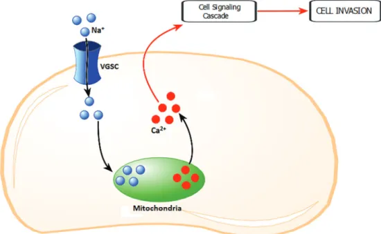
Ca2+and Na+crosstalk
A third possibility is related to the regulation of intracellular Ca2+through VGSC. In excitable and
non-excitable cells, influx of Na+may result in an increase of
intracellular Ca2+levels through the activation of
voltage-gated Ca2+ channels (49). In addition to the plasma
membrane, voltage-gated Ca2+channels are present in
internal membranes such as podosomes and endosomes of cancer cells and macrophages (13,50,51). In THP-1
macro-present in vesicular membranes are gated and/or whether they interact with VGSC present in the plasma membrane (9). In vascular endothelial cells the elevation of intracel-lular Ca2+requires Na+influx and in turn activates PKC
and extracellular signal-regulated kinase (ERK)1/2, poten-tiating angiogenic functions including proliferation, differ-entiation and adhesion (52).
Back to VGSC
In cancer cells, there is no specific pattern for subunits, since differentasubunits are expressed in different types of cancer. For breast cancer, the subtype most commonly found so far is the Nav1.5, which is encoded by the gene SCN5Aand is found in two different forms: 1) the neonatal (nNav1.5), and 2) the adult splicing variant (9–11). An
alternative splicing can occur between two different exons present in exon 6 ofSCN5A(5’exon and 3’exon) and the difference will be the presence or absence of an aspartate residue at position 7 in the exon 6 (3). Through studies in rat brain, it was possible to conclude that the transcription containing the 5’ exon was more common at birth but was quickly replaced by the 3’exon. This splicing pattern
Figure 4. Regulation of intracellular Ca2+ through voltage-gated Na+ channels (VGSC). After the activation of VGSC, a quick
has been found in studies with Nav1.1, Nav1.2, Nav1.3, Nav1.5, Nav1.6 and Nav1.7 (53–58).
In a study to determine which Na+ channels have
functional expression in different types of cancer cell lines and cancer primary cultures, the neonatal Nav1.5 channel isoform was found in breast cancer cells with high meta-static potential. This protein was identified in biopsy sam-ples and its increased expression could be associated with patients with lymph node metastasis. This indicates that the neonatal isoform is significantly up-regulated in breast cancer cells and potentiates cell processes in-volved in metastasis (15).
Another observation was that the majority of expressed functional channels were resistant to TTX, a specific Na+
channel blocker (15). Experiments performed with breast, prostate and lung cancers using TTX resulted in the sup-pression of a variety of behaviors specific to highly metas-tatic cells, such as invasion (14,59,60), lateral motility (61), adhesion (62), migration (45), galvanotaxis (63) and endo-cytosis (64). However, the importance and function of
bsubunits in increased cell aggressiveness remains to be explored (23).
The expression of b subunits and their participation in cell migration and adhesion in two cell types, MDA-MB-231 and MCF-7, demonstrated that MCF-7 cells had higher expression levels of proteins encoded bySCN1B, SCN2B and SCN4B genes compared to MDA-MB-231 cells. However, for both cell lines the most common sub-unit wasb1. Silencingb1 expression resulted in decreased cell adhesion and higher migration in 3D cultures for MCF-7 cells (23).
The same study showed that overexpression of b1 subunit in MDA-MB-231 cells increased Na+current, the
length of cell processes and the intercellular adhesion, in addition to reducing the lateral motility and proliferation. Considering all these findings, the authors were able to conclude that the expression ofb1 enhances cell adhe-sion and reduces the migration of cells in breast cancer. It is important to note that the effects on the adhe-sive capacity of these cells can occur independently of changes in membrane excitability, confirming that
b subunits have the ability to operate in the absence of theasubunit (23).
In summary, thebsubunits appear to have a role in the regulation of several cellular processes including migra-tion, adhesion, cell proliferation and resistance to apop-tosis (65–68). Moreover, these functions appear to have
opposite effects to the regulation carried on byasubunits (23). As we have seen previously,asubunits have greater expression in highly metastatic cells and can increase the ability of invasion and migration.bsubunits are expressed in cells with little metastatic capacity and are able to modu-late Na+influx, increase cell adhesion and the extension
processes, and also regulate different activities such as migration and a subunit mRNA expression (Figure 5) (11,65).
Therapeutic approaches
Each cancer should be considered individually when deciding on the most appropriate treatment procedure. Usually, a combination of treatments is used aiming at the cure or to prolong survival while improving the patient’s quality of life. The most commonly used treatments are surgical removal of the tumor, chemotherapy and radio-therapy. Surgery is the most effective treatment when the total removal of the tumor is possible, but in most cases, it is combined with chemotherapy and/or radiotherapy (17). Chemotherapy is the utilization of various drugs with the objective of killing the cancer cells, although it also causes side effects on normal cells. The mechanism of action of chemotherapic drugs is by the interference in the cell cycle and in cell proliferation. Metabolic differences and faster proliferation rate make cancer cells more sus-ceptible to the drugs. However, normal cells with fast rates of renewal are also affected and generate side effects as hair loss, anemia, immunologic depression, nausea, vomiting, dizziness, weakness and others (69). The dose needed to achieve a balance between the maximal toxic effect to malignant cells with the minimal effect on normal cells is the challenge in chemotherapy.
Radiotherapy employs the emission of ionizing radia-tion that releases free electrons in affected tissues and causes alterations in the DNA, triggering different cell signaling and causing tumor destruction. The effective-ness of this treatment depends on various factors such as tumor sensitivity to radiation, tumor location and the amount of radiation. The adverse effects of this treatment also result from the injury to normal cells, but since the treatment is very localized it is usually well tolerated by patients when the principles of total dose per treatment and fractionated application are respected (69).
The important role that VGSC seems to have in meta-static cells and unsatisfactory clinical results of regular treatments, make these ion channels a real possibility for the development of new methods of diagnosis and perhaps a more effective therapeutic approach. Among the pharmacological tools presently available to target these molecules are drugs that block the functioning of ion channels. One main blocker that has been studied is TTX, a well-known Na+channel blocker (9).
Eacha subunit isoform has a particular sensitivity to TTX, in addition to other pharmacological and physiologi-cal properties. The Nav1.1, Nav1.4, Nav1.6 and Nav1.7 Na+channels are part of a group that exhibits the greatest
sensitivity to this toxin, since a nanomolar (nM) concen-tration is necessary to block these channels. As for the Nav1.5, Nav1.8 and Nav1.9 Na+channels, the necessary
phenytoin, examples of other Na+channel blockers, are
now being tested for use in cancer treatment. Thus far, the results with these drugs show an inhibition of metastatic behavior in in vitro experiments, reducing invasion and metastasis (13,28,70–72).
Another form of therapy is genetic silencing, which uses a technique where double-stranded RNAs (siRNA) are synthesized artificially. The ideal target of siRNAs are genes that are expressed or abnormally regulated only in the tumor cells, or genes specifically involved in cell prolif-eration, angiogenesis and/or metastasis (73).
This approach is still being tested for different diseases with variable results (74,75). The U.S. Food and Drug Administration approved a new antineoplastic siRNA for clinical phase I studies against solid tumors (76). In breast cancer, the in vitro study for gene silencing of nNav1.5 showed very good results. The interruption of the function and expression of these channels in MDA-MB-231 breast cancer cells caused a significant suppression in the migra-tory capacity (about 50%) of these cells (77). However, the reproduction of these resultsin vivo is much more com-plicated and the clinical use of this new treatment depends on the stability of these molecules and the non-suppression of other targets (3).
Conclusion and future perspectives
Studies on cancer and ion channels started in the 1990s with prostate cancer, followed by a series of investigations focusing on the pathophysiological role of ion channel expression in different types of cancer. Afterwards, more studies were performed with differ-ent cell types, and upregulation of voltage-gated Na+
channels have been described in cells presenting high metastatic potential. Over the years, new molecular biol-ogy techniques have been used to investigate ion chan-nels, revealing the presence of different splice variants and giving a better understanding of the underlying mechanisms responsible for the different behaviors of metastatic cells.
The association between breast cancer and Na+
channels, especially voltage-gated channels, was shown to have great importance for cellular events that increase tumors aggressiveness, mainly proliferation, migration, loss of adhesion and galvanotaxis. Some of the proposed theories on how the VGSC relate to the increased aggres-siveness of tumors refute all the current knowledge on Na+homeostasis and raise the questions: do cancer cells
normal cells? Can we identifying the mechanisms of a tumor cell looking at the normal cell or should we accept that we are facing the unknown, where cellular structures might have different functions and relations in cancer cells?
Besides being distressful, these differences can also encourage the search of a better understanding for cancer pathophysiology. Further research should be conducted to unravel mechanisms involving VGSC, NHE-1 and pH. Also of importance is determining if the properties associated with bsubunits are or not dependent on the asubunits expression.
At this point, no ligand is available that can be used selec-tively for Nav1.5 channels, which is one of the main targets for new anti-cancer therapeutic approaches. However, new toxins are discovered every day, increasing the existing arsenal that would lead to new and more effective drugs.
Acknowledgments
This study receivedfinancial support from the Fundac¸ão de Amparo à Pesquisa de Minas Gerais (FAPEMIG) and Fundac¸ão Mineira de Educac¸ão e Cultura (FUMEC).
References
1. Kim S. New and emerging factors in tumorigenesis: an over-view.Cancer Manag Res2015; 7: 225–239, doi: 10.2147/ CMAR.S47797.
2. Kimbung S, Loman N, Hedenfalk I. Clinical and molecular complexity of breast cancer metastases.Semin Cancer Biol 2015; 35: 85–95, doi: 10.1016/j.semcancer.2015.08.009. 3. Onkal R, Djamgoz MB. Molecular pharmacology of
voltage-gated sodium channel expression in metastatic disease: clinical potential of neonatal Nav1.5 in breast cancer.Eur J Pharmacol2009; 625: 206–219, doi: 10.1016/j.ejphar.2009. 08.040.
4. Roger S, Potier M, Vandier C, Besson P, Le Guennec JY. Voltage-gated sodium channels: New targets in cancer therapy?Curr Pharm Des2006; 12: 3681–3695, doi: 10.2174/ 138161206778522047.
5. Campbell TM, Main MJ, Fitzgerald EM. Functional expres-sion of the voltage-gated Na+-channel Nav1.7 is necessary for EGF-mediated invasion in human non-small cell lung cancer cells. J Cell Sci2013; 126 (Part 21): 4939–4949, doi: 10.1242/jcs.130013.
6. Gillet L, Roger S, Besson P, Lecaille F, Gore J, Bougnoux P, et al. Voltage-gated Sodium channel activity promotes cys-teine cathepsin-dependent invasiveness and colony growth of human cancer cell.J Biol Chem2009; 284: 8680–8691, doi: 10.1074/jbc.M806891200.
7. Hernadez-Plata E, Ortiz CS, Marquina-Castillo B, Medina-Martinez I, Alfaro A, Berumen J, et al. Overexpression of Nav1.6 channels is associated with the invasion capacity of human cervical cancer.Int J Cancer2012; 130: 2013–2023, doi: 10.1002/ijc.26210.
8. Litan A, Langhans SA. Cancer as a channelopathy: ion channels and pumps in tumor development and progres-sion.Front Cell Neurosci 2015; 9: 86, doi: 10.3389/fncel. 2015.00086.
9. Brackenbury WJ. Voltage-gated sodium channels and meta-static disease.Channels 2012; 6: 352–361, doi: 10.4161/ chan.21910.
10. Rao VR, Perez-Neut M, Kaja S, Gentile S. Voltage-gated ion channels in cancer cell proliferation. Cancers 2015, 7: 849–875. 11. Roger S, Gillet L, Le Guennec JY, Besson P. Voltage-gated sodium channels and cancer: is excitability their primary role? Front Pharmacol 2015; 6: 152, doi: 10.3389/fphar. 2015.00152.
12. Gillet L, Roger S, Bougnoux P, Le Guennec JY, Besson P. Beneficial effects of omega-3 long-chain fatty acids in breast
cancer and cardiovascular diseases: voltage-gated sodium channels as a common feature?Biochimie2011; 93: 4–6, doi: 10.1016/j.biochi.2010.02.005.
13. Yang M, Kozminski DJ, Wold LA, Modak R, Calhoun JD, Isom LL, et al. Therapeutic potential for phenytoin: targeting Nav1.5 sodium channels to reduce migration and invasion in metastatic breast cancer.Breast Cancer Res Treat 2012; 134: 603–615, doi: 10.1007/s10549-012-2102-9.
14. Roger S, Besson P, Le Guennec JY. Involvement of a novel fast inward sodium current in the invasion capacity of a breast cancer cell line.Biochim Biophys Acta2003; 1616: 107–111, doi: 10.1016/j.bbamem.2003.07.001.
15. Fraser SP, Diss JKJ, Chioni A, Mycielska ME, Pan H, Yamaci RF, et al. Voltage-gated sodium channel expression and potentiation of human breast cancer metastasis. Clin Cancer Res2005; 11: 5381–5389, doi: 10.1158/1078-0432. CCR-05-0327.
16. GLOBOCAN. Estimated cancer incidence, mortality and prevalence worldwide in 2012. http://globocan.iarc.fr/Pages/ fact_sheets_cancer.aspx. Accessed June 29, 2016. 17. World Health Organization. Cancer. http://www.who.int/
mediacentre/factsheets/fs297/en/. Accessed June 23, 2016. 18. World Health Organization. Are the number of cancer cases increasing or decreasing in the world? http://www.who.int/ features/qa/15/en/index.html. Accessed June 20, 2016. 19. Hanahan D, Weinberg RA. Hallmarks of cancer: the next
generation. Cell 2011; 144: 646-674, doi: 10.1016/j.cell. 2011.02.013.
20. INCA. O que é o câncer? http://www1.inca.gov.br/conteu-do_view.asp?id=322. Accessed June 16, 2016.
21. Hille B.Ionic channels of excitable Membranes. 3rd edn. Sunderland, Sinauer Associates Inc. 2001.
22. Schönherr R. Clinical relevance of ion channels for diagnosis and therapy of cancer.J Membr Biol2005; 205: 175–184, doi: 10.1007/s00232-005-0782-3.
23. Chioni A, Brackenbury WJ, Calhoun JD, Isom LL, Djamgoz MBA. A novel adhesion molecule in human breast cancer cells: Voltage-gated Na+channelb1 subunit.Int J Biochem Cell Biol 2009; 41: 1216–1227, doi: 10.1016/j.biocel.2008.11.001. 24. Kruger LC, Isom LL. Voltage-gated Na+channels: not just
for conduction.Cold Spring Harb Perspect Biol2016; 8. 25. Catterall WA, Goldin AL, Waxman SG. Nomenclature and
2014; 13: 264, doi: 10.1186/1476-4598-13-264.
29. Bourguignon LY, Singleton PA, Diedrich F, Stern R, Gilad E. CD44 interaction with Na+-H+exchanger (NHE1) creates acidic microenvironments leading to hyaluronidase-2 and cathepsin B activation and breast cancer tumor cell invasion.J Biol Chem 2004; 279: 26991–7007, doi: 10.1074/jbc.M311838200. 30. Busco G, Cardone RA, Greco MR, Bellizzi A, Colella M,
Antelmi E, et al. NHE1 promotes invadopodial ECM proteo-lysis through acidification of the peri-invadopodial space. FASEB J2010; 24: 3903–3915, doi: 10.1096/fj.09-149518. 31. Brisson L, Gillet L, Calaghan S, Besson P, Le Guennec JY,
Roger S, et al. NaV1.5 enhances breast cancer cell invasive-ness by increasing NHE1-dependent H(+) efflux in caveolae. Oncogene2011; 30: 2070–2076, doi: 10.1038/onc.2010.574. 32. Cardone RA, Casavola V, Reshkin SJ. The role of disturbed pH dynamics and the Na+/H+exchanger in metastasis. Nat. Rev. Cancer2005; 5: 786–795, doi: 10.1038/nrc1713. 33. Brisson L, Driffort V, Benoist L, Poet M, Counillon L, Antelmi E, et al. Nav1.5 Na+channels allosterically regulate the NHE-1 exchanger and promote the activity of breast cancer cell invadopodia.J Cell Sci2013; 126 (Part21): 4835–4842, doi: 10.1242/jcs.123901.
34. Klumperman J, Raposo G. The complex ultrastructure of the endolysosomal system. Cold Spring Harb Perspect Biol 2014; 6: a016857, doi: 10.1101/cshperspect.a016857. 35. Carrithers MD, Dib-Hajj S, Carrithers LM, Tokmoulina G,
Pypaert M, Jonas EA, et al. Expression of the voltage-gated sodium channel NaV1.5 in the macrophage late endosome regulates endosomal acidification. J Immunol. 2007; 178: 7822–7832, doi: 10.4049/jimmunol.178.12.7822.
36. Black JA, Dib-Hajj S, Cohen S, Hinson AW, Waxman SG. Glial cells have heart: rH1 Na+channel mRNA and protein in spinal cord astrocytes.Glia1998; 23: 200–208, doi: 10.1002/ (SICI)1098-1136(199807)23:3o200::AID-GLIA343.0.CO;2-8. 37. Scott CC, Vacca F, Gruenberg J. Endosome maturation, transport and functions.Semin Cell Dev Biol2014; 31: 2–10, doi: 10.1016/j.semcdb.2014.03.034.
38. Olson OC, Joyce JA. Cysteine cathepsin proteases: regula-tors of cancer progression and therapeutic response. Nat Rev Cancer2015; 15: 712–729, doi: 10.1038/nrc4027. 39. Pislar A, Nanut MP, Kos J. Lysosomal cysteine peptidases
-Molecules signaling tumor cell death and survival.Semin Cancer Biol2015; 35: 168–179, doi: 10.1016/j.semcancer. 2015.08.001.
40. Repnik U, Cesen MH, Turk B. The endolysosomal system in cell death and survival. Cold Spring Harb Perspect Biol 2013; 5: a008755, doi: 10.1101/cshperspect.a008755. 41. Demaurex N. pH homeostasis of cellular organelles.News
Physiol Sci2002; 17: 1–5.
42. Casey JR, Grinstein S, Orlowski J. Sensors and regulators of intracellular pH.Nat Rev Mol Cell Biol2010; 11: 50–61, doi: 10.1038/nrm2820.
343–356, doi: 10.1113/jphysiol.2006.106906.
46. Mycielska ME, Palmer CP, Brackenbury WJ, Djamgoz MB. Expression of Na+-dependent citrate transport in a strongly metastatic human prostate cancer PC-3M cell line: regula-tion by voltage-gated Na+channel activity.J Physiol2005; 563 (Part 2): 393–408, doi: 10.1113/jphysiol.2004.079491. 47. Lopez-Santiago LF, Meadows LS, Ernst SJ, Chen C,
Malhotra JD, McEwen DP, et al. Sodium channel Scn1b null mice exhibit prolonged QT and RR intervals. J Mol Cell Cardiol2007; 43: 636–647, doi: 10.1016/j.yjmcc.2007.07.062. 48. House CD, Vaske CJ, Schwartz AM, Obias V, Frank B, Luu T, et al. Voltage-gated Na+channel SCN5A is a key regulator of a gene transcriptional network that controls colon cancer invasion.Cancer Res 2010; 70: 6957–6967, doi: 10.1158/ 0008-5472.CAN-10-1169.
49. Fekete A, Franklin L, Ikemoto T, Rózsa B, Lendvai B, Sylvester Vizi E, et al. Mechanism of the persistent sodium current activator veratridine-evoked Ca elevation: implication for epilepsy.J Neurochem2009; 111: 745–756, doi: 10.1111/ j.1471-4159.2009.06368.x.
50. Carrithers MD, Chatterjee G, Carrithers LM, Offoha R, Iheagwara U, Rahner C, et al. Regulation of podosome formation in macrophages by a splice variant of the sodium channel SCN8A. J Biol Chem 2009; 284: 8114–8126, doi: 10.1074/jbc.M801892200.
51. Carrithers MD, Dib-Hajj S, Carrithers LM, Tokmoulina G, Pypaert M, Jonas EA, et al. Expression of the voltage-gated sodium channel NaV1.5 in the macrophage late endosome regulates endosomal acidification. J Immunol 2007; 178: 7822–7832, doi: 10.4049/jimmunol.178.12.7822.
52. Andrikopoulos P, Fraser SP, Patterson L, Ahmad Z, Burcu H, Ottaviani D, et al. Angiogenic functions of voltage-gated Na+
Channels in human endothelial cells: modulation of vascular endothelial growth factor (VEGF) signaling.J Biol Chem2011; 286: 16846–1660, doi: 10.1074/jbc.M110.187559.
53. Copley RR. Evolutionary convergence of alternative splicing in ion channels.Trends Genet2004; 20: 171–176, doi: 10.1016/ j.tig.2004.02.001.
54. Gustafson TA, Clevinger EC, Oneill TJ, Yarowsky PJ, Krueger BK. Mutually exclusive exon splicing of type-III brain sodium channel-alpha subunit RNA generates devel-opmentally-regulated isoforms in rat-brain. J Biol Chem 1993; 268: 18648–18653.
55. Sarao R, Gupta SK, Auld VJ, Dunn RJ. Developmentally regulated alternative RNA splicing of rat brain sodium channel mRNAs.Nucleic Acids Res1991; 19: 5673–5679, doi: 10.1093/nar/19.20.5673.
57. Plummer NW, Galt J, Jones JM, Burgess DL, Sprunger LK, Kohrman DC, et al. Exon organization, coding sequence, physical mapping, and polymorphic intragenic markers for the human neuronal sodium channel gene SCN8A. Geno-mics1998; 54: 287–296, doi: 10.1006/geno.1998.5550. 58. Raymond CK, Castle J, Garrett-Engele P, Armour CD, Kan
Z, Tsinoremas N, et al. Expression of alternatively spliced sodium channel alpha subunit genes: unique splicing patterns are observed in dorsal root ganglia.J Biol Chem 2004; 279: 46234–46241, doi: 10.1074/jbc.M406387200. 59. Laniado ME, Lalani EN, Fraser SP, Grimes JA, Bhangal G,
Djamgoz MB, et al. Expression and functional analysis of voltage-activated Na+channels in human prostate cancer cell lines and their contribution to invasionin vitro. Am J Pathol1997; 150: 1213–1221.
60. Bennett ES, Smith BA, Harper JM. Voltage-gated Na+ channels confer invasive properties on human prostate cancer cells.Pflugers Arch2004; 447: 908–914, doi: 10.1007/s00424-003-1205-x.
61. Fraser SP, Salvador V, Manning EA, Mizal J, Altun S, Raza M, et al. Contribution of functional voltage-gated Na+ channel expression to cell behaviors involved in the metastatic cascade in rat prostate cancer: I. lateral motility. J Cell Physiol2003; 195: 479–487, doi: 10.1002/jcp.10312. 62. Palmer CP, Mycielska ME, Burcu H, Osman K, Collins T, Beckerman R, et al. Single cell adhesion measuring appa-ratus (SCAMA): application to cancer cell lines of different metastatic potential and voltage-gated Na+channel expres-sion. Eur Biophys J 2008; 37: 359–368, doi: 10.1007/ s00249-007-0219-2.
63. Djamgoz MBA, Mycielska M, Madeja Z, Fraser SP, Korohoda W. Directional movement of rat prostate cancer cells in direct-current electric field: involvement of voltage gated Na+ channel activity.J Cell Sci2001; 114 (Part 14): 2697–2705. 64. Onganer PU, Seckl MJ, Djamgoz MB. Neuronal
character-istics of small-cell lung cancer. Br J Cancer 2005; 93: 1197–1201, doi: 10.1038/sj.bjc.6602857.
65. Brackenbury WJ, Isom LL. Na+ channels b subunits: overachievers of the ion channel family.Front Pharmacol 2011; 2: 53, doi: 10.3389/fphar.2011.00053.
66. Malhotra JD, Kazen-Gillespie K, Hortsch M, Isom LL. Sodium channelbsubunits mediate homophilic cell adhe-sion and recruit ankyrin to points of cell-cell contact.J Biol Chem2000; 275: 11383–11388, doi: 10.1074/jbc.275.15. 11383.
67. Brackenbury WJ, Davis TH, Chen C, Slat EA, Detrow MJ, Dickendesher TL, et al. Voltage-gated Na+ channel b1 subunit-mediated neurite outgrowth requires fyn kinase and contributes to central nervous system developmentin vivo. J Neurosci2008; 28: 3246–3256, doi: 10.1523/JNEUROSCI. 5446-07.2008.
68. Kazarinova-Noyes K, Malhotra JD, McEwen DP, Mattei LN, Berglund EO, Ranscht B, et al. Contactin associates with Na+ channels and increases their functional expession. J Neurosci2001; 21: 7517–7525.
69. INCA. Tratamento ao câncer. http://www2.inca.gov.br/wps/ wcm/connect/cancer/site/tratamento. Accessed July 2, 2016. 70. Nelson M, Yang M, Dowle AA, Thomas JR, Brackenbury WJ. The sodium channel-blocking antiepileptic drug pheny-toin inhibits breast tumor growth and metastasis.Mol Cancer 2015; 14: 13, doi: 10.1186/s12943-014-0277-x.
71. Roger S, Le Guennec JY, Besson P. Particular sensitivity to calcium channel blockers of the fast inward voltage-dependent sodium current involved in the invasive proper-ties of a metastatic breast cancer cell line.Br J Pharmacol 2004; 141: 610–615, doi: 10.1038/sj.bjp.0705649. 72. Martin F, Ufodiama C, Watt I, Bland M, Brackenbury WJ.
Therapeutic value of voltage-gated sodium channel inhibi-tors in breast, colorectal, and prostate cancer: a systematic review.Front Pharmacol2015; 6: 273, doi: 10.3389/fphar. 2015.00273.
73. Huang C, Li M, Chen C, Yao Q. Small interfering RNA therapy in cancer: mechanism, potential targets, and clinical applications.Expert Opin Ther Targets2008; 12: 637–645, doi: 10.1517/14728222.12.5.637.
74. Wall NR, Shi Y. Small RNA: can RNA interference be exploited for therapy? Lancet2003; 362: 1401–1403, doi: 10.1016/S0140-6736(03)14637-5.
75. Masiero M, Nardo G, Indraccolo S, Favaro E. RNA inter-ference: implications for cancer treatment.Mol Aspects Med 2007; 28: 143–166, doi: 10.1016/j.mam.2006.12.004. 76. Behlke MA. Chemical modification of siRNAs forin vivouse.
Oligonucleotides2008; 18: 305–319, doi: 10.1089/oli.2008. 0164.
77. Brackenbury WJ, Djamgoz MB. Nerve growth factor enhances voltage-gated Na+channel activity and Transwell migration in Mat-LyLu rat prostate cancer cell line.J Cell Physiol2007; 210: 602–608, doi: 10.1002/jcp.20846.