Negative inotropic and chronotropic
effects on the guinea pig atrium of
extracts obtained from
Averrhoa
carambola
L. leaves
1Laboratório de Biofísica do Coração, Departamento de Fisiologia,
Centro de Ciências Biológicas e da Saúde, Universidade Federal de Sergipe, Aracaju, SE, Brasil
2Laboratório deTecnologia Farmacêutica “Prof. Delby Fernandes de Medeiros“, Universidade Federal da Paraíba, João Pessoa, PB, Brasil
C.M.L. Vasconcelos1,2, M.S.Araújo1, B.A. Silva2 and E.A. Conde-Garcia1
Abstract
It has been reported that star fruit can lead to a fatal outcome in uremic patients. The intoxication syndrome consists of hiccups, mental confusion, dizziness, and vomiting. On the other hand, folk medicine uses teas and infusions of carambola leaves to treat headache, vomiting, cough, insomnia, and diabetes. This moti-vated us to determine if Averrhoa carambola can act on the contractility and automaticity of the guinea pig heart. We mea-sured the atrial isometric force in stimulated left atria and deter-mined the chronotropic changes in spontaneously beating right atria. The carambola leaf extracts (1.5 mg/ml) abolished the contrac-tile force in a concentration-dependent manner. Among the crude, methanolic, ethanolic, aqueous, and acetic extracts, the aqueous one was the most potent (EC50 = 520 ± 94 µg/ml; flavonoids and
tannins are the main constituents; Na+ and K+ contents in 1.0 mg/
ml of aqueous extract were 0.12 ± 0.016 and 1.19 ± 0.15 mM, respectively). The aqueous extract abolished the positive Bowditch staircase phenomenon and reduced the inotropic response to CaCl2
(0.17-8.22 mM), events that are dependent on the cellular Ca2+
inward current. The adrenergic, muscarinic or opioid membrane receptors do not seem to participate in the mechanism of action of the cardioactive substance(s). In spontaneously beating atria, the aqueous extract promoted a negative chronotropic effect that was antagonized by 0.1 µM isoproterenol bitartrate. With this agonist, the EC50 of the aqueous extract increased from 133 ± 58 to 650 ± 100
µg/ml. These data regarding the effect of A. carambola on guinea pig atrial contractility and automaticity indicate an L-type Ca2+
channel blockade.
Correspondence
E.A. Conde-Garcia
Laboratório de Biofísica do Coração Departamento de Fisiologia, CCBS Universidade Federal de Sergipe 49100-000 Aracaju, SE Brasil
Fax: +55-79-246-3377 E-mail: [email protected]
Research supported by ELETROBRAS (No. 23113.009351/03-67), FAP/SE (No. FAP-SE/FUNTEC/FNS/No. 01/ 2003), and CNPq.
Received March 3, 2004 Accepted February 16, 2005
Key words
•Averrhoa carambola L.
•Star fruit •Inotropism •Chronotropism •Guinea pig atrium •Ca2+ channel blockade
Introduction
Averrhoa carambola L. (star fruit,
‘carambola’ in Brazil) is a plant of the
for treating headache, vomiting, cough, in-somnia, hypertension, and diabetes (1-5). The literature reports that uremic patients develop severe and acute intoxication after eating carambola or drinking its juice, with intractable hiccups, sudden onset of limb numbness, muscle weakness, conscious-ness disturbance, and seizure (6,7). These neurological events can be treated only by submitting the patients to hemodialysis. In spite of this therapeutic effort, many cases show a poor outcome which result in death (8-10). A water-soluble neurotoxin was iso-lated from star fruit (11). This protein in-duced convulsion in rats by acting on L-glutamate release, as demonstrated in synap-tosomes prepared from the rat cerebral cor-tex (8). These clinical and experimental ef-fects motivated us to test some of the A.
carambola extracts on the mammalian heart
because folk medicine extensively uses teas and infusions obtained from carambola leaves. The objective of the present study was to investigate the myocardial effects produced
by A. carambola leaf extracts on left atrium
contractility and automaticity of spontane-ously beating isolated guinea pig right atria.
Material and Methods
Extract preparation
Averrhoa carambola leaves were collected
on the campus of the Federal University of Sergipe (Brazil) from healthy and agrotoxic-free plants. The botanical identification was performed by comparing the vegetal parts with those deposited in the Herbarium of the Federal University of Pernambuco, Brazil (Voucher No. 24,720). The crude extract was obtained by macerating dry leaves in a water:ethanol mixture (6:4, v/v) for 10 days at 27 ± 3ºC. The solvents were then filtered and evaporated in a rotary evaporator (Tecnal TE 210, Piracicaba, SP, Brazil) and a crude extract was obtained. Several extracts were also obtained by submit-ting another set of dry leaves to a hot extraction
with hexane, chloroform, acetone, ethanol, methanol, water, and acetic acid using a Soxhlet apparatus. Each extract was concentrated in a rotary evaporator and stored at 27 ± 3ºC in a dry environment. The water-insoluble residue of each extract was determined to permit correct adjustment of the extract concentra-tion in the organ bath.
Phytochemistry screening
The phytochemistry screening was per-formed on the A. carambola aqueousextract (AEx) according to the techniques proposed by Domínguez (12).
Measurement of Na+ and K+
The Na+ and K+ contents of the AEx were determined by flame photometry (Instituto de Tecnologia e Pesquisa de Sergipe, Governo do Estado de Sergipe, Aracaju, SE, Brazil).
Aqueous extract fractionation
Five grams of the AEx was loaded on a 4 x 80 cm silica gel 60 chromatographic column (Sigma-Aldrich, St. Louis, MO, USA). The column was eluted with a mixture of methanol and water with increasing polarity (100:0, 95:5, 90:10, 80:20, 50:50, and 0:100, v/v).
Inotropic effect of Averrhoa carambola
Itu, SP, Brasil and HAAKE FJ, BW 76227, Karlsruhe, Germany) and oxygenated with a gas mixture consisting of 95% O2 and 5% CO2. The atria were stretched to 10 mN and submitted to field stimulation of 2 Hz. The stimulation pulses (400 V and 0.5 ms) were provided by a Digitimer 3072 Stimulator, which was controlled by a Digitimer D4030 Pro-grammer (Welwyn, Garden City, Hertford-shire, England). The atrial force was recorded with an isometric force transducer (HP FTA 10-1 Sunborn, Chicago, IL, USA) coupled to a thermal paper polygraph (HP8805B, HP7754A/B). The electrical signals were also stored in a computer to be processed off-line (DATAQ DI400, DI 205, WINDAQ PRO Acquisition, Windaq EX Calculate, Akron, OH, USA).
To analyze the atrial contractile force data stored in the computer, fifty successive contractions were selected from each ex-perimental procedure (control, test, and washout) and their parameters were deter-mined and averaged with the EG1v1 soft-ware (Conde-Garcia, Patent deposit No. 00051104 Instituto Nacional de Propriedade Industrial - INPI, Ministério do Desenvolvi-mento, Indústria e Comércio Exterior, Brasília, DF, Brasil, also deposited at the Cartório do 10º Ofício, Títulos e Documentos e Pessoas Jurídicas, Aracaju, SE, Brazil).
Determination of the relative potencies of the extracts
To determine the EC50 (concentration needed to produce 50% of the maximum effect) of each A. carambola extract, the force amplitudes obtained with different ex-tract concentration in the organ bath were fitted by curves calculated by the Hill-Langmuir equation:
where: E = effect of the extract on the contractile force amplitude; Emax = maximum effect of the extract on the contractile force amplitude; C = extract concentration in the organ bath; EC50 = extract concentration needed to produce 50% of the maximum effect; n = Hill constant.
Averrhoa carambolavs membrane receptors
To determine whether opioid, muscarinic or ß-adrenergic membrane receptors could be involved in the mechanism of action of A.
carambola, the inotropic effect produced by
the AEx was tested in artificially paced guinea pig left atria both in a control situation and after blocking each one of the specific mem-brane receptor. For this procedure, the fol-lowing drugs were added to the organ bath: 1.5 µM atropine sulfate, 0.7 µM propranolol hydrochloride, or 120 µM naloxone hydro-chloride.
Chronotropic effect of Averrhoa carambola
To test the effect of the extract on the atrial pacemaker activity, some experiments were carried out in spontaneously beating guinea pig right atria. The biological prepara-tions remained immersed in a modified Tyrode solution (13). The preparation, previously stretched to a resting tension of 5 mN, was maintained at 27 ± 0.1ºC. The atrial rate was determined by measuring the interval be-tween successive contractions. For this pro-cedure, fifty contractile force curves were processed with the EG1v1 software. Con-centration-response curves for the A.
ca-rambola AEx (0.005 to 1.6 mg/ml) were
obtained in a control situation or after adding 0.1 µM isoproterenol bitartrate to the bath.
Averrhoa carambolavs inward Ca2+ current
The cellular inward Ca2+ current was studied using the Nayler and Merrillees pro-tocol (14) applied to guinea pigs reserpinized E
E C
EC
C
EC
n
n
=
+
max 50
50
(5 mg/kg, ip) 24 h before the experiment. This experimental protocol is based on the Bowditch positive staircase, a phenomenon that is dependent on the cellular inward Ca2+ current. The experiments were carried out in five successive phases: 1) the left atrium was maintained under a low frequency of stimu-lation (0.2 Hz, control); 2) the stimustimu-lation rate was abruptly increased to 0.33, 0.66, 1.0, 1.33, or 1.66 Hz for 50 s; 3) the stimulation was interrupted for 40 s; finally, 5) the control rate (0.2 Hz) was reinitiated. This experimental sequence was followed before and after adding the A. carambola AEx to the organ bath.
In addition to the Nayler and Merrillees protocol, concentration-response curves ob-tained by progressively adding CaCl2 to the organ bath were used to study calcium entry in cardiac cells. The experiments were carried out in guinea pig left atria previously stretched to a resting tension of 4.9 mN, maintained at 27ºC, and under electrical stimulation of 1.5 Hz. The calcium concentration in the Tyrode solution was increased to the following values: 0.17, 0.34, 0.685, 1.37, 2.05, 2.74, 4.11, 5.48, 6.85, and 8.22 mM and the atrial force was recorded as described above.
Determination of oxalic acid
The Hodgkinson colorimetric method modified by Rassi and Damas (15) was used to determine the oxalic acid content in 1.0 mg/ml AEx of carambola.
Statistical analysis
Data are reported as means ± SEM. The Student t-test for unpaired data was used to determine differences between means. The results were considered to be statistically different when P was less than 0.05 (Statistica for Windows, Tulsa, OK, USA).
Drugs
The following drugs were used: 1.5 µM atropine sulfate, 0.3 µM acetylcholine chlo-ride, 0.7 µM propranolol hydrochlochlo-ride, 1 µM and 0.1 µM isoproterenol bitartrate, 120 µM naloxone hydrochloride, and 5 mg/kg reserpine. Drugs were purchased from Sigma-Aldrich, except naloxone (Rhodia Farma Ltda., São Paulo, SP, Brazil) and reserpine (kindly supplied by Laboratório Gross S.A., Rio de Janeiro, RJ, Brazil).
Results
The yield of each A. carambola extract (g/100 g of dry leaves) was: crude (15.0), hexane (2.36), chloroform (1.75), acetone (5.16), ethanol (13.4), methanol (2.92), aque-ous (7.8), and acetic acid (18).
Figure 1 shows the negative inotropic effect produced by the A. carambola crude extract on the guinea pig left atrium. The effect, clearly dependent on extract concen-tration, disappeared when A. carambola was removed from the bath (washout).
To calculate the relative potency of the A.
carambola extracts, the atrial
force-concen-tration data were fitted with Hill-Langmuir curves. The results showed that the AEx was the most potent (EC50 = 520 ± 94 µg/ml, N = 4, 10 trials, Hill constant = 3, P < 0.001 when compared to the ethanol, methanol, acetic acid, or crude extract). The following results were obtained for the remaining extracts: etha-nol (EC50 = 700 ± 65 µg/ml, N = 3, Hill constant = 2), methanol (EC50 = 1200 ± 171 µg/ml, N = 3, Hill constant = 3), acetic acid (EC50 = 1400 Figure 1. Negative inotropic
± 285 µg/ml, N = 3, Hill constant = 2), and crude extract (EC50 = 1400 ± 283 µg/ml, N = 8, 14 trials, Hill constant = 2). Figure 2 depicts the concentration-effect curves for the aque-ous and crude extract.
The phytochemical screening of the AEx revealed the presence of the following com-ponents: 1) saponin [foam (-)]; 2) alkaloid [Bouchardat (-), Mayer (-), Dragendorff (-), tungstosilicic acid (-)]; 3) steroid [acetic anhydride plus sulfuric acid (+)]; 4) tannin [gelatin (+), FeCl3 (+++)], and 5) flavonoid [magnesium tape (++), fluorescence (+++)]. The oxalic acid concentration of this extract was only 21 µg/ml.
Flame photometry revealed that the Na+ and K+ contents of the AEx were 0.12 ± 0.016 mM and 1.19 ± 0.15 mM, respective-ly. To determine whether this K+ concentra-tion could be responsible for the negative inotropic effect of AEx, some experiments were carried out by progressively increasing the K+ concentration in the bath from 5 to 7 mM and the atrial force was measured. This concentration range was chosen because the maximum depressant effect of AEx was reached when it was concentrated at 1.5 mg/ ml in the organ chamber. This concentration increased the potassium content of Tyrode solution from 5 to 6.78 mM. In the presence of 7 mM K+, the atrial force amplitude de-creased by only 10%. This change is not compatible with the negative inotropic effect of the AEx because when the extract was used at 1.5 mg/ml - which is equivalent to a total K+ concentration of 6.78 mM in the bath solution - the atrial force decreased by more than 99% of its control value.
To determine the possible involvement of membrane receptors in the negative inotrop-ic effect of A. carambola, experiments were performed by using a fraction (FAEx) ob-tained by eluting the AEx with a 1:1 methanol:water (v/v) mixture through a silica gel 60 column. This FAEx was more potent than the carambola AEx (EC50 = 140 ± 7 µg/ ml, N = 3, 5 trials, P < 0.001), suggesting that
Figure 3. A, Atropine sulfate (Atrop, 1.5 µM); B, propranolol hydrochloride (Prop, 0.7 µM), or C, naloxone hydrochloride (Nx, 120 µM), although blocking the muscarinic, ß-adrener-gic and opioid receptors, respectively, did not modify the depressor effect produced by the Averrhoa carambola aqueous fraction (FAEx, 0.6 mg/ml), suggesting that none of them participate in the mechanism of action of this extract on the guinea pig atrium. ISO: isoproterenol bitartrate (1 µM); Ach = acetylcholine chloride (0.3 µM; 27 ± 0.1ºC, stimuli: 2 Hz, 400 V, 0.5 ms).
the active principle was concentrated in it. The inotropic effect of FAEx was deter-mined both in the control solution and after blocking membrane receptors. Since the con-tractile effect remained unchanged after the receptor blockade (Figure 3), we assume
B
C
Figure 2. Hill-Langmuir curves constructed to determine the relative potency of aqueous (squares) and crude (triangles) Averrhoa carambola extracts. The extracts were aqueous (EC50 = 520 ± 94 µg/ml, Hill constant = 3, N = 4 atria, 10 trials) and crude (EC50 = 1400 ± 283 µg/ml, Hill constant = 2, N = 8 atria, 14 trials). The experi-ments were carried out in guin-ea pig left atria (stimuli: 2 Hz, 400 V, 0.5 ms; 27 ± 0.1ºC, rest-ing tension: 10 mN). The EC50 were significantly different (P < 0.001, paired Student t-test).
that none of them mediated the negative response promoted by FAEx (Figure 4, N = 3 atria).
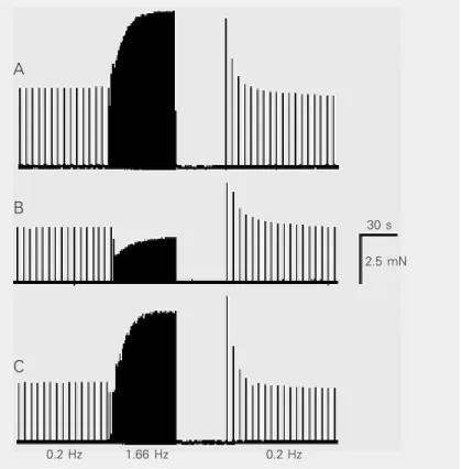
Figure 5 depicts the Bowditch phenome-non obtained with the Nayler and Merrillees protocol. In this case, the stimulation rates were: 0.2 Hz in the control, 1.66 Hz during the overdrive, and 0.2 Hz after the stimula-tion pause. The upper panel shows that during the overdrive the atrial force increased from 5.25 to 9.5 mN (atrial force overshoot equal to 80.9%). The middle panel shows the result obtained after adding 1.0 mg/ml of AEx to the bath. Note that, during overdrive, instead of an atrial force overshoot, a slight decrease in atrial force (13.5%) from 3.7 to 3.2 mN was observed. This effect, however, disappeared during washout (lower panel) and the Bowditch phenomenon was restored to its control behavior (from 4.1 to 8 mN, 95.1% of the control atrial force).
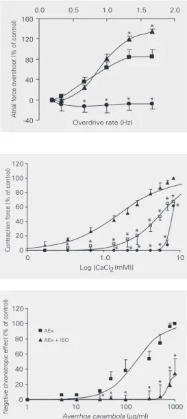
Figure 6 shows that, insofar as the over-drive rate was increased from 0.2 to 1.66 Hz (N = 3 hearts, 27 ± 0.1ºC), the atrial force overshoot progressively increased to 84% (control solution). When the overdrive rate was increased with AEx (1.0 mg/ml) in the bath, the atrial force, instead of increasing, decreased by about 16%. Nevertheless, this effect was not dependent on the stimulation rate applied. These effects rapidly disap-peared during washout and the preparations recovered their control contractile ability.
The inotropic effect induced by cumula-tive addition of CaCl2 (0.17 to 8.22 mM) to the organ bath fluid is depicted in Figure 7. AEx caused both a progressive shift of the log concentration-response curve for CaCl2 to the right and a reduction of the maximum effect of the AEx (2.0 mg/ml) from 100 to 64 ± 8%. In the control solution the EC50 of CaCl2 was 1.7 ± 0.3 mM but with AEx at concentrations of 1.2 and 2.0 mg/ml, the EC50 changed to 4.2 ± 0.4 and 7.2 ± 0.9 mM, respectively (P < 0.05).
In the spontaneously beating atria, AEx promoted a negative chronotropic effect that Figure 4. Atrial force reduction of 82% induced by 0.6 mg/ml Averrhoa carambola
aqueous fraction (FAEx). The experiments were carried out in the guinea pig left atrium using at least 3 atria to test each of the receptors (27 ± 0.1ºC, stimuli: 2 Hz, 400 V, 0.5 ms).The blockade of adrenergic, cholinergic or opioid membrane receptors promoted by propranolol hydrochloride (Prop, 0.7 µM), atropine sulfate (Atrop, 1.5 µM) or naloxone hydrochloride (Nx, 120 µM) did not modify (P > 0.05, unpaired Student t-test) the depressor effect of the FAEx on myocardial contractility.
Figure 5. Effect of the Averrhoa carambola aqueous extract (AEx) on the mem-brane calcium inward current studied by the Bowditch positive staircase phenom-enon which was obtained by increasing the atrial rate from 0.2 to 1.66 Hz. A, Atrial contractile force in control solution. During the stimulus overdrive the atrial force increased from 5.25 to 9.5 mN (80.9%). B, Atrium submitted to 1.0 mg/ml AEx. In this case, during overdrive, the atrial force declined from 3.7 mN to 3.2 mN (13.5%). C, Atrial force behavior during washout. In this experimental phase, the atrial force increased from 4.1 to 8 mN (95.1%) when the preparation was in overdrive. The experiment was carried out at 27 ± 0.1ºC (stimuli: 0.2 or 1.66 Hz, 400 V, 0.5 ms).
0 20 40 60 80 100 120
Control FAEx Prop Atrop Nx
Atrial contractile force
(% of control)
A
B
C
30 s
2.5 mN
was dependent on the extract concentration (Figure 8). At the maximum effect, which was obtained with a concentration of 1.6 mg/ ml, the spontaneous atrial rate decreased by about 99% (P < 0.001, N = 4 atria, 5 trials), leading some preparation to complete asys-tole. By adding isoproterenol bitartrate (0.1 µM) to the organ bath, the concentration-effect curve was shifted to the right, its EC50 increased from 133 ± 58 to 650 ± 100 µg/ml (P < 0.001, N = 4 atria), and the relative efficacy decreased from 100 to 33.4 ± 14% (P < 0.001, N = 4 atria).
Figure 6. Effects of stimulation rate on the amount of force over the control one (Bowditch phenomenon). Squares: control curve; circles: test curve obtained with 1.0 mg/ml Averrhoa carambola aqueous extract; tri-angles: washout curve. The experiments were carried out in guinea pig left atria (N = 4 atria, 27 ± 0.1ºC). *P < 0.001 compared to the control curve (paired Student t -test).
Figure 7. Inhibition by the aqueous extract of Averrhoa carambola of the positive inotropic effect induced by increasing CaCl2 concentrations in the left driven atria. CaCl2 was added cumulatively to the organ bath. Points are means ± SD of 6 atria (14 assays). Triangles: control; squares: 1.2 mg/ml aqueous extract; circles: 2.0 mg/ml aqueous extract. Concentration-response curves were significantly shifted to the right (*P < 0.05, paired Student t-test) and the relative efficacy decreased from 100 to 64 ± 8% (27 ± 0.1ºC, 1.5 Hz).
Figure 8. Concentration-response curves for the nega-tive chronotropic effect promoted by the aqueous extract (AEx) of Averrhoa carambola in the absence (EC50 = 133 ± 58 µg/ml) or in the presence of 0.1 µM isoproterenol (ISO, EC50 = 650 ± 100 µg/ml). With ISO, the concentration-response curve was shifted to the right (*P < 0.01, paired Student t-test)) and the relative efficacy decreased from 100 to 33.4 ± 14%, P < 0.01 (27 ± 0.1ºC, N = 4 atria, 5 trials).
Discussion
The results presented here demonstrated that A. carambola promotes a decrease in cardiac contractility in the guinea-pig atria. This effect was observed when using the crude, ethanol, methanol, or acetic acid ex-tracts. The AEx was the most potent, indi-cating that active substance(s) is(are) water soluble and polar. Despite the well-defined negative effect of A. carambola on myocar-dial contractility, the atrial force promptly recovered to control values during washout.
* * *
* * *
* * *
* * *
*
In heart muscle, stimulation of opioid (µ,
κ, and δ) or muscarinic (M2) receptors may mediate a reduction in cardiac contractility (16-19), a result that can also be obtained by activating the ß3-adrenoceptor. This effect involves the nitric oxide synthase pathway (20,21). In spite of these possibilities, our results showed that neither the opioid nor the muscarinic or adrenergic receptors seem to participate in the inotropic effects of AEx because no changes were observed in the atrial contractile response after the atrium had been treated with opioid, muscarinic or adrenergic antagonists.
It is well known that in cardiac muscle the higher the stimulation rate, the stronger the atrial forces. The mechanism underlying this process is an enhanced Ca2+ membrane in-flux (10). In contrast, however, when AEx was added to the bath, the Bowditch positive staircase phenomenon disappeared and fur-thermore during the stimulation overdrive the atrial force remained 16% below the control amplitude value. This huge change strongly suggests that the AEx could be acting on the guinea pig atrium by blocking the membrane L-type Ca2+ channels.
In the spontaneously beating atria, AEx produced a concentration-dependent nega-tive chronotropic effect. It has been well established that the slow diastolic depolariza-tion, which can be observed between two successive action potentials of pacemaker myocardial cells (e.g., sinoatrial cells), is a phenomenon that, according to Lipsius et al. (22), involves several kinds of membrane ionic currents such as: 1) a slow inward Na+ current, If, the so-called ‘funny current’ that is induced by cell hyperpolarization, 2) a temporal decrease of the outward K+ current due to a time-dependent decay of the mem-brane K+ conductance, 3) a low background K+ outward current, 4) an inward Na+/Ca2+ exchange current, and 5) an inward T-type and L-type Ca2+ current. The individual con-tribution of these currents to the pacemaker functioning is, however, controversial. To
decrease the atrial rate, as in fact the A.
carambola extract does, one or even many of
these currents should be altered. It seems less probable that If could be involved with the extract mechanism of action because intracellular recordings of the cardiac cell transmembrane potential showed a slight depolarization (10 mV) when 500 µg/ml of crude extract was added to the extracellular medium (data not shown). Thus, the effect
of A. carambola does not seem to be related
to an acetylcholine-like mechanism because, if this were the case, an increase of the outward K+ current would occur, leading to cellular hyperpolarization. Apart from the electrical change, this mechanism also ap-pears to be an implausible hypothesis since blockade of the muscarinic receptors did not interfere with the effects of the extract. Supporting this assumption, we observed that the AEx increased the electrocardio-graphic QT interval in isolated hearts (data not shown). This result suggests that the extract may, in fact, block some of the membrane K+ channels, an effect opposite to that of acetylcholine.
The Na+/Ca2+ exchange can either ex-trude Ca2+ (as an inward I
Na/Ca current) or bring Ca2+ into the cell (as an outward I
Na/Ca current) (23). The inward current of the Na+/ Ca2+ exchanger is an important event for generating the pacemaker action potential. Unfortunately, we have no data to conclude precisely about the importance of such cur-rent on the mechanism of action of A.
carambola.
known to be dependent on the cellular Ca2+ influx through the L-type membrane Ca2+ channels (24). The rightward displacement of the CaCl2 produced by the AEx also suggests that the calcium inward current is reduced by the action of the extract on the myocardium. The Ca2+ inward current is a crucial element for the excitation-contrac-tion coupling due to its pivotal importance for triggering Ca2+ release from the sarcoplas-mic reticulum, the so-called Ca2+-induced Ca2+ release (25).
According to the Napralet™ data base (Natural Products Alert, College of Phar-macy, University of Illinois, Chicago, IL, USA), many substances have been isolated from and identified in A. carambola. Among them there are four flavonoids (chrysanthe-mum, cyanin, isoquercitrin, rutin) isolated from flowers, and several terpenoids (ses-quiterpenes, monoterpenes, and one triter-pene, i.e., lupeol) isolated from fruit and bark. Gentisic acid and some alkaloids were isolated from the leaves of the plant. Unfor-tunately, there is no report dealing with the effect of these substances on heart tissue. Some papers have reported that carambola
fruits are rich in oxalates. However, in 1.0 mg/ml of leaf carambola AEx we could only find 21 µg/ml of oxalic acid. At this low concentration neither oxalic acid nor sodium oxalate were able to change atrial contractil-ity (data not shown), suggesting that such agents cannot be the main depressant com-pounds present in the carambola leaf ex-tracts.
The present study, which was carried out in the left and right guinea pig atria, demon-strated that the A. carambola AEx is an agent that strongly depresses the heart rate and the myocardial contractile force. Although the active compound has not been identified, its action on the L-type Ca2+ channels is impor-tant to explain the mechanism of action of this plant on the mammalian atrial myocar-dium.
Acknowledgments
The authors thank Professor José Maria Barbosa Filho (LTF/UFPB, Paraíba, Brazil) and Mr. Raimundo Nonato Silva Filho for their assistance in improving this paper.
References
1. Oliveira VPV, Cavalcanti FS, Bezerra CLF & Pinto JL (1989). Plantas medicinais: considerações ecológicas e uso popular. Salusvita, 8: 49-58.
2. Pio-Correa M (1931). Dicionário de Plantas Úteis do Brasil. Ministé-rio da Agricultura, Brasília, DF, Brasil.
3. Muir CK & Lam CK (1980). Depressant action of Averrhoa carambo-la. Medical Journal of Malaysia, 34: 279-280.
4. Martha RCD, Poubel J, Ferreira LCL, Lima RS, Borrás MR, Costa PRC & Roland IA (2000). Atividade hipoglicêmica de Averrhoa carambola L. usada em Manaus como antidiabético. NewsLab, 38: 142-148.
5. Padmawinata K & Hoyaranda E (1980). The effect of the juice of Averrhoa carambola fruit and the aqueous extract of Persia ameri-cana leaves on rat blood pressure. 4th Asian Symposium on Medicinal Plants and Spices, Bangkok, Thailand, 159 (Abstract). 6. Neto MM, Costa JA, Garcia-Cairasco N, Netto JC, Nakagawa B &
Dantas M (2003). Intoxication by star fruit (Averrhoa carambola) in 32 uremic patients: treatment and outcome. Nephrology, Dialysis, Transplantation, 18: 120-125.
7. Tse K-C, Yip P-S, Lam M-F, Choy B-Y, Li F-K, Lui S-L, Lo W-K, Chan
T-M & Lai K-N (2003). Star fruit intoxication in uremic patients: case series and review of the literature. Internal Medicine Journal, 33: 314-316.
8. Neto MM, Robl F & Netto JC (1998). Intoxication by star fruit (Averrhoa carambola) in six dialysis patients. Nephrology, Dialysis, Transplantation, 13: 570-572.
9. Chang J-M, Hwang S-J, Kuo H-T, Tsai J-C, Guh JY, Chen H-C, Tsai J-H & Lai Y-H (2000). Fatal outcome after ingestion of star fruit (Averrhoa carambola) in uremic patients. American Journal of Kid-ney Diseases, 35: 189-193.
10. Chang CT, Chen YC, Fang JT & Huang CC (2002). Star fruit (Averrhoa carambola) intoxication: an important cause of con-sciousness disturbance in patients with renal failure. Renal Failure, 24: 379-382.
Experimental, Caxambu, MG, Brazil, August 25-28, Abstract 12.193.
12. Domínguez XA (1973). Métodos de Investigación Fitoquímica. Editorial Limusa, México City, México.
13. Dorigo P, Gaion RM, Bergamin M, Giacometti A, Valentini E & Moragno I (1990). Comparison between the cardiac effects in-duced by muzolimine and furosemide in guinea-pig atria. Cardio-vascular Drugs and Therapy, 4: 1477-1486.
14. Nayler WG & Merrillees NCR (1971). Cellular exchange of calcium. In: Harris P & Opie LH (Editors), Calcium and the Heart. Academic Press, London and New York.
15. Rassi IE & Damas MJ (1983). Método colorimétrico para determi-nação de oxalúria. Revista Brasileira de Patologia Clínica, 19: 171-174.
16. Wong TM & Shan J (2001). Modulation of sympathetic actions on the heart by opioid receptor stimulation. Journal of Biomedical Science, 8: 299-306.
17. Micol JA & Laorden ML (1994). Effects of µ-, δ- and κ-agonists on isolated right atria of the rat. Neuropeptides, 26: 365-370. 18. Giessler C, Dhein S, Pönicke K & Brodde OE (1999). Muscarinic
receptors in the failing human heart. European Journal of Pharma-cology, 375: 197-202.
19. Shi H, Yang B, Xu D, Wang H & Wang Z (2003). Electrophysiologi-cal characterization of cardiac muscarinic acetylcholine receptors:
different subtypes mediate different K+ currents. Cellular
Physiol-ogy and Biochemistry, 13: 59-74.
20. Kitamura T, Onishi K, Dohi K, Okinaka T, Isaka N & Nakano T (2000). The negative inotropic effect of ß3-adrenoceptor stimulation in the beating guinea pig heart. Journal of Cardiovascular Pharmacology, 35: 786-790.
21. Gauthier C, Leblais V, Kobzik L, Trochu J-N, Khandoudi N, Bril A, Ballingand J-L & Le Marec H (1998). The negative inotropic effect of ß3-adrenoceptor stimulation is mediated by activation of a nitric oxide synthase pathway in human ventricle. Journal of Clinical Investigation, 102: 1377-1384.
22. Lipsius SL, Hüser J & Blatter LA (2001). Intracellular Ca2+ release sparks atrial pacemaker activity. News in Physiological Sciences, 16: 101-106.
23. Bers DM (2002). Cardiac excitation-contraction coupling. Nature, 415: 198-204.
24. Grossman A & Furchgott RF (1964). The effects of frequency of stimulation and calcium concentration on 45Ca exchange and con-tractility in the isolated guinea pig auricle. Journal of Pharmacology and Experimental Therapeutics, 143: 120-130.