BIFLAGELLATE SPERMATOZOON STRUCTURE OF THE
HERMAPHRODITE FISH
Satanoperca jurupari
(HECKEL, 1840) (TELEOSTEI, CICHLIDAE) FROM THE
AMAZON RIVER
MATOS, E.,1 SANTOS, M. N. S.2 and AZEVEDO, C.3
1Faculdade de Ciências Agrárias do Pará, Av. Tancredo Neves, 2501, CEP 66077-530, Belém, PA, Brazil 2Centro de Ciências Biológicas, Universidade Federal do Pará, Belém, PA, Brazil
3Department of Cell Biology, Institute of Biomedical Sciences and CIMAR, University of Oporto, Porto, Portugal Correspondence to: Edilson Matos, Faculdade de Ciências Agrárias do Pará, Av. Tancredo Neves, 2501,
CEP 66077-530, Belém, PA, Brazil, e-mail: ed.matos@bol.com.br
Received October 4, 2001 – Accepted December 18, 2001 – Distributed November 30, 2002
(With 11 figures)
ABSTRACT
The ultrastructural features of the sperm were studied in the hermaphroditic teleost Satanoperca jurupari HECKEL, 1840 from Amazon River. Spermatocytes, spermatids and sperm develop in the testicular cysts among the different oocyte stages. Different stages of early spermatocyte develop-ment, mainly the ones with synaptonemal complexes were often observed. The mature spermatozoa belong to the introsperm type, with a short head (~ 3 µm long and 1.3 µm wide) without acrosome, short midpiece (~ 1.2 µm long and 1.8 µm wide) containing several mitochondria surrounding two centrioles and forming a mitochondrial collar. They have two flagella (each ~15 µm long) each of which has a common 9 + 2 microtubular pattern. Each flagellum has two opposite lateral cytoplas-mic extensions that begin about 3 µm the midpiece still close to the end piece of flagellum. Key words: hermaphrodite, teleost, Satanoperca jurupari, ultrastructure, sperm.
RESUMO
Estrutura do espermatozóide biflagelado do peixe hermafrodita, Satanoperca jurupari
Heckel, 1840 (Teleosteo, Cichlidae) do rio Amazonas
Foram estudados aspectos ultra-estruturais do espermatozóide do teleósteo hermafrodita Satanoperca jurupari (HECKEL, 1840) do rio Amazonas. Em diferentes oocistos testiculares encontram-se fases evolutivas de espermatócitos, espermátides e espermatozóides. Nos estádios mais jovens, nos espermatócitos, foram observados os respectivos complexos sinaptonêmicos. O espermatozóide maduro do tipo “introsperm” apresenta cabeça pequena de ~ 3 µm de comprimento e 1,3 µm de largura, sem acrosoma, com uma pequena peça intermediária de ~ 1,2 µm de comprimento e 1,8 µm de largura, contendo algumas mitocôndrias esféricas, circundando os dois centríolos e formando um colar mitocondrial. Esse espermatozóide apresenta 2 flagelos, cada um com ~ 15 µm de comprimento e com a formação microtubular comum de 9 + 2. Cada flagelo tem 2 extensões citoplasmáticas lateralmente opostas, sendo formadas cerca de 3 µm abaixo da peça intermediária, acompanhando o flagelo até a parte final.
genesis producing sperm with two flagella were only described in few teleost families as Bagridae (Yasuzumy, 1971), Batrachoididae (Hoffman, 1963), Myctophidae (Mattei & Mattei, 1976), Apogonidae (Mattei & Mattei, 1984) and Zoarcidae (Yao et al., 1995). On the other hand, the presence of the two lateral cytoplasmic extensions (or ribbons) in opposite insertion on each flagellum has been described in few teleost species (Billard, 1970) as well as in other fish groups (Mattei, 1970; Matos & Azevedo, 1989). Despite a well and detailed date on sperma-togenesis, the majority of teleost spermatozoa remains unexamined lacking data to allow the establishment of phylogenetic relationships amongst the teleost fishes (Mattei, 1991; Gwo et al., 1993).
The present study provides an ultrastructural description of the spermiogenesis and mature spermatozoon of the teleost Satanoperca jurupari that is a simultaneous hermaphrodite species and undergoes internal auto fertilization.
MATERIAL AND METHODS
Fragments of the ovotestis of the herma-phrodite fish, Satanoperca jurupari Heckel, 1840 (Teleostei, Cichlidae), collected in the estuarine region of the Amazon River near Belém, Brazil, were used for light (LM) and transmission electron microscopical (TEM) studies. For LM study, smears of the ovotestis without fixation or fixed with buffered 2% glutaraldehyde were observed by Nomarski differential interference-contrast optics (DIC). For TEM, small fragments of ovotestis were fixed for 3 h in 3% glutaraldehyde in 0.2M sodium cacodylate buffer (pH 7.2) at 4oC, washed in the
same buffer for 2-4 h at 4°C, and postfixed in 2%
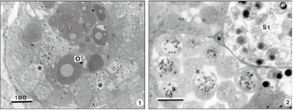
arranged, elongate sac-like follicles. Each follicle contains both developing eggs and sperm. The ovary portions are located in the distal zone of the follicles while the testicular cysts are located in the proximal zone in close contact with the ovotestis lumen (Figs. 1 and 2). Some free mature sperm were found among oocytes and within the lumen of the ovotestis.
The primary spermatocytes are irregular shaped cells (Fig. 3). Their nuclei are spherical, containing the synaptonemal complexes. In the cytoplasm there are a few circular mitochondrias and the other cytoplasmic organelles (Fig. 3).
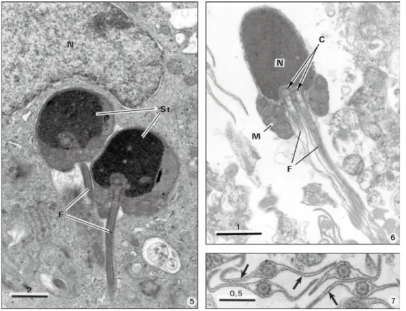
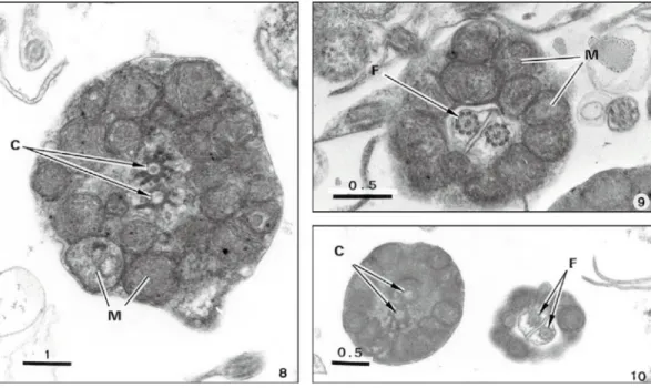
Near oocytes we observe some spermatids in different phases of maturation (Fig. 4), some of which in close contact with Sertolli cells (Fig. 5). The latter, containing a prominent nucleus, are located at the periphery of the testis cyst (Fig. 5). The mature spermatozoa consist of a short head containing a nucleus but without acrosome, a midpiece and two tails (Figs. 6 and 11). The electron dense sperm nucleus presents fewer than two forms. Most of the nuclei were cylindrical 3 µm long and 1.3 µm wide and with a rounded an-terior end (Fig. 6). Some other spermatozoa contain a circular nucleus with 3.0-3.4 µm in diameter. The midpiece consists of a collar ring-shaped mitochondrion containing several mitochondria situated in different levels, and two centrioles (Figs. 6, 8-10).
Fig. 1 — Satanoperca jurupari hermaphrodite teleostean fish. Semithin section of the ovotestis showing different stages of the spermatogenesis (*) surrounding several oocyts (O). Fig. 2 — Semithin section of two cysts containing spermato-cytes (*) and spermatids (St).
Fig. 3 — Ultrathin section of spermatocytes showing the synaptonemal complexs (arrows). Fig. 4 — Ultrathin section of
Fig. 5 — Ultrathin section showing a Sertoli cell with a large nucleus (N) containing two middle spermatids (St), each showing one of the two flagella (F). Fig. 6 — Ultrathin longitudinal section of a spermatozoon showing a dense nucleus (N) with-out acrosome, two centrioles (C) each one in continuity with two flagela (F) the mitochondrial collar (M) forming the midpiece surrounded the basal portion of the nucleus centriolar region and the begin of the flagella. Fig. 7 — Ultrathin transverse section of four flagella showing a pair of lateral ribbons (arrows) and the axoneme composed of the classic 9 + 2 micro-tubular patterns in each.
They extend along most of the length of the tail. The largest extensions, including the axoneme has 3 µm in diameter (Fig. 7). The axoneme exhibits a common 9 + 2 microtubular pattern (Figs. 7 and 9) along of the tail length.
DISCUSSION
The morphological structure exhibited by sperms of the teleost Satanoperca jurupari described here show similarity to those described in most of the teleostean species in which the absence of acrosome is a common character (Mattei, 1970). Although they belong to the primitive type they show some unusual features. To date all the fish spermatozoa examined show some structural
homogeneity which supports the idea that the ultrastructural features of spermatozoon can be useful for taxonomic and phylogenetic studies (Baccetti et al., 1984; Mattei, 1991; Mattei & Mattei, 1984; Gwo et al., 1993, 1996).
Figs. 8-10 — Ultrathin transverse sections of three sequential portions of the midpiece showing the centrioles (C) surrounded by mitochondria (M) of the mitochondrial collar and the two flagella (F).
from Brazil to be examined. Examination of other cichlids will be necessary to determine whether the primitive features of S. jurupari spermatozoa are common to this family (Mattei, 1991).
This investigations will also furnish the information needed for determining the phylo-genetic position to the Cichlids on the basis of spermatozoan structure which is known to be a valuable indicator of phylogenetic relationships in numerous groups (Mattei, 1991; Jamieson, 1991; Gwoet al., 1993).
Acknowledgments— We thank the excelent technical assistence of Mrs Laura Corral and the ichonographic work of Mr J. Carvalheiro. This work was partially supported by A. Almeida Foundation, Porto, Portugal and FCAP-Belém, Pará, Brazil.
REFERENCES
BACCETTI, B. & AFZELIUS, B. A., 1976, In: A. Wolsky (ed.),The biology of the sperm cell. Basel, S., Karger, pp. 1-254.
BACCETTI, B., BURRINI, A. G., CALLAINI, G., GIBERTINI, G., MAZZINI, M. & ZERUNIAN, S., 1984, Fish germinal cells. I. Comparative spermatology of seven cyprinid species.
Gamete Res.,10: 373-396.
BILLARD, R., 1970, Ultrastructure comparée de spermato-zoide de quelques poissons téléostéens, pp. 71-79. In: B. Baccetti (ed.), Comparative spermatology. Academic Press, New York.
GWO, J. C., GWO, H. H. & CHANG, S. L., 1993, Ultras-tructure of the spermatozoon of the teleost fish
Acanthopagrus schlegeli (Perciformes; Spanidae). J. Morphol.,216: 29-33.
chez un poisson téléostéen de la famille des Apogonides.
J. Ultrastruct. Re., 88: 223-228.
MATTEI, X., 1970, Spermiogenèse comparée des Poissons. pp. 57-69. In: B. Baccetti (ed.), Comparative spermatology. Academic Press, New York.
MATTEI, X., 1991, Spermatozoon ultrastructure and its systematic implications in fishes. Can. J. Zool.,69: 3038-3055.
MATTEI, X. & MATTEI, C., 1976, Spermatozoides à deux flagelles de type 9 + 0 chez Lampanyctus sp. (Poisson Myctophidae).J. Microscopie Biol. Cell.,25: 187-188. ROUSE, G. W. & JAMIESON, B. G. U., 1987, An ultras-tructural study of the spermatozoa of the polychaetes
Eurythoe complanata (Amphinomidae), Clymenella sp. and
Micromaldane sp. (Maldanidae), with definition of sperm types in relation to reproductive biology. J. Submicrosc. Cytol., 19: 573-584.
STANLEY, H. P., 1965, Electron microscopic observations on the biflagellate spermatids the teleost fish Porichtys notatus.Anat. Rec., 151: 477.
STOSS, J., 1983, Fish gamete preservation and spermatozoon physiology, pp. 305-350. In: W. S. Hoar, D. J. Randall & E. M. Donaldson (eds.), Fish physiology. v. 9, Academic Press, New York.
YAO, Z., EMERSON, C. J. & CRIM, L. W., 1995, Ultrastructure of the spermatozoa and eggs of the ocean Pout (Macrozoarces americanus L.), an internally fertilizing marine fish. Molecular Reproduction and Develop., 42: 58-64.