Original Article
ANTIMICROBIAL AND ANTIOXIDANT ACTIVITIES OF SELECTED PLANTS USED BY
POPULATIONS FROM JURUENA VALLEY, LEGAL AMAZON, BRAZIL
LARISSA IRENE DA SILVA
a, ARUNACHALAM KARUPPUSAMY
a, FABIO MIYAJIMA
bc, IVANA MARIA POVOA
VIOLANTE
a, ISANETE GERALDINI COSTA BIESKI
ad, SIKIRU OLAITAN BALOGUN
ad, DOMINGOS TABAJARA DE
OLIVEIRA MARTINS
a*aArea of Pharmacology, Department of Basic Health in Sciences, Faculty of Medicine, Federal University of Mato Grosso, Cuiabá, Mato Grosso, Brazil, bDepartment of Pharmacology, Translational Medicine Institute, University of Liverpool, United Kingdom, cDepartment of Physiology and Pharmacology, Federal University of Ceara, Brazil, dCurso de Farmácia, Faculdade de Noroeste de Mato Grosso, Associação
Juinense de Ensino Superior (AJES), Juína, Mato Grosso, Brazil Email: taba@terra.com.br
Received: 14 Jan 2017 Revised and Accepted: 31 Mar 2017
ABSTRACT
Objective: The purpose of this study was to evaluate selected Brazilian plants from Juruena valley region of Mato Grosso, for their in vitro
antimicrobial and antioxidant activities.
Methods: The powder obtained from different parts of the twenty-six (26) plants were macerated in hydroethanolic solution to obtain the extracts. The hydroethanolic extracts were tested for their in vitro antimicrobial activity by determining the MIC using broth microdilution. The 2, 2-diphenyl-1-picrylhydrazyl (DPPH) radical, ferric reducing antioxidant power (FRAP) and nitric oxide (NO) methods were used for the determination of antioxidant activities. Correlation between classes of secondary metabolites and antioxidant activity was assessed.
Results: Phanera glabra extract (HEPg) showed broad antibacterial spectrum, presenting the best activity against Klebsiella pneumoniae. Hevea microphylla extract (HEHm) presented a narrow spectrum of antibacterial activity with strong effect against Shigella flexneri. The only plant with broad spectrum antifungal activity was Bertholletia excelsa (HEBe), with moderate activity against strains of Aspergillus and Candida. The following extracts were prominent regarding their activities in the DPPH and FRAP assays-HEBe, Cariniana rubra (HECr) and in the FRAP assay alone, Cedrela odorata (HECo) and HEPg. None of the extracts was active in the NO assay. A significant association was observed between DPPH activity and the total phenolic contents.
Conclusion: Our results justified the use of some of the investigated plants in the Brazilian ethnomedicine. The antibacterial activities of these plants are bacteriostatic in nature. These findings support that a number of investigated plants could be a valuable source of new antioxidant and antimicrobial compounds that can potentially deliver novel mechanisms of actions. However, further studies are required.
Keywords: Medicinal plants, Antifungal, Antibacterial, Phytochemical analysis
© 2017 The Authors. Published by Innovare Academic Sciences Pvt Ltd. This is an open access article under the CC BY license (http://creativecommons.org/licenses/by/4.0/) DOI: http://dx.doi.org/10.22159/ijpps.2017v9i5.17086
INTRODUCTION
The usefulness of medicinal plants in the management, cure, and prevention of diseases, as described by local people since time immemorial, have aroused both commercial and scientific interests [1]. Several epidemiological studies have endorsed the importance of high consumption of secondary plant products found in fruits and vegetables, for the modulation of degenerative diseases. Brazil possess the largest floristic diversity on Earth, as it contains six continental biomes; the Amazon rainforest, the Brazilian Cerrado, the Caatinga, the Atlantic forest, the Pantanal, and the Pampas. The Amazon rainforest being the most noteworthy, since it is the largest tropical forest in the world, and whose diversity of plant species constitutes an endless source for the research of phytotherapies and new molecules with biological activities [2].
The state of Mato Grosso (MT), the largest agricultural producer in Brazil, contains three important biogeographical regions (Amazon rainforest, Cerrado (Brazilian savannah) and Pantanal) and a rich ethnocultural diversity, represented by 42 indigenous groups (42,538 individuals) and traditional quilombola, cabocla and riverine communities Amazon rainforest, Cerrado (Brazilian savannah) and Pantanal [3].
The region of Juruena valley which encompasses seven important cities, namely Juína, Castanheira, Brasnorte, Juruena, Aripuanã, Cotriguaçu and Colniza, is situated in the northern part of Mato Grosso, within the officially recognized Legal Amazon region. The region inhabitants, as at the time of the study, were 139,524, which
represent 4.6 % of the population of the state. This region is entirely constituted of the Amazon rainforest biome, covering a total area of 111,849.70 km2 and despite being safeguarded by numerous laws,
suffers intense exploitation by pastoral activities [4].
Despite the great advances achieved by science, infectious diseases are still among the ten main causes of death worldwide, and it is estimated that the so-called ‘super-microorganisms’ alone will be responsible for over 10 million deaths by 2050, making imperative the search for alternative treatments to infectious diseases which do not respond to most modern antibiotics [5].
These growing needs for more effective and safer antimicrobial agents have stimulated the renewal of multidisciplinary investigation on natural products, in which new approaches, combined with traditional techniques, are potentiating the tracking of substances which present antimicrobial activities, as well as the identification of the molecular targets responsible for their effects, many of which present novel mechanisms of action [6].
atherosclerosis, diabetes, carcinogenesis, and inflammatory diseases, just to mention but few [7].
Among the active compounds sought-after in plants, special focus is given to those that show antioxidant activity, being capable of inactivating ROS, which are intimately related to the aetiology of numerous illnesses [9].
In this context, polyphenols constitute one of the largest and widely distributed groups of natural products in the plant kingdom [10]. They are capable of scavenging ROS and may have therapeutic health effects for a variety of illnesses, including antimicrobial properties [11]. More recently, specific molecular targets for various polyphenols have been described and have resulted in renewed scientific interests in the polyphenols as therapeutic agents [9-10]. The methods used for estimation of antioxidant capacities of the plant extracts were based on their simplicity, low cost, and reproducibility. In this case, we employed the three commonly utilized antioxidant assays, namely DPPH, FRAP and NO scavenging assays to verify the antioxidant potential of each extract [12]. Thus, the preliminary in vitro screening of antimicrobial activity of plants popularly used to treat infections serves as a guide for the selection of substantial candidates for future phytochemical and pharmacological research aimed at the development of new antimicrobial prototypes.
Based on our recent ethnopharmacological surveys of medicinal plants from the state of Mato Grosso, Brazil [1-13] and literature review [14], we carried out screening of selected plants for their antibacterial, antioxidative and antiradical activities. We also evaluated the linear correlation between the selected secondary metabolites classes present in the extracts to their in vitro
antioxidant and anti-radical activities.
MATERIALS AND METHODS
Plant material
The 26 species of plants utilized in the bioassays were selected through an ethnobotanical survey carried out between the years 2010-2013 as reported by Bieski et al. [1]. The study involved (n= sampling number) 365 informants selected based on statistical sampling, calculated per the total population of 139,524 inhabitants of the 7 municipals of Juruena valley, north-western Mato Grosso, in the Legal Amazon region [1]. The access to samples of the genetic patrimony was authorized by Conselho Nacional de Desenvolvimento Científico e Tecnológico (CNPq), under the number 010728/2013-9.
The selection of medicinal plants mentioned by the population of Juruena valley for the treatment of infections was made after ethnobotanical data analysis and literature review. The ethnobotanical survey data was obtained via the application of a semi-structured questionnaire, containing the descriptors: infection, antibiotic, pneumonia, bronchitis, gastritis, diarrhoea, vaginal discharge, syphilis, impetigo, conjunctivitis, venereal disease, gonorrhoea, boils, inflammation, fever, wound, wound healing, fungi, mycoses, skin spots, mouth sore, thrush, itching, nail infection, chilblains, and athlete’s foot [1-11]. Voucher specimens were deposited and identified at the UFMT Herbarium, under the supervision of a botanist and curator Prof. Germano Guarim Neto.
Microbial organisms
All the microorganisms used were representative from American Type Culture Collection (ATCC) and obtained through Oswaldo Cruz Foundation (Fiocruz, Rio de Janeiro, Brazil), namely: Enterococcus faecalis (29212), Staphylococcus aureus (25923), Staphylococcus epidermidis (12228), Streptococcus pyogenes (19615), Bacillus subitilis (6633), Escherichia coli (25922), Klebsiella pneumoniae
(13883), Pseudomonas aeruginosa (27853), Salmonella typhimurium
(14028), Shigella flexneri (12022), Helicobacter pylori (43504 (VacA and CagA positive), Candida albicans (10231), fluconazole-resistant
C. albicans (64550), C. parapsilosis (22019), C. tropicalis (750), Cryptococcus neoformans (32045), C. glabrata (9030), Aspergillus fumigatus (46640), A. niger (10535), A. parasiticus (15517), A.
terreus (7860), Penicillium verrucosum (10513), Trichophyton mentagrophytes (9533), T. rubrum (28189) and Microsporum gypseum (14383).
Chemicals and reagents
2,2-diphenyl-1-picrylhydrazyl (DPPH), brain heart infusion media (BHI), and Müller-Hinton agar, Skirrow’s medium, amphotericin B, foetal bovine serum (FBS), Sabourad broth, clarithromycin, methanol, trichloroacetic acid, ascorbic acid, potassium phosphate buffer, potassium ferricyanide, ferric chloride, quercetin, nitroprusside, phosphate buffer saline, Griess, gallic acid, sodium carbonate, rutin, acetic acid, aluminium chloride, acetate were obtained from Sigma-Aldrich Co., St. Louis, MO, USA. Other chemicals/reagents used were: Ethanol, chloridric acid (Tedia Company, Inc., CA, USA).
Preparation of plant extracts
Bark, stem bark, branches, leaves, roots, or whole plant were collected, cleaned and dried at room temperature and then ground in a knife mill (Tecnal® Te 625, São Paulo, BR), standardized with a 40 sieve. The resulting powder of each plant was macerated in 70 % ethanol/water solution (1:3 v/v), during 7 d at 24 °C with daily shaking for homogenization. After filtration, the material was concentrated in rotary evaporator (MA 120, Marconi®, São Paulo, BR), under reduced pressure of 600 mmHg and temperature of 40±1 °C. The residual solvent was evaporated in an oven (MA035/5, Marconi®, São Paulo, BR) at 40±1 °C and the hydroethanolic extracts therefore obtained were freeze-dried (LJJ06V, JJ Científica®, São Paulo, BR) and kept in amber flask at 4 °C. At the time of the assays, the extracts’ stock solutions were dissolved in 2 % DMSO.
Antimicrobial activity test
Microdilution in broth
The minimum inhibitory concentration (MIC) was determined by microdilution in broth as described in Clinical and Laboratory Standards Institute (CLSI) guidelines [15, 16]. Bacterial isolates were cultured in brain heart infusion media (BHI), except for H. pylori
where Skirrow’s supplement was added to BHI medium (vancomycin 10 µg/ml, trimethoprim 5 µg/ml, polymyxin B 2.5 U/ml), amphotericin B (5 µg/ml) and foetal bovine serum (FBS) 10 %. Microdilution in Sabourad broth was employed for the MIC investigation of fungi.
The hydroethanolic extracts were solubilized in culture media for concentrations ranging from 800-6.25 µg/ml. The bacterial inoculum was adjusted to the concentration of 1 x 10⁸CFU/ml (0.5 in
McFarland scale), except for H. pylori, which was adjusted to the concentration of 6 x 10⁸ CFU/ml (2 on McFarland scale). Filamentous
fungi and yeast-like fungi were adjusted to the concentration of 5 x 10³ CFU/ml. Clarithromycin (50-0.195 µg/ml) was used as the
standard antibacterial drug and amphotericin B (32-0.125 µg/ml) as the standard antifungal drug.
These microorganisms were incubated at 37 °C; the bacteria for 1-2 d, apart from H. pylori, which remained incubated for 3-5 d. The yeast-like fungi were incubated for 2-3 d, the filamentous fungi for 3 d and the dermatophyte filamentous fungi were incubated for between 3 and 21 d.
MICs were determined based on the turbidity of microplate wells, read in a spectrophotometer at 450 nm (Thermo ScientificTM,
MultiskanTM, New Hampshire, USA), and confirmatory assessment
was conducted using resazurin as an indicator of redox. MIC was considered the concentration that completely impedes microbial growth.
MIC values<100 µg/ml, 100 ≤ MIC ≤ 500 µg/ml, 500<MIC ≤ 800 µg/ml, and MIC>800 µg/ml were considered of good, moderate, weak and without activity, respectively based on the modified classification of Holetz et al. [17].
Determination of the MBC and MFC
per the method of Mbah et al. [18]. An aliquot (10 µl) of bacterial cells from the MIC test plate was subcultured on solid Müller-Hinton agar by making streaks on the surface of the agar and incubating at 37 °C for 1-2 d, and for plates where there was no visible fungal growth, the same was plated on Sabouraud agar and was incubated for 2 d in the case of yeast-like fungi, 3 d for filamentous fungi or 3-21 d for the dermatophytes. The concentration of the wells inoculated with MIC which showed no bacterial growth was recorded as MBC and those that presented bacterial growth were considered bacterio-static. In the case of the fungi, test concentration without growth at all is considered as the MFC. All assays were carried out in independent triplicates.
In vitro antioxidant activity
DPPH assay
The antiradical activity of the extracts was evaluated using DPPH [19]. In this assay, compounds that present antioxidant activity act as electron donors, causing the reduction or neutralization of the DPPH radical that can be verified through the decrease in absorbance. In a 96-well microplate, 100 µl of different concentrations of the extracts (800-6.25 µg/ml), 100 µl of 50 μM DPPH, both diluted in methanol/water solution (50 %). After 30 min, the reading was carried out in a microplate reader at a wavelength of 540 nm. Ascorbic acid (100-0.39 µg/ml) was used as the standard.
FRAP assay
To determine the antioxidant activity of the extracts based on their capacity to reduce ferric ion (Fe+3)into ferrous ion (Fe+2) [20],
extracts at concentrations ranging from 800-6.25 µg/ml and ascorbic acid (50-0.39 µg/ml) were dissolved in 50 % aqueous methanol solution and FRAP assays were conducted. In each well of the 96 wells plate, 30 µl of each plant extract concentration or ascorbic acid was added. Subsequently, 40 µl each of potassium phosphate buffer (0.2 M, pH 7.2) and potassium ferricyanide (1 % w/v) were added. The reaction mixtures were incubated at 50 °C for 20 min. After the incubation period, 40 µl of trichloroacetic acid (10% w/v), 150 µl of distilled water and 30 µl ferric chloride (0.1% w/v) were added, followed by a second incubation at room temperature for 30 min in the dark. Absorbance was measured at 630 nm using a microplate reader.
NO assay
Nitrite (NO2-)formation inhibition was used for the evaluation of the
antioxidant potential of the extracts, per the modified method of Sreejayan and Rao [21]. 100 µl of each extract (800-6.25 µg/ml) or quercetin (50-0.39 µg/ml) were added to a 96-well microplate, containing sodium nitroprusside at 5 mmol in phosphate buffer saline. After incubation at 25 °C for 150 min, 100 µl of Griess reagent were added to each well and absorbance measurements carried out in a microplate reader at 540 nm.
Quantitative analysis of selected metabolic classes
Total phenolic content
Total phenolic content was quantified using the Folin–Ciocalteu method [22]. Methanolic solutions (0.2 ml) of the extracts (1 mg/ml, w/v) or the standard used (Gallic acid, 1.0-0.1 mg/ml, w/v) were mixed with Folin-Ciocalteu reagent (0.5 ml of 10 %, v/v), sodium carbonate (1 ml of 75 %, w/v) and Milli-Q water (8.3 ml). The mixture was gently agitated and kept for 30 min in the dark. The absorbance was measured at 760 nm using a UV–visible spectrophotometer (Genesys 5, Spectronic®, Texas, USA). Total phenolic was determined by interpolation of the absorbance of the samples against a calibration curve constructed with different concentrations of gallic acid in Milli-Q water (y = 0.0747x+0.01471, with R2 = 0.9874). The result was expressed as mg tannic acid
equivalents (TAE) per gram of the extracts (mg TAE/g).
Flavonoid content
Determination of flavonoid content was estimated by a colorimetric method based on the formation of the flavonoid-aluminium complex
[22] with slight modifications. Briefly, 0.5 ml of methanolic solutions of the extracts (1 mg/ml w/v) or standard (rutin, 10-1 mg/ml w/v) was mixed with an aqueous solution of 0.5 ml of 60 % acetic acid, 2 ml methanolic solution of 20 % pyridine (v/v), 1 ml of 5% aluminium chloride (w/v) and 6 ml of Milli-Q water. The mixture was gently stirred and kept for 30 min in the dark and its absorbance was then measured at 420 nm using a spectrophotometer. The total flavonoid contents were determined by extrapolating the absorbance of the samples against a calibration curve constructed with different concentrations of the rutin standard (y = 0.0419x+0.0044, with R2 = 0.9977) and expressed as
milligrammes of rutin equivalents (RE) per gram of the extracts (mg RE/g).
Coumarins content
To determine the concentration of coumarins, the colorimetric method [22] was employed. Briefly, to the extracts (1 mg/ml) 1 ml of distilled water, 0.5 ml of 5 % solution of lead acetate and 7 ml of distilled water. Then, 2 ml of this solution was transferred to tubes containing 8 ml of 0.1 M chloridric acid. The solution was kept in the dark for 30 min and the absorbance was measured using a UV-light spectrophotometer at 320 nm, using distilled water as a blank. The results were expressed in mg of coumarin per gram of the extract (mg CE/g), using the equation of the standard curve: y = 0.0935x+0.0708, with R2 = 0.9927.
Correlation of antioxidant against phenolic contents
To assess the relationship between total phenolic, flavonoids or coumarins contents and antioxidant or antiradical capacities (except NO assay, in which extracts had no IC50) of the extracts, a linear
correlation analysis was performed using the IC50 of each extract.
Data analysis
For the organization and analysis of the date, the GraphPad Prism software version 5.01 was employed. The antimicrobial results were expressed in terms of MIC and the results of the antioxidant assays were expressed in terms of IC50 obtained from a linear regression
curve, relating the percentage of antioxidant activity to the tested concentrations. The results of the quantitative analysis of some selected metabolic classes were expressed in terms of mean±SD, calculated through the standard curve obtained for each representative of the analysed metabolic class. Shapiro-Wilk test was conducted to evaluate if the samples originated from a normal population. For the study of the correlation between the antioxidant activity of the extracts in the DPPH and FRAP assays with Total phenols and Flavonoids concentrations, Pearson's correlation coefficient was used, since it measures the degree of linear correlation and the quantitative variables were from a normal population. In the case of correlation between the coumarins with antioxidant activity against DPPH and FRAP, Spearman correlation coefficient was used, which is a measure of non-parametric correlation. If so, the presence or absence of correlation was evaluated using Spearman or Pearson test correlation test. Values of
p<0.05 were considered significant.
RESULTS AND DISCUSSION
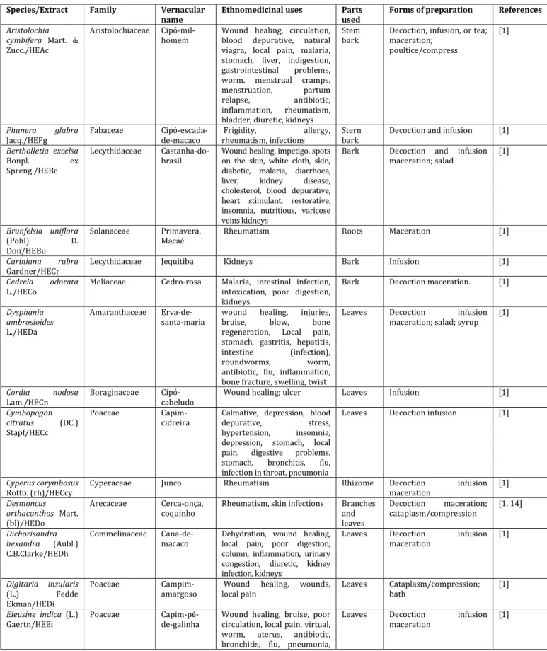
The choice of maceration of the plant powder in a hydroethanolic solvent in the present study was based on many factors. The use of this type of extract in ethnopharmacological and ethnobotanical related studies and others, particularly for plants having more than one form of preparations (see table 1) has been a standard practice.
polarity is close to that of water, its allows for better conservation of the material being extracted at a milder temperature (40 °C) in the preparation of lyophilized extract, and its extract more efficiently the active pharmacological constituents [24]. Table 1 shows a summary of the ethnomedicinal uses of the plants and the form of preparations of each plant.
Antibacterial activity
The antibacterial effects of the extracts are presented in table 2. Among the 26 tested extracts, 18 were active against at least one bacterial strain of the tested panel. Three hydroethanolic extracts presented broad spectra of activity, namely HEPg, H. coronarium
(HEHc) and HEHm.
Table 1: A summary of ethnomedicinal uses of the plants screened for the antimicrobial activities, forms of preparations and related references
Species/Extract Family Vernacular
name
Ethnomedicinal uses Parts
used
Forms of preparation References
Aristolochia cymbifera Mart. & Zucc./HEAc
Aristolochiaceae Cipó-mil-homem
Wound healing, circulation, blood depurative, natural viagra, local pain, malaria, stomach, liver, indigestion, gastrointestinal problems, worm, menstrual cramps, menstruation, partum relapse, antibiotic, inflammation, rheumatism, bladder, diuretic, kidneys
Stem bark
Decoction, infusion, or tea; maceration;
poultice/compress
[1]
Phanera glabra
Jacq./HEPg
Fabaceae Cipó-escada-de-macaco
Frigidity, allergy, rheumatism, infections
Stern bark
Decoction and infusion [1]
Bertholletia excelsa
Bonpl. ex Spreng./HEBe
Lecythidaceae Castanha-do-brasil
Wound healing, impetigo, spots on the skin, white cloth, skin, diabetic, malaria, diarrhoea, liver, kidney disease, cholesterol, blood depurative, heart stimulant, restorative, insomnia, nutritious, varicose veins kidneys
Bark Decoction and infusion maceration; salad
[1]
Brunfelsia uniflora
(Pohl) D. Don/HEBu
Solanaceae Primavera, Macaé
Rheumatism Roots Maceration [1]
Cariniana rubra
Gardner/HECr
Lecythidaceae Jequitiba Kidneys Bark Infusion [1]
Cedrela odorata
L./HECo
Meliaceae Cedro-rosa Malaria, intestinal infection, intoxication, poor digestion, kidneys
Bark Decoction maceration. [1]
Dysphania ambrosioides
L./HEDa
Amaranthaceae Erva-de-santa-maria
wound healing, injuries, bruise, blow, bone regeneration, Local pain, stomach, gastritis, hepatitis, intestine (infection), roundworms, worm, antibiotic, flu, inflammation, bone fracture, swelling, twist
Leaves Decoction infusion maceration; salad; syrup
[1]
Cordia nodosa
Lam./HECn
Boraginaceae Cipó-cabeludo
Wound healing; ulcer Leaves Infusion [1]
Cymbopogon citratus (DC.) Stapf/HECc
Poaceae Capim-cidreira
Calmative, depression, blood depurative, stress, hypertension, insomnia, depression, stomach, local pain, digestive problems, stomach, bronchitis, flu, infection in throat, pneumonia
Leaves Decoction infusion [1]
Cyperus corymbosus
Rottb. (rh)/HECcy
Cyperaceae Junco Rheumatism Rhizome Decoction infusion maceration
[1]
Desmoncus orthacanthos Mart. (bl)/HEDo
Arecaceae Cerca-onça, coquinho
Rheumatism, skin infections Branches and leaves
Decoction maceration; cataplasm/compression
[1, 14]
Dichorisandra hexandra (Aubl.) C.B.Clarke/HEDh
Commelinaceae Cana-de-macaco
Dehydration, wound healing, local pain, poor digestion, column, inflammation, urinary congestion, diuretic, kidney infection, kidneys
Leaves Decoction infusion maceration
[1]
Digitaria insularis
(L.) Fedde Ekman/HEDi
Poaceae Campim-amargoso
Wound healing, wounds, local pain
Leaves Cataplasm/compression; bath
[1]
Eleusine indica (L.) Gaertn/HEEi
Poaceae Capim-pé-de-galinha
Wound healing, bruise, poor circulation, local pain, virtual, worm, uterus, antibiotic, bronchitis, flu, pneumonia,
Leaves Decoction infusion maceration
respiratory problems, cough, kidneys
Gossypium hirsutum
L./HEGi
Malvaceae Algodão-roxo
Infection in the ovaries, uterine infection, postpartum, throat, kidneys
Leaves Decoction infusion maceration, bath
[1]
Hedychium coronarium J. Koenig/HEHc
Zingiberaceae Gengibre-do-mato
Rheumatism Rhizome Decoction [1]
Hevea microphylla
Ule/HEHm
Euphorbiaceae Barriguda Ulcer Branches Maceration [1]
Jacaranda cuspidifolia
Mart./HEJc
Bignoniaceae Caroba Wound healing; blood depurative, kidney stones
Branches and leaves
Decoction, infusion maceration, bath
[1]
Manihot esculenta
Crantz/HEMt
Euphorbiaceae Mandioca Itching, antibiotic Leaves Infusion bath [1]
Maytenus ilicifolia
Mart. ex Reissek/HEMi
Moraceae Espinheira-santa
Wound healing, stomach, gastritis, antibiotic, inflammation, kidneys
Bark Infusion [1]
Parodiolyra micrantha (Kunth) Davidse and Zuloaga/HEPm
Poaceae Bambuzinho Stomach, kidneys Leaves Decoction infusion [1]
Philodendron acutatum
Schott/HEPa
Araceae Cipó-imbé Erysipelas, back pain Whole plant
Decoction
cataplasm/compression [1]
Renealmia alpinia (Rottb.) Maas/HERa
Zingiberaceae Pacová, Gengibre-do-mato
Rheumatism Rhizome Maceration [1]
Smilax brasiliensis
Spreng./HESb
Smilacaceae Japecanga Wound healing, blood depurative, inflammation, rheumatism
Branches and leaves
Decoction [1]
Spondias mombin L./HESm
Anacardiaceae Cajazinho, caju-açu-da-mata
Wound healing, diarrhoea Leaves Decoction infusion bath [1]
Trema micrantha (L.) Blume/HETm
Cannabaceae Grandiuva, periquiteiro
Insect bite, inflammation, heartburn, ulcer
Bark
Table 2: Evaluation of the in vitro antibacterial activity of the hydroethanolic extracts of medicinal plants used for the treatment of infections
Species/extract aMinimum inhibitory concentration (MIC, µg/ml)
Gram-negative Gram-positive
Ec Kp Pa St Sf Hp Ef Sa Sp Se Bs
Aristolochia cymbifera Mart. and Zucc. (bl)/HEAc >80 0 >80 0 >80 0 >80 0 >80 0
400 >80 0 >80 0 >80 0 >80 0 >80 0
Phanera glabra (Jacq.) Vaz (sb) / HEPg 800 25 800 800 800 >80 0
200 >80 0
400 800 800
Bertholletia excelsa Bonpl. (b)/HEBe >80 0 >80 0 >80 0 >80 0 >80 0
200 >80 0
400 200 >80 0
>80 0
Brunfelsia uniflora (Pohl) D. Don (r)/HEBu >80 0 >80 0 >80 0 >80 0 >80 0 >80 0 >80 0 >80 0 >80 0 >80 0 >80 0
Cariniana rubra Gardner ex Miers (b)/HECr >80 0 >80 0 >80 0 >80 0 >80 0
100 400 200 400 >80 0
>80 0
Cedrela odorata L. (b)/HECo >80 0 >80 0 >80 0 >80 0
800 >80 0
>80 0
400 >80 0
>80 0
>80 0
Dysphania ambrosioides L. (l)/HEDa >80 0 >80 0 >80 0 >80 0 >80 0
200 400 >80 0
400 400 >80 0
Cordia nodosa Lam. (l)/HECn >80 0
>80 0
>80 0
200 50 >80 0
>80 0
>80 0
400 100 >80 0
Cymbopogon citratus (DC.) Stapf (l)/HECc >80 0 >80 0 >80 0 >80 0 >80 0 >80 0 >80 0 >80 0 >80 0 >80 0 >80 0
Cyperus corymbosus Rottb. (rh)/HECcy >80 0 >80 0 >80 0 >80 0 >80 0 >80 0 >80 0 >80 0 >80 0 >80 0 >80 0
Desmoncus orthacanthos Mart. (bl)/HEDo >80 0 >80 0 >80 0 >80 0
25 >80 0
>80 0
>80 0
200 >80 0
>80 0
Dichorisandra hexandra (Aubl.) C. B. Clarke (l)/HEDh >80 0 >80 0 >80 0 >80 0
800 >80 0 >80 0 >80 0 >80 0 >80 0 >80 0
Digitaria insularis (L.) Fedde (l)/HEDi >80 0
>80 0
>80 0
800 200 >80 0
200 >80 0
200 >80 0
>80 0
Eleusine indica (L.) Gaertn (l)/HEEi >80 0 >80 0 >80 0 >80 0 >80 0 >80 0 >80 0 >80 0 >80 0 >80 0 >80 0
0 0 0 0 0 0 0 0 0 0
Hedychium coronarium J. Koenig (rh)/HEHc >80 0
200 >80 0
>80 0
100 >80 0
>80 0
25 100 200 >80 0
Hevea microphylla Ule (b)/HEHm 400 400 >80 0
>80 0
400 >80 0
200 400 >80 0
>80 0
>80 0
Jacaranda cuspidifolia Mart. (bl)/HEJc >80 0
>80 0
>80 0
>80 0
>80 0
400 200 >80 0
>80 0
>80 0
>80 0
Manihot esculenta Crantz (l)/HEMt >80 0
>80 0
>80 0
>80 0
>80 0
>80 0
>80 0
>80 0
>80 0
>80 0
>80 0
Maytenus ilicifolia Mart. ex Reissek (b) HEMi >80 0
>80 0
>80 0
>80 0
>80 0
>80 0
>80 0
800 >80 0
>80 0
>80 0
Parodiolyra micrantha (Kunth) Davidse & Zuloaga (l)/HEPm
>80 0
>80 0
>80 0
>80 0
>80 0
>80 0
>80 0
>80 0
>80 0
>80 0
>80 0
Philodendron acutatum Schott (wp) HEPa >80 0
>80 0
>80 0
>80 0
>80 0
>80 0
200 400 >80 0
>80 0
>80 0
Renealmia alpinia (Rottb.) Maas (rh)/HERa >80 0
>80 0
>80 0
>80 0
>80 0
>80 0
400 >80 0
100 800 >80 0
Smilax brasiliensis Spreng. (bl)/HESb >80 0
>80 0
>80 0
>80 0
>80 0
>80 0
400 >80 0
>80 0
>80 0
>80 0
Spondias mombin L. (l)/HESm >80 0
>80 0
>80 0
>80 0
>80 0
200 200 400 800 >80 0
>80 0
Trema micrantha (L.) Blume (bl)/HETm >80 0
>80 0
>80 0
>80 0
>80 0
>80 0
200 >80 0
>80 0
>80 0
>80 0 Standard drug
Clarithromycin 0.39 0.39 6.25 0.78 0.78 0.39 0.39 0.39 0.39 0.39 0.39 aObtained by broth microdilution, (b): bark; (bl): branches and leaves; (l) leaves; (rh): rhizome; (r): root (sb): stem bark; (wp): whole plant; Ec:
Escherichia coli, Kp: Klebsiella pneumoniae, Pa: Pseudomonas aeruginosa, St: Salmonella typhimurium, Sf: Shigella flexneri, Hp: Helicobacter pylori, Ef:
Enterococcus faecalis, Sa: Staphylococcus aureus, Se: Staphylococcus epidermidis, Sp: Streptococcus pyogenes, Bs: Bacillus subtilis, Results expressed as mean of three independent assays.
HEPg demonstrated activity against 9 of the 11 bacteria tested, presenting good antibacterial activity against K. pneumoniae (MIC = 25 µg/ml), moderate against E. faecalis (MIC = 200 µg/ml) and S. pyogenes (MIC = 400 µg/ml), and weak activity (MIC = 800 µg/ml) against E. coli, P. aeruginosa, S. typhimurium, S. flexneri, S. epidermidis
and B. subtilis. Although the genus Bauhinia is used popularly in the treatment of infections and antimicrobial biological activities have been confirmed for some of its species [25], no studies were found concerning P. glabra antibacterial activity. It is interesting to note that HEPg was more active against K. pneumoniae, one of the most common enterobacteria known to cause hospital infections, especially in immunosuppressed patients [26], suggesting its potential in treating infections caused by this bacterium.
HEHc showed good (MIC = 25 µg/ml) and moderate (MIC = 100 µg/ml and 200 µg/ml) activities against S. aureus; S. flexneri and S. pyogenes; K. pneumoniae and S. epidermidis, respectively. Most studies found in the literature refer to the antimicrobial activities of the essential oil obtained from the rhizome of the plant and the authors employed bacterial strains different from those in this work, apart from S. aureus with MIC = 4.2 mg/ml [27].
In addition, Joshi [28] observed MICs of 7.8 µg/ml against E. coli and
S. aureus and 31.13 µg/ml for S. flexneri. These values are 168 folds greater (S. aureus), 3 (S. aureus), and 13 (S. flexneri) times lower, respectively. No activity was observed for HEHc in our study against
E. coli. Concerning the activity of the essential oil of H. coronarium in the studies of Suksathan et al., using disc diffusion method, activities against S. aureus, B. subtilis and E. coli (inhibition zone = 13; 15 and 9 mm, respectively) were recorded. In the same vein, HEHc demonstrated activity (inhibition zones>10 mm)against S. aureus, S. epidermidis, E. coli and P. aeruginosa for the aqueous and methanolic extract of the leaves and rhizome of H. coronarium [29-30]. However, in our study, HEHc was inactive against strains of E. coli, P. aeruginosa and B. subtilis.
Reuk-ngam et al. [31], showed the antimicrobial activity of coronarin D, isolated from the rhizomes of H. coronarium, with a more potent antibacterial activity than observed for HEHc, with MIC 2 times lower for S. aureus and S. epidermidis (MIC = 12.5 µg/ml).No activity was seen against E. faecalis by HEHc in our study. These results suggest that coronarin D, may be responsible, at least in part, for the antibacterial activity of this plant. However, there is a need for phytochemical studies to confirm the presence of coronarin D in HEHc.
According to Gobbo-Neto and Lopes [32], there are several factors which may be responsible for the variations in the activity of a plant, among which are variations in the seasons of collections, temperature, water availability, ultraviolet radiation, nutrients availability, atmospheric pollution, mechanical damage, and pathogen attacks. These external differences invariably influence the composition and levels of important secondary metabolites of the plant materials employed in these different studies, in addition to the variations related to methodological approaches in the antimicrobial assays [33]. Moderate antibacterial activity by HEHm was detected against E. faecalis (MIC = 200 µg/ml), E. coli, K. pneumoniae, S. flexneri and S. aureus (MIC = 400 µg/ml). These microorganisms are notably related to intestinal diseases, urinary infections, and pneumonia, as well as infections of heart tissue, meningitis, septicaemia, mainly in immunocompromised patients [5]. In literature, most articles found concerning the genus Hevea refers to preliminary investigations of
H. brasiliensis against clinical isolates of E. coli, K. pneumoniae, S. aureus and B. cereus [34]. However, to the best of our knowledge, there are no reports on the antimicrobial activity of H. microphylla. Based on these results it will be interesting to carry out further pharmacological and phytochemical studies with HEHm.
Although the spectrum of action of HECn is not as broad as that of HEPg, HEHcand HEHm, it deserves attention for showing activity against two important pathogenic bacteria, namely S. flexneri (MIC = 25 µg/ml), an enterobacteria responsible for between 80-165 million cases of severe dysentery and about 600 thousand deaths annually, and S. epidermidis (MIC = 100 µg/ml), a frequent pathogenic agent in invasive procedures in hospital environment [5-35]. There are no previous antibacterial studies with HECn, however activity has been reported for some of its fractions against S. aureus
and S. epidermidis (inhibition halos = 8 and 15 mm, respectively), but at extremely high concentrations (125-1000 mg/ml) which according to Holetz et al. criteria, at these concentrations the fractions would be considered inactive, thus corroborating our findings. However, there is no report concerning its activity against
S. flexneri until now [36-17]. The potent activity of a hydroethanolic extract of Desmoncus orthacanthos (HEDo) was observed only against S. flexneri (MIC = 25 µg/ml). We were unable to make the comparison as there does not exist in the literature information on its in vitro antibacterial activity.
100 µg/ml) by HERa, but it was inactive against all other bacteria. This represents the first report of antibacterial activity of HERa against this strain, which is associated to a variety of infections, such as erysipelas, necrotizing fasciitis, biofilm formation and especially in bacteraemia cases [37]. In this context, this extract represents phytotherapeutic potential against S. pyogenes, however, further pharmacological, and phytochemical investigations are warranted. Extracts that presented activity against H. pylori deserved to be highlighted, as this bacterium is regarded to be the main etiological agent of chronic gastritis, gastroduodenal ulcer, adenocarcinoma, and gastric lymphomas [38]. Among the extracts are the hydroethanolic extracts of the bark ofHECr and HEBe, the leaves of
D. ambrosioides (HEDa) and S. mombin (HESm).
HECr showed moderate activity against H. pylori with MIC = 100 µg/ml, while HEBe, HEDa and HESm also present same moderate activity but at a higher MIC of 200 µg/ml. There are no reports of anti-H. pylori activity for these extracts and therefore these plants may represent potential phytotherapeutic agents in the treatment of infections caused by H. pylori. Lima Neto et al. [39] reported that HECr showed activity against S. aureus, E. faecalis, E. coli and P. aeruginosa (MIC = 250--1000 µg/ml), however, the same effect could not be demonstrated in our study.
Liu et al. [40] related that the essential oil of C. ambrosioides
presents activity (MIC = 640 µg/ml) against resistant strains of H. pylori, but at this concentration we considered this activity to be weak, based on the criteria that we employed.
In addition to the anti-H. pylori activity, HESm presented moderate activity against E. faecalis and S. aureus (MIC = 200 and 400 µg/ml, respectively) and weak activity against S. pyogenes (MIC = 800
µg/ml). Maduka et al. [41] showed that the ethanolic extracts of the leaves and bark of S. mombin showed activity against E. coli
(inhibition halos of 20 % for both extracts), K. pneumoniae and P. aeruginosa (inhibition halos of 40 and 20 %, respectively) and S. aureus (inhibition halos of 60 %). Regarding the latter strain, our results corroborate that of Maduka et al. [41]. In our work, HESm showed no activity against E. coli, P. aeruginosa and K. pneumoniae,
which may be caused by the methods employed (solid medium compared to liquid medium) or even to seasonal variations (e. g. harvest location) [33].
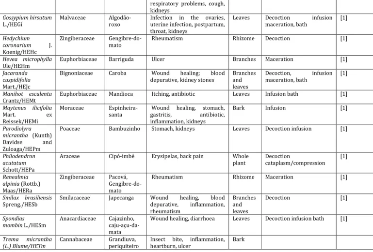
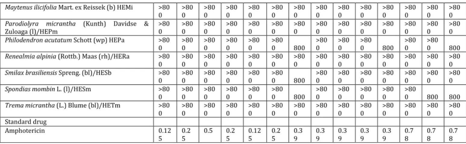
Antifungal activity
The results of the effects of the extract on the fungal growth are displayed in table 3. Out of the 26 tested extracts, 14 (53.8 %) showed activity against at least one fungal strain tested.
In this anti-fungal activity screening, HEBe was the most active compared to the hydroethanolic extract of A. cymbifera (HEAc), HEDo, HECr and HECo.
HEBe was the only extract that presented broad antifungal spectrum, with moderate activity against A. terreus (MIC = 100 µg/ml), A. fumigatus (MIC = 200 µg/ml), C. glabrata and C. neoformans (MIC = 400 µg/ml), and weak activity against C. albicans and C. tropicalis (MIC = 800 µg/ml). There is no report of HEBe antifungal activity until now, though Martins et al. [42] demonstrated that the pure oil (v = 500-1500 µl) obtained from the nuts of B. excelsa was active against toxicogenic strains of A. parasiticus, an effect that was not observed for
HEBe in this study. However, it is worth noting that the concentration tested by Martins et al. [42], if classified according to Holetz et al. [17], would be considered inactive.
Table 3: Evaluation of in vitro antifungal activity of the hydroethanolic extracts of medicinal plants used for the treatment of infections
Species/Extract aMinimum inhibitory concentration (MIC, µg/ml)
yeast-like fungi Filamentous
Aspergillus Dermatophytes
Cal Ca6 Cp Ct Cg Cn Af An Ap At Pv Tm Tr Mg
Aristolochia cymbifera Mart. & Zucc. (bl)/HEAc >80
0 >80 0 >80 0 >80 0 >80 0 >80 0 >80
0 200
>80 0 >80 0 >80 0 >80 0 >80 0 >80 0
Phanera glabra (Jacq.) Vaz / HEPg >80
0 >80 0 >80 0 >80 0 >80 0 >80
0 200
>80 0 >80 0 >80 0 >80 0 >80 0 >80 0 >80 0
Bertholletia excelsa Bonpl. (b)/HEBe 800 >80
0 >80
0 800
400
400 200
>80 0
>80
0 100
>80 0 >80 0 >80 0 >80 0
Brunfelsia uniflora (Pohl) D. Don (r)/HEBu >80
0 >80 0 >80 0 >80 0 >80 0 >80 0 >80 0 >80 0 >80 0 >80 0 >80 0 >80 0 >80 0 >80 0
Cariniana rubra Gardner ex Miers (b)/HECr >80
0 >80 0 >80 0 >80 0 >80 0 >80
0 200
>80 0 >80 0 >80 0 >80 0 >80 0 >80 0 >80 0
Cedrela odorata L. (b)/HECo >80
0 >80 0 >80 0 >80 0 >80 0 >80 0 >80 0 >80 0 >80 0 >80
0 800
>80
0 800 400
Dysphania ambrosioides (L.) Mosyakin &
Clemants/HEDa >80 0 >80 0 >80 0 >80 0 >80 0 >80 0 >80 0 >80 0 >80 0 >80 0 >80 0 >80 0 >80 0 >80 0
Cordia nodosa Lam. (l)/HECn >80
0 >80 0 >80 0 >80 0 >80 0 >80 0 >80 0 >80 0 >80
0 800
>80 0 >80 0 >80 0 >80 0
Cymbopogon citratus (DC.) Stapfc (l)/HECcy >80
0 >80 0 >80 0 >80 0 >80 0 >80 0 >80 0 >80 0 >80 0 >80 0 >80 0 >80 0 >80 0 >80 0
Cyperus corymbosus Rottb. (rh)/HECc >80
0 >80 0 >80 0 >80 0 >80 0 >80 0 >80
0 800
>80 0 >80 0 >80 0 >80 0 >80 0 >80 0
Desmoncus orthacanthos Mart. (bl)/HEDo >80
0 >80 0 >80 0 >80 0 >80 0 >80 0 >80
0 400
>80 0 >80 0 >80 0 >80 0 >80 0 >80 0
Dichorisandra hexandra (Aubl.) C. B. Clarke
(l)/HEDh >80 0 >80 0 >80 0 >80 0 >80 0 >80 0 >80 0 >80 0 >80 0 >80
0 800
>80 0 >80 0 >80 0
Digitaria insularis (L.) Fedde (l)/HEDi >80
0 >80 0 >80 0 >80 0 >80 0 >80 0 >80 0 >80 0 >80 0 >80 0 >80 0 >80 0 >80 0 >80 0
Eleusine indica (L.) Gaertn (l)/HEEi >80
0 >80 0 >80 0 >80 0 >80 0 >80 0 >80
0 800
>80
0 800
>80 0 >80 0 >80 0 >80 0
Gossypium hirsutum L. (l)/HEGi >80
0 >80 0 >80 0 >80 0 >80 0 >80 0 >80
0 800
>80 0 >80 0 >80 0 >80 0 >80 0 >80 0
Hedychium coronarium J. Koenig (rh)/HEHc >80
0 >80 0 >80 0 >80 0 >80 0 >80 0 >80 0 >80 0 >80 0 >80 0 >80 0 >80 0 >80 0 >80 0
Hevea microphylla Ule (b)/HEHm >80
0 >80 0 >80 0 >80 0 >80 0 >80 0 >80 0 >80 0 >80 0 >80 0 >80 0 >80 0 >80 0 >80 0
Jacaranda cuspidifolia Mart. (bl)/HEJc >80
0 >80 0 >80 0 >80 0 >80 0 >80 0 >80 0 >80 0 >80 0 >80 0 >80 0 >80 0 >80 0 >80 0
Manihot esculenta Crantz (l)/HEMt >80
Maytenus ilicifolia Mart. ex Reissek (b) HEMi >80 0 >80 0 >80 0 >80 0 >80 0 >80 0 >80 0 >80 0 >80 0 >80 0 >80 0 >80 0 >80 0 >80 0
Parodiolyra micrantha (Kunth) Davidse &
Zuloaga (l)/HEPm >80 0 >80 0 >80 0 >80 0 >80 0 >80 0 >80 0 >80 0 >80 0 >80 0 >80 0 >80 0 >80 0 >80 0
Philodendron acutatum Schott (wp) HEPa >80
0 >80 0 >80 0 >80 0 >80 0 >80
0 800
>80 0
>80 0
>80
0 800
>80 0
>80
0 800
Renealmia alpinia (Rottb.) Maas (rh)/HERa >80
0 >80 0 >80 0 >80 0 >80 0 >80 0 >80 0 >80 0 >80 0 >80 0 >80 0 >80 0 >80 0 >80 0
Smilax brasiliensis Spreng. (bl)/HESb >80
0 >80 0 >80 0 >80 0 >80 0 >80
0 800
>80 0 >80 0 >80 0 >80 0 >80 0 >80 0 >80 0
Spondias mombin L. (l)/HESm >80
0 >80 0 >80 0 >80 0 >80 0 >80
0 800
>80 0 >80 0 >80 0 >80 0 >80
0 800 800
Trema micrantha (L.) Blume (bl)/HETm >80
0 >80 0 >80 0 >80 0 >80 0 >80 0 >80 0 >80 0 >80 0 >80 0 >80 0 >80 0 >80 0 >80 0 Standard drug
Amphotericin 0.12
5 0.2 5
0.5 0.2
5 0.12 5 0.2 5 0.3 9 0.3 9 0.3 9 0.3 9 0.3 9 0.7 8 0.7 8 0.7 8
aObtained by broth microdilution, (b): bark; (bl): branches and leaves; (l) leaves; (rh): rhizome; (r): root (sb): stem bark; (wp): whole plant; Ca1:
Candida albicans, Ca6: Candida albicans fluconazole-resistente, Cp: Candida parapsilosis, Ct: Candida tropicalis, Cn: Cryptococcus neoformans, Cg: Candida glabrata, Af: Aspergillus fumigatus, An: Aspergillus niger,Ap: Aspergillus parasiticus, At: Aspergillus terreus, Pv: Penicillium verrucosum, Tm: Trichophyton mentagrophytes, Tr: Trichophyton rubrum, Mg: Microsporum gypseum, Results expressed as mean of three independent assays.
However, this above-mentioned study differs from ours in the chemical constituents of the plant (essential oil compared to the hydroethanolic extract), the part of the plant that was tested (nuts against the bark) and the methodology applied (disc diffusion against broth microdilution) [43].
The yeast-like fungi of the Candida genus are the most common hospital infectious agents. C. glabrata has reduced sensitivity to most conventional antifungal drugs and is associated with skin, mucosal and systemic infections [44]. The infections caused by C. neoformans occur in in-patients who are at advanced state of human immunodeficiency virus (HIV) infection.
It is estimated that one million people are infected with cryptococcal meningitis in the world [45]. Finally, there are the filamentous fungi that are highly lethal, among which is the genus Aspergillus, known to include opportunistic species that are frequently encountered in intensive care units and surgical centres, responsible for the cause of endocarditis and pulmonary infections [46]. Based on our results, HEBe may be a potential candidate in the treatment of fungal illnesses, although further pharmacological and phytochemical studies for the clarification of its mechanism of action and identification of the compounds responsible for this effect.
HEAc and HEDo and HEPg and HECr exhibited a narrow spectrum of action and moderate antifungal activity only against A. niger (MIC = 200 and 400 µg/ml) and A. fumigatus (MIC = 200 µg/ml), respectively. It was not possible to do a comparison for these extracts as there are no reports of their antifungal activities. Contrary to the reported antifungal activity of the methanolic extract of A. cymbifera against C. albicans (IC50 = 49.66 µg/ml) by Tempone
et al. [47], HEAc had no detected activity against this strain in the present study. This inactivity may be related to seasonality (harvest time and location) or even to the genetic composition of the plant (variety) which also directly affects the levels of compounds responsible for the antifungal activity [33].
A. fumigatus and A. niger are two of the most common causes of bronchopulmonary aspergillosis, which is currently one of the
infections with the highest mortality rates, with the rate as high as 50-90 %, even after the administration of antifungal therapy, in immunocompromised patients [48, 49]. Therefore, these extracts may serve as alternatives in the treatment of opportunistic infections, especially in cases of nosocomial infections.
Among the tested extracts, only HECo showed moderate activity against the dermatophyte M. gypseum (MIC = 400 µg/ml) and weak activity against P. verrucosum and T. rubrum (MIC = 800 µg/ml). The only report in literature about antifungal activity of Cedrela odorata
by Idu et al. [50] reported that the ethanolic and chloroformic extracts of the leaves and bark essential oil of C. odorata showed activity against C. albicans (MIC = 50 mg/ml), P. notatum and Mucor mucedo (MIC = 100 mg/ml, for both), however, the tested
concentrations were very high, and, for this reason, we considered it as inactive according to the criteria used in the present study. Dermatophytoses are the most frequent fungal infections in the world, affecting various age-groups and causing a reduction on the patient’s quality of life, not to mention the financial aspect of the medical treatments [51]. M. gypseum is responsible for 20 % of the dermatophytic infections and the main problems concerning their eradication are the increase in the resistant strains, shortages in a variety of the antifungal classes and toxicity associated with the treatment [52]. Therefore, HECo deserves further experimental evaluation with the intent of developing it as a prototype phytotherapeutic option to dermatophytosis treatment. This should include steps to concentrate its pharmacological activity and or isolation of phytoconstituents responsible for its anti-fungal action.
Antioxidant activity and their correlations to the polyphenol contents
Kintzios et al. [53] stressed the importance of identifying agents involved in sequestering ROS and prevention of their deleterious actions on cells and tissues. Among these substances, three metabolic classes are prominent, namely-phenols, flavonoids and coumarins, which contain substances known to possess antioxidant, antiradical, and antimicrobial activities [54].
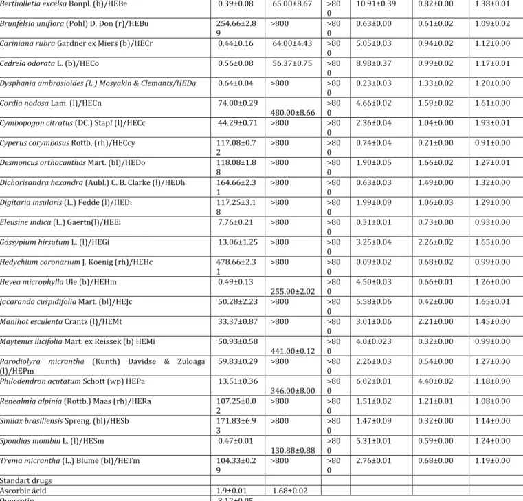
Table 4: Evaluation of the in vitro antioxidant activity and the concentrations of total phenolic compounds, total flavonoids, and coumarins of the hydroethanolic extracts of medicinal plants used for the treatment of infections
Species/extract DPPH FRAP NO Total
phenols
Flavonoids Coumarins
IC50±SD, μg/ml mg
TAE/g±SD
mg RE/g±SD
mg CE/g±SD
Aristolochia cymbifera Mart. and Zucc. (bl)/HEAc 96.91±0.72 >800 >80 0
3.67±0.09 1.21±0.00 1.36±0.01
Phanera glabra (Jacq.) Vaz / HEPg 4.67±0.21 60.76±1.52 >80 0
Bertholletia excelsa Bonpl. (b)/HEBe 0.39±0.08 65.00±8.67 >80 0
10.91±0.39 0.82±0.00 1.38±0.01
Brunfelsia uniflora (Pohl) D. Don (r)/HEBu 254.66±2.8 9
>800 >80 0
0.63±0.00 0.61±0.02 1.09±0.02
Cariniana rubra Gardner ex Miers (b)/HECr 0.44±0.16 64.00±4.43 >80 0
5.05±0.03 0.94±0.02 1.12±0.00
Cedrela odorata L. (b)/HECo 0.56±0.08 56.37±0.75 >80 0
8.98±0.37 0.99±0.02 1.17±0.01
Dysphania ambrosioides (L.) Mosyakin & Clemants/HEDa 0.64±0.04 >800 >80 0
0.23±0.03 1.33±0.02 1.20±0.00
Cordia nodosa Lam. (l)/HECn 74.00±0.29
480.00±8.66 >80 0
4.66±0.02 1.59±0.02 1.61±0.00
Cymbopogon citratus (DC.) Stapf (l)/HECc 44.29±0.71 >800 >80 0
2.36±0.04 1.04±0.00 1.93±0.01
Cyperus corymbosus Rottb. (rh)/HECcy 117.08±0.7 2
>800 >80 0
0.74±0.04 0.21±0.00 0.91±0.00
Desmoncus orthacanthos Mart. (bl)/HEDo 118.08±1.8 8
>800 >80 0
1.90±0.05 1.66±0.02 1.27±0.01
Dichorisandra hexandra (Aubl.) C. B. Clarke (l)/HEDh 164.66±2.3 1
>800 >80 0
0.63±0.03 1.49±0.00 1.32±0.00
Digitaria insularis (L.) Fedde (l)/HEDi 117.25±3.1
8
>800 >80 0
1.99±0.09 1.06±0.03 1.29±0.00
Eleusine indica (L.) Gaertn(l)/HEEi 7.76±0.21 >800 >80 0
0.31±0.01 0.73±0.00 0.93±0.00
Gossypium hirsutum L. (l)/HEGi 13.06±1.25 >800 >80 0
3.25±0.04 2.26±0.02 1.65±0.00
Hedychium coronarium J. Koenig (rh)/HEHc 478.66±2.3 1
>800 >80 0
0.09±0.02 0.68±0.02 0.99±0.00
Hevea microphylla Ule (b)/HEHm 0.49±0.13
255.00±2.02 >80 0
4.50±0.03 0.66±0.01 1.26±0.00
Jacaranda cuspidifolia Mart. (bl)/HEJc 50.28±2.23 >800 >80 0
5.58±0.06 0.42±0.00 1.65±0.01
Manihot esculenta Crantz (l)/HEMt 33.37±0.87 >800 >80 0
3.01±0.06 2.21±0.00 1.45±0.00
Maytenus ilicifolia Mart. ex Reissek (b) HEMi 50.93±0.58
441.00±0.12 >80 0
4.0±0.023 0.32±0.00 0.99±0.00
Parodiolyra micrantha (Kunth) Davidse & Zuloaga (l)/HEPm
59.83±0.29 >800 >80 0
2.26±0.03 0.54±0.00 1.27±0.00
Philodendron acutatum Schott (wp) HEPa 13.51±0.36
346.00±8.00 >80 0
6.02±0.01 4.40±0.02 1.18±0.00
Renealmia alpinia (Rottb.) Maas (rh)/HERa 107.25±0.0 2
>800 >80 0
1.51±0.02 1.21±0.01 1.08±0.00
Smilax brasiliensis Spreng. (bl)/HESb 171.83±6.9 3
>800 >80 0
1.47±0.09 0.32±0.00 1.14±0.00
Spondias mombin L. (l)/HESm 0.47±0.01
130.88±0.88 >80 0
5.31±0.01 0.59±0.00 1.24±0.00
Trema micrantha (L.) Blume (bl)/HETm 104.33±0.2 9
>800 >80 0
2.76±0.01 0.68±0.00 1.19±0.00
Standart drugs
Ascorbic ácid 1.9±0.01 1.68±0.02
Quercetin 3.12±0.05
Results expressed as IC50±SDfor the antioxidant activity, for the quantification of polyphenols in mg of tannic acid per gramm of sample (mg TAE/g)
for total phenolic compounds; mg of rutine per gramm of sample (mg RE/g) for total flavonoids; mg of coumarin per gramm of sample (mg CE/g) for coumarins. (b): bark; (bl): branches and leaves; (l) leaves; (rh): rhizome; (r): root (sb): stern bark; (wp): whole plant; quercetin. The assays were carried out in triplicate.
The methods used for estimation of the total antioxidant capacities were selected based on their simplicity, low cost, and reproducibility and since we evaluated the antioxidant potentials of the different plant's extracts since there is no single, widely-acceptable assay method applicable to a reasonable variety of compounds in plasma and plant matrices. Although, in vitro
methods used in assessing radical scavenging activity are artificial radicals and do not reproduce in vivo situation, they are however, useful in the evaluation of the antioxidant activity in a rapid, easy, and inexpensive way. Besides, it is important to note that, a valid
in vitro assay is an invaluable tool for clinical studies if it is combined with bioavailability data and valid oxidative stress biomarker assays [55, 56].
In this case, we employed the three commonly utilized antioxidant assays, namely DPPH, FRAP and NO scavenging assays to verify the
antioxidant potential of each extracts. Where considered active in these models only the extracts whose values of inhibition/capture of ROS allowed for the calculation of IC50±SD.
Table 4 displays the results of the antioxidant activity evaluation in vitro, as well as the concentrations of certain classes of polyphenols on the hydroethanolic extracts.
For further discussion of the antioxidant results, we selected, in the case of DPPH, only extracts with IC50 lower than the standard
antioxidant, ascorbic acid, while in the case of FRAP, we discussed only extracts with IC50 equal or less than 100 µg/ml.
It was noted that all 26 extracts exhibited, to a lesser or greater extent, the capacity to reduce DPPH radical, whereas, in the FRAP assay, only 9 extracts were capable of reducing Fe+3. None of the
tested concentrations. However, quercetin, the standard drug for this assay, presented intense antioxidant activity with IC50 =
3.12±0.05 µg/ml in all the in vitro antioxidant models tested. The content of total phenols in the hydroethanolic extract varied between 0.09-10.91 mg TAE/g, the highest content being in the HEBe (10.91±0.08 mg TAE/g), HECo (8.98±0.08 mg TAE/g) and HEPa (6.02±0.01 mg TAE/g), while the lowest were present in HEHc (0.09±0.02 mg TAE/g), HEDa (0.23±0.37 mg TAE/g) and HEEi (0.31±0.01 mg TAE/g).
Flavonoid contents varied between 0.32-4.40 mg RE/g, the highest contents being in HEPa (4.40±0.02 mg RE/g), HEGh (2.26±0.02 mg RE/g), HEMe (2.21±0.00 mg RE/g) and HEDo (1.66±0.02 mg RE/g), while the lowest were present in HECcy (0.21±0.00 mg RE/g), HESb (0.32±0.00 mg RE/g), HEMi (0.32±0.00 mg RE/g) and HEPm (0.54±0.00 mg RE/g).
As for the coumarin contents, the variation was between 0.91-1.93 mg EC/g, the highest contents being in HECci (1.93±0.01 mg EC/g), HEGh (1.65±0.00 mg EC/g) and HECn (1.61±0.00 mg EC/g) while the lowest concentration were encountered in HECcy (0.91±0.00 mg EC/g), HEEi (0.93±0.00 mg EC/g), HEHc and HEMi (0.99±0.00 mg EC/g).
The outstanding free radicals scavenging and antioxidant activities of the following extracts are notable: HEBe (IC50 = 0.39±0.08 and
65.00±8.67 µg/ml), HECr (IC50 = 0.44±0.16 and 64.00±4.43 µg/ml)
and HECo(IC50 = 0.56±0.08 and 56.37±0.75µg/ml) for the DPPH and
FRAP antioxidant models, respectively.
HESm(IC50 = 0.47±0.01 µg/ml), HEHm(IC50 = 0.49±0.13 µg/ml) and
HECa (IC50 = 0.64±0.04 µg/ml) exhibited activity only in the DPPH,
whereas HEPg (IC50 = 60.76±1.52 µg/ml) was active only in the FRAP.
It is worth mentioning that HEBe, HECr, HECo, HEHSm, HEHm and HEDa presented IC50 values lower than that of the standard compound
ascorbic acid (IC50 = 1.90±0.01 µg/ml) in the DPPH model, while in the
FRAP assay, HEBe, HECr, HECo and HEPg displayed IC50 greater than
the standard drug (IC50 = 1.68±0.02 µg/mll).
John and Shahidi reported antioxidant activity for the methanolic, ethanolic and acetone fractions of B. excelsa nuts in DPPH assay, however, with values of IC50 1000 times larger than those of HEBe [57].
The same authors showed that soluble and insoluble phenolic extracts of the kernel, nut, and shell, reduce ferric ion (0.21-59.20 µmol ascorbic acid eq/g), with activity equivalent to that obtained for HEBe (IC50 = 35.22 µmoles ascorbic acid eq/ml), indicating that
the extracts of B. excelsa have antioxidant capacity in both models,
being more active in the DPPH assay. In this screening, HEBe presented the highest contents of total phenolic compounds. Buratto
et al. [57, 58] reported that the antioxidant activity of the nuts of B. excelsa is related to the presence of these compounds, suggesting that the antioxidant activity of HEBe may be due to the presence of this metabolic class.
The DPPH scavenging activity of HECr was close to that of HEBe. Lima et al. [59], reported radical scavenging activity in DPPH assay for the ethanolic extract of C. rubra, but at concentrations 3 times higher than the ones obtained in this study. These authors described the presence of phenolic compounds (β-sitosterol, stigmasterol, amyrin and arjunolic acid) demonstrably antioxidant in the methanolic extract of C. rubra. This is the first report on the antioxidant activity of HECr using FRAP assay. These results indicate that phenolic compounds present in polar extracts of the bark of C. rubra, may be responsible, at least in part, for the antioxidant
activity of the plant.
The DPPH scavenging activity of HECo was observed and it also yielded the lowest FRAP value. Lima [60] showed that the methanolic bark extract of C. odorata possessed antioxidant capacity in both models, but with about 60 and 20 times less active than HECo in the DPPH and FRAP assays, respectively. Rashed [61] also verified that the methanolic extract as well as the fractions of this plant as possessing DPPH scavenging activities, however, at higher concentrations (IC50 = 1 mg/ml). Giordani et al. [62] described that
HECo presents among its constituents, gallic acid and gallocatechin, and attributed the antioxidant activity of the extract to their presence. The higher antioxidant activity observed for HECo in this study compared to the literature may be due to the differences in the preparation techniques, which may result in variation in the content and type of phenolic compounds present in it.
HESm showed antioxidant activity only on DPPH assay. Akinmoladun et al. demonstrated that the methanolic extract of the leaves of S. mombin and its fractions presented antioxidant activity against DPPH radical but at concentrations 20 times higher than those of HESm. Akinmoladun et al. and Igwe et al. [63-65] stated that the methanolic extract of the leaves of S. mombin is rich in flavonoid and phenolic contents, metabolic classes known to be rich in antioxidant compounds also present in HESm.
HEHm presented antioxidant activity on DPPH assay, this being the first report of this activity for H. microphylla, Fernandes [66], demonstrated that the methanolic extract of the endosperm of Hevea
sp. is rich in phenols also present in HEHm (apart from flavonoids and coumarins), which together could be responsible for the antioxidant effect of HEHm.
As with HEHm, HECa showed antioxidant activity on DPPH assay. Barros et al. [67] reported that the methanolic extract and the infusion inflorescences and upper leaves of D. ambrosioides capture the DPPH radical, but with IC50 values (0.62 and 0.49 mg/ml,
respectively) around 1000 times larger than that of HEDa. As for FRAP, the same authors report that both extracts were active, but at concentrations nearly 1000 times higher than those used in the present study with HEDa. In addition to the phenolic compounds found by Barros et al. [67], HEDa also presented flavonoids and coumarins, possibly related to the extract’s antioxidant activity. HEPg showed antioxidant activity only in FRAP assay, this being the first report of the activity for P. glabra, though there have been descriptions of antioxidant activity for some species belonging to this same genus, and particularly ascribed to the presence of phenolic compounds [68].
None of the hydroethanolic extracts tested yielded IC50 in the NO
assay, although ten of the extracts inhibited the formation of this gas at percentages ranging from 10-33 %.
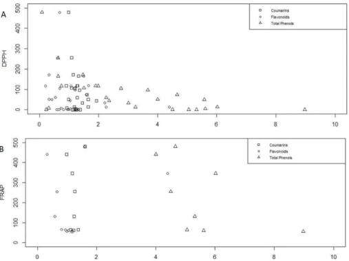
All the tested hydroethanolic extracts contained, to some extent, total phenols,flavonoids and coumarins. Since none of the extracts presented substantial effect in the NO assay, thus, the correlation studies between antioxidant activity and presence of a determined metabolic class only involved the extracts that were active in DPPH and FRAP assays. As seen in table 5 and fig. 1, there is a moderate negative linear correlation between the IC50 values on DPPH (1A) (the lower the IC50
value, the higher antioxidant activity observed) and the total phenolic contents (r =-0.53, p = 0.004), indicating that the antioxidant activity of
these extracts is directly related to their phenolic content.
Table 5: Evaluation of the in vitro correlation between the in vitro antioxidant activity and the concentrations of coumarins, total phenolics and flavonoid classes
Secondary metabolite Correlation coefficient
DPPH centralize p-value FRAP p-value
aTotal phenols -0.53 0.004** -0.56 0.112
aFlavonoids -0.21 0.300 0.29 0.435
bCoumarins -0.17 0.392 0.25 0.516