233 Gun S et al. Complete renal fusion in a child
Radiol Bras. 2012 Jul/Ago;45(4):233–234
Complete renal fusion in a child with recurrent urinary tract
infection
*
Fusão renal completa em criança com infecção recorrente do trato urinário
Saul Gun1,Guilherme Lippi Ciantelli2, Marília Akemi Uzuelle Takahashi2, Alexandre Mineto Brabo2, Lívea Athayde de Morais2, Caio Barros Figueiredo2
Cake kidney, a rare anomaly of the urinary tract, may be diagnosed at any age range. During the investigation of recurrent urinary tract infection in a 12-year-old child, contrast-enhanced computed tomography demonstrated the presence of a right-sided ectopic kidney, with renal fusion, drained by two ureters. Prophylactic treatment with nitrofurantoin was instituted, and the patient currently remains asymptomatic.
Keywords: Kidney; Malformation; Cake kidney.
O rim em bolo é uma rara anormalidade do trato urinário que pode ser diagnosticada em qualquer faixa etária. Durante investigação de infecção urinária recorrente em criança de 12 anos, foi revelada em tomografia computadorizada contrastada a presença de rim direito ectópico, com fusão renal, drenado por dois ureteres. Foi iniciado tratamento profilático com nitrofurantoína e o paciente se encontra assintomático.
Unitermos: Rim; Malformação; Rim em bolo.
Abstract
Resumo
* Study developed at Faculdade de Ciências Médicas e da Saúde da Pontifícia Universidade Católica de São Paulo (FCMS/ PUC-SP), Sorocaba, SP, Brazil.
1. PhD, Associate Professor, Department of Surgery, Facul-dade de Medicina de Sorocaba, Urologist at Conjunto Hospita-lar de Sorocaba, Sorocaba, SP, Brazil.
2. Graduate Students (6th year) of Medicine, Faculdade de Ciências Médicas e da Saúde da Pontifícia Universidade Cató-lica de São Paulo (FCMS/PUC-SP), Sorocaba, SP, Brazil.
Mailing Address: Guilherme Lippi Ciantelli. Avenida Moreira César, 39, ap. 112, Centro. Sorocaba, SP, Brazil, 18010-010. E-mail: [email protected]
Received January 27, 2012. Accepted after revision March 23, 2012.
Gun S,Ciantelli GL, Takahashi MAU, Brabo AA, Morais LA, Figueiredo CB. Complete renal fusion in a child with recurrent urinary tract infection. Radiol Bras. 2012 Jul/Ago;45(4):233–234.
0100-3984 © Colégio Brasileiro de Radiologia e Diagnóstico por Imagem CASE REPORT
because of acute pyelonephritis. A previous pelvic ultrasound report brought by the patient demonstrated a mass in the right kidney and absence of the left kidney.
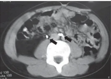
Contrast-enhanced computed tomogra-phy of the pelvis was requested and dem-onstrated right-sided renal ectopia in the lower pelvis and presence of a fused cake kidney (Figure 1) drained by two distinct ureters (Figure 2), without any further al-teration.
Considering the possibility of coexist-ing abnormalities, supplementary imagcoexist-ing ters diagnosed at a tertiary health care unit
specialized in urology.
CASE REPORT
A 12-year-old male patient with a his-tory of recurrent urinary tract infections since childhood was referred to the urology unit of a tertiary school hospital for inves-tigation of a mass on right kidney topogra-phy discovered on a ultrasonogratopogra-phy ex-amination. In his city of origin, the patient had already been hospitalized three times,
INTRODUCTION
Cake kidney is a rare congenital abnor-mality of the genitourinary tract, with barely more than twenty cases described in the literature(1). The term “cake kidney” or
fused pelvic kidney was defined by Glenn in 1958 as “an abnormality in which all the renal system tissue are fused into a single mass lying at the bottom of the pelvis and which two ureters drain separately into the vesical trigone”(2). The early diagnosis and
the recognition of potential complications associated with such abnormality constitute relevant factors to prevent permanent renal injury(3).
In the present paper, the authors report a case of cake kidney drained by two
234
Gun S et al. Complete renal fusion in a child
Radiol Bras. 2012 Jul/Ago;45(4):233–234 studies were requested but no other
alter-ation was found. Additionally, creatinine testing (0.6 mg/dl) and DMSA renal scan were performed and did not demonstrate renal function compromise.
The patient is currently undergoing pro-phylactic treatment with nitrofurantoin and has not presented further episode of urinary tract infection and is asymptomatic since.
DISCUSSION
Cake kidney is a congenital abnormal-ity defined as a complete fusion of both kidneys, representing only 2% of all renal fusion cases. Such abnormality may be di-agnosed at any age range and likewise other renal fusion abnormalities, is most fre-quently found in men at a 2–3:1 ratio(4).
Such abnormality occurs at the early phases of the embryonic development. Under normal conditions, two masses of metanephrogenic tissue existing in the lower pelvis develop until taking their de-finitive positioning in the lumbar region after complex movements involving lateral and ascending migration, axial deflection and internal rotation. It is believed that during the formation of a cake kidney, nephrogenic blastemas are compressed by the umbilical arteries at the beginning of the cranial migration of ureteral buds, which could be the cause of the fusion. The fused kidneys do not ascend as they should in a normal development, remaining in an ectopic pelvic position(5).
Partial renal fusion is more frequently found, being principally represented by
horseshoe kidney and crossed fused renal ectopia. Horseshoe kidneys correspond to 90% of all the renal abnormalities, with an incidence of up to 0.25%(1,4).
Anatomically, the cake kidney presents a lobulated anterior aspect while the pos-terior facet is smooth and homogeneous(2). The renal pelvis is located anteriorly to the kidney and except for some few cases, there are two ureters which drain into the bladder in the normal anatomical regions of the vesical trigone(6). Such congenital abnormalities may present some histologi-cal alterations, namely: immature glom-eruli; cystic changes; widening and dilata-tion of tubules; or even evidences of chronic renal disease(3). In other cases, there may be signs of infarct or ischemia secondary to a blood supply abnormality(4). This renal fusion abnormality can re-main asymptomatic, or even be detected only in an autopsy exam(1,5).
The presence of a cake kidney does not indicate a poor prognosis with kidney mal-function or possible progressive deteriora-tion of its funcdeteriora-tion. The follow up is impor-tant for the patient for early diagnosis of complications such as: obstruction, calcu-lus, infection, hematuria and uremia. These conditions also can be present in other uri-nary tract fusions. Besides, it´s important to exclude other congenital abnormalities and perform constant evaluation of the re-nal function, which reduces the chances of associated morbidity and mortality(1,3,4).
The vascular supply to the kidneys is compatible with their migration, and the ar-terial irrigation can be provided directly
from the aorta in a region proximal to its bifurcation or from the common iliac arter-ies and the venous drainage usually occurs towards the distal segment of the inferior vena cava or to the common iliac veins(1,3).
Such anomalous blood irrigation is consid-ered a risk factor for renal vascular compro-mise due to pelvic trauma, vascular disease, pregnancy and atherosclerosis(7).
Typically, the cake kidney coexists with other anomalies, such as: abnormal testicu-lar descent, Tetralogy of Fallot, vaginal atresia, sacral agenesis, caudal regression syndrome, bifid spine and anal abnormali-ties(6).
Such renal fusion anomaly may remain asymptomatic or even be detected at au-topsy(1,5). But, in some cases, there may be
infections secondary to the obstruction and calculosis or localized pain resulting from the renal vessels’ traction caused by the weight of the organ; in other cases, a mis-diagnosis of renal tumor may lead to unnec-essary nephrectomy(5).
The finding of a cake kidney is not in-dicative of a worse prognosis, renal dys-function, or possible progressive renal function deterioration(1,3,4), but it is
impor-tant that the patient is followed-up to allow the early diagnosis of possible complica-tions such as obstruction, calculosis, infec-tion, hematuria and uremia, which may also be present in other urinary tract fusions. Besides, ruling out other concomitant con-genital abnormalities and perform constant evaluation of the renal function is impor-tant to reduce the associated morbidity.
REFERENCES
1. Calado AA, Macedo Jr A, Srougi M. Cake kidney drained by single ureter. Int Braz J Urol. 2004; 30:321–2.
2. Glenn JF. Fused pelvic kidney. J Urol. 1958;80:7– 9.
3. Türqkvatan A, Demir D, Olçer T, et al. Cake kid-ney: MDCT urography for diagnosis. Clin Imag-ing. 2006;30:420–2.
4. Kaufman MH, Findlater GS. An unusual case of complete renal fusion giving rise to a ‘cake’ or ‘lump’ kidney. J Anat. 2001;198(Pt 4):501–4. 5. Srivastava RN, Singh M, Ghai OP, et al. Complete
renal fusion (“cake”/“lump” kidney). Br J Urol. 1971;43:391–4.
6. Goren E, Eidelman A. Pelvic cake kidney drained by single ureter. Urology. 1987;30:492–3. 7. Brock JW 3rd, Braren V, Phillips K, et al. Caudal
regression with cake kidney and a single ureter: a case report. J Urol. 1983;130:535–6.