262 Rev Assoc Med Bras 2011; 57(3):262-265
IMAGEM EM MEDICINA
Renal replacement lipomatosis and xanthogranulomatous pyelonephritis:
differential diagnosis
Renal replacement lipomatosis (RRL) is a relatively uncommon entity, although misdiagnosis - mainly with xanthogranulomatous pyelonephritis (XGP) - due to lack of awareness by urologists, radiologists, and pathologists may be responsible for underreporting1,2.
We illustrate a case of RRL that was initially misdi-agnosed as XGP, and compare it with a classic case of XGP, underscoring the similarities and the diferences between them.
PATIENT 1
A 63 year-old morbid obese (BMI = 52 kg/m2) female
was admitted to the hospital with right lank pain. She had a medical history of open cholecystectomy and in-ferior median laparotomy for gynaecological surgery. Investigation revealed urinary tract infection with E. coli
and Klebsiella sp. Creatinine clearance was 80 mL/min
and hemogram was unremarkable.
Plain radiography demonstrated a right renal
stag-FREDERICO R. ROMERO1, ROBERTO PILATI2, MARIA FERNANDA SALES FERREIRA CABOCLO3, ANTÔNIODE PÁDUA GOMES SILVA4,
MARCO AURÉLIO CRAVO5, THADEU BRENNY FILHO2
1 PhD in General Surgery at Universidade Federal do Paraná; Urologist at Hospital São Vicente de Curitiba, Curitiba, PR 2 MD, Urologist, Hospital São Vicente de Curitiba, Curitiba, PR
3 MD, Radiologist, Hospital São Vicente de Curitiba, Curitiba, PR
4 MD, Pathologist, Citopar - Centro de Citopatologia Paraná Ltda., Curitiba, PR 5 MD, Pathologist, Consulpat - Laboratório de Patologia e Citologia, Curitiba, PR
Study conducted at Hospital São Vicente de Curitiba; Citopar - Centro de Citopatologia Paraná Ltda; Consulpat - Laboratório de Patologia e Citologia, Curitiba, PR
Correspondence to:Frederico R. Romero – Rua Emiliano Perneta, 653 – apto. 41, Curitiba – PR – CEP: 80420-080 – [email protected]
horn calculus. Computed tomography (CT) scan showed an enlarged right kidney with hydronephrosis, paren-chymal atrophy, and calculi located in the right renal pel-vis, with marked fatty proliferation within the right renal sinus (Figure 1A). he case was initially misdiagnosed as XGP, and transperitoneal laparoscopic nephrectomy was ofered to the patient.
During surgery, large amount of perirenal and hilar fat was identiied. Renal artery and vein were dissected from surrounding fat tissue, and sequentially clipped with Hem-o-lok clips. here were no perirenal adhesions or iniltration, commonly observed when approaching XGP. he kidney was dissected free within Gerota’s fascia and removed through the previous median laparotomy inci-sion. Operative time was 180 minutes and estimated blood loss was 300 mL. he specimen was 10 x 8 x 7 cm in size, and was pathologically diagnosed as renal replacement ly-pomatosis (Figure 2). Postoperative course was unevent-ful and the patient was discharged home at day number 3.
Figure 1 – (A) CT scan demonstrating an enlarged right kidney with hydronephrosis, parenchymal atrophy, and calculi located in the right renal pelvis, with marked fatty proliferation within the right renal sinus (arrows). (B) CT scan showing multiple low attenuation areas with peripheral parenchyma enhancement (arrows), thickening of Gerota’s fascia, and densiication of perirenal and periureteral fat.
(A) (B)
263
RENALREPLACEMENTLIPOMATOSISANDXANTHOGRANULOMATOUSPYELONEPHRITIS: DIFFERENTIALDIAGNOSIS
Rev Assoc Med Bras 2011; 57(3):262-265 PATIENT 2
A 51 year-old obese (BMI = 32 kg/m2) female was
ad-mitted to the hospital with right lank pain. She had a history of weight loss of 20 kg in the last 12 months, and open surgical drainage for a right lank abscess ive months earlier that persisted with small amount of puru-lent discharge until one month previously. Investigation revealed urinary tract infection with E. coli.
Plain radiography demonstrated a right renal stag-horn calculus. A CT scan showed an enlarged right kid-ney with hydronephrosis, calculi located in the right re-nal pelvis, multiple low attenuation areas with peripheral parenchyma enhancement, thickening of Gerota’s fascia, and densiication of perirenal and periureteral fat, all of which suggested the diagnosis of XGP. here were no re-sidual perinephric collections (Figure 1B).
During right-sided transperitoneal laparoscopic ne-phrectomy, there were intense perirenal adhesions, with irm attachments between Gerota’s fascia and the ante-rior aspect of infeante-rior vena cava (IVC). While trying to identify the plane between the IVC and the ureter at the lower pole of the kidney, we inadvertently entered the interaortocaval space and, without realizing it, lited the IVC anteriorly. Dissection over this area resulted in ac-cidental tearing of a lumbar vein, which was managed laparoscopically.
Renal artery and vein were not distinctly identiied. All vascular structures were controlled with Hem-o-lok clips and harmonic scalpel. he kidney was dissected free inside Gerota’s fascia, with small purulent discharge
during dissection of the upper pole. he specimen was removed through the posterior lumbar incision used for abscess drainage. Operative time was 150 minutes. Esti-mated blood loss was 500 mL, and the patient received two units of blood intraoperatively. he specimen was 11 x 9 x 8 cm in size, with a ibrous, lobulated, yellow-tan capsule. Pathology conirmed the diagnosis of xan-thogranulomatous pyelonephritis (Figure 3). Postopera-tively, she received cetriaxone and metronidazole for 7 days, with no complications except for one episode of unexplained fever, and was discharged home on post-operative day number 8.
DISCUSSION
Renal replacement lypomatosis and xanthogranuloma-tous pyelonephritis have similar etiopathogenic, clini-cal and radiologiclini-cal features. Both are characterized by atrophy and destruction of renal parenchyma, oten as-sociated with unilateral chronic renal infection, hydro-nephrosis or pyohydro-nephrosis, and calculous disease. he main diference between them is that in RRL, irst report-ed by Brown in 18613,the atrophic renal parenchyma is
replaced by fatty tissue proliferation3-6. In XGP, initially
described as staphylomycosis in 1916 by Schlogenhaufer, xanthoma cells (lipid-laden macrophages) iniltrate and substitute necrotic renal tissue resulting in a lipomatous degeneration4,7.
RRL, renal sinus lipomatosis, and ibrolipomatosis of the kidney represent a spectrum of changes in which normal renal sinus and perirenal fat increase in amount
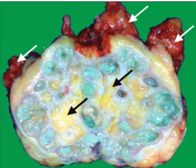
Figure 2 – Cross-section of the specimen revealed an ex-tremely atrophied renal parenchyma, and a large lobulated bright-yellow mass with calculi illing the dilated pelvicalyce-al system. Note the similarity between the fat tissue inside the renal pelvis and the perirrenal fat (white arrows), which may distinguish renal replacement lypomatosis from xantho-granulomatous pyelonephritis.
264
IMAGEMEMMEDICINA
Rev Assoc Med Bras 2011; 57(3):262-265
and replace the renal parenchyma. Renal sinus lipoma-tosis, the mildest form, is associated with obesity, renal atrophy of varying causes (e.g. aging and atherosclero-sis), Cushing’s syndrome or the use of exogenous ste-roids4-6. his mild form infrequently produces symptoms
because of the absence of caliceal obstruction5, and is a
common inding at autopsy4. Invasion of adipose cells
from the peripelvic fat into the kidney occurs along the blood vessels in the renal sinus3. At the other end of the
spectrum is RRL, where the entire renal parenchyma is replaced with adipose tissue, usually secondary to calcu-lous disease and longstanding inlammatory/infectious disease (e.g. renal tuberculosis)4,6. In this group there is
no invasion from without. Proliferation of fat cells in the interstitium of the kidney is mediated by connective tissue cells3.
Pathogenesis of XGP is not fully understood. It be-gins within the pelvis and calyces and subsequently ex-tends into and destroys renal parenchymal and adjacent tissues. All theories agree that the primary factors in-volved in the development of XGP are bacterial infec-tion, obstruction and calculous disease. Other possible interrelated factors include venous occlusion and hem-orrhage, abnormal lipid metabolism, lymphatic block-age, failure of antimicrobial therapy, altered immuno-logic competence, and renal ischemia7.
RRL and XGP usually occur between the ith and seventh decade of life. he patient may be asymptomatic or present a varied clinical picture related to the primary disease, the most frequent manifestations being urinary tract infections, lank pain, weight loss, hematuria, fe-ver, and palpable mass6-8.
CT scan is the most valuable method for diferentiat-ing RRL and XGP8. In XGP, it demonstrates a large
reni-form mass with a central staghorn calculus, and peripheral enhancement that may correspond to compressed resid-ual parenchyma or a capsule of inlammatory tissue8-10.
Renal parenchyma is replaced by multiple low attenua-tion (-15 to 29 HU) areas with radial distribuattenua-tion, which represent dilated calyces and abscess cavities illed with pus and debris, frequently described as a “bunch of grapes” or a “bear claw”8,10.True fat density is usually not
seen8. Air inside the urinary tract and perinephric
ex-tension to adjacent organs are also indicative of XGP9,10.
In RRL, the characteristic distribution of adipose mass within the renal sinus and perirenal space, with areas of negative attenuation values similar to those of adipose tissue (conirmed by a value of -20 HU or lower) help in the diagnosis5,6. Even though the CT scan illustrated in
Figure 1A is characteristic of RRL, we missed the diag-nosis because of our lack of knowledge about this clini-cal entity.
During surgery, although the fat in renal lipomatosis is tougher and more ibrous than normal fat3, severe
ad-hesions and iniltration observed in XGP are usually not present in RRL. Further, the renal hilum may not be dis-tinguished within the dense perihilar ibrosis present in XGP, while it is relatively easily dissected in renal lipo-matosis. his is especially important when approaching the kidney by laparoscopy. XGP is considered a relative contraindication to laparoscopy because of increased diiculty, higher morbimortality, and greater conver-sion rates. However, patients diagnosed with XGP and described as having no adhesions, iniltrations or ibro-sis during laparoscopic nephrectomy should be criti-cally investigated as having RRL misdiagnosed as XGP, as in the irst patient illustrated herein.
Pathologically, the kidney is usually enlarged and presents with a gross ibrofatty appearance. When the specimen is opened, only a thin rim or shell of atro-phied renal parenchyma if found, with bright-yellow fat tissue in the renal sinus that is similar to the perirenal fat in RRL, and a pale-yellow fatty tissue in XGP. His-tologically, there is increase of lipid-laden macrophages (xanthoma cells) inside the renal parenchyma in XGP, whereas RRL contains large fat cells outside the renal parenchyma2,4,5, with sharp demarcation between the
adipose tissue and the renal parenchyma, showing that there is no real invasion of the kidney by fat but merely replacement of fat as it atrophies1,3,4.
Additionally though very rare, XGP and RLL may coexist. Other focal fatty lesions such as lipomas, an-giomyolipomas, and liposarcomas must be considered. These lesions, however, are usually not associated with parenchymal atrophy or staghorn calculi, and frequent-ly produce a mass effect on the intrarenal collecting system2,5.
Renal replacement lipomatosis should always be kept in mind by clinicians, urologists, and radiologists when evaluating a patient with suspicion of xanthogran-ulomatous pyelonephritis. Speciic imaging, operative, and pathological diferences may provide clues for the diferential diagnosis.
REFERENCES
1. Shah VB, Rupani AB, Deokar MS, Pathak HR. Idiopathic renal replacement lipomatosis: a case report and review of literature. In-dian J Pathol Microbiol. 2009;52:552-3.
2. Xu Y, Liu RL, Zhang ZH, Zhao WM, Yang QC. Renal replacement lipomatosis. Eur Surg Res. 2006;38:385-7.
3. Dukes CE. he pathology of renal lipomatosis. Proc Royal Soc Med. 1938;31:1361-4.
4. Ambos MA, Bosniak MA, Gordon R, Madayag MA. Replacement lipomatosis of the kidney. AJR Am J Roentgenol. 1978;130:1087-91.
5. Kocaoglu M, Bozlar U, Sanal HT, Guvenc I. Replacement lipo-matosis: CT and MRI indings of a rare renal mass. Br J Radiol. 2007;80:e287-9.
265
RENALREPLACEMENTLIPOMATOSISANDXANTHOGRANULOMATOUSPYELONEPHRITIS: DIFFERENTIALDIAGNOSIS
Rev Assoc Med Bras 2011; 57(3):262-265 7. Sharma S, Jhobta A, Goyal D, Surya M, Sumala, Negi A. Ureteral
involvement in xanthogranulomatous pyelonephritis - rare mani-festation. Ind J Radiol Imag. 2006;16:243-5.
8. D’Ippolito G, Tokechi D, Shigueoka DC, Ajzen S. Tomographic aspects of xanthogranulomatous pyelonephritis and related com-plications. São Paulo Med J. 1996;114:1091-6.
9. Calisir C, Can C, Kebapci M. Renal replacement lipomatosis: mul-tidetector-row computed tomography indings in one case. Acta Radiol. 2007;48:242-5.