Received from Department of Anesthesia and Department of Obstetrics and Gynecology, Mount Sinai Hospital, University of Toronto.
1. MD, PhD, Obstetric Anesthesia Fellow 2. MD, Anesthesia Fellow
3. BA, Assistant
4. MD, Professor, Department of Obstetrics and Gynecology
5. MD, PhD, FANZCA, FRCPC, Professor of Anesthesia and Obstetrics and Gynecology, University of Toronto
Submitted on June 11, 2010. Approved on June 14, 2010. Correpondence to: Dr. Jose C.A.Carvalho
Department of Anesthesia and Pain Management Mount Sinai Hospital 600 University Avenue, Room 781 Toronto, ON M5G 1X5 Canada E-mail: [email protected]
SCIENTIFIC ARTICLE
Non-Invasive Hemodynamic Assessment of Non-pregnant,
Healthy Pregnant and Preeclamptic Women using
Bio-Reactance
Yayoi Ohashi
1, Hisham Ibrahim
2, Louis Furtado
3, John Kingdom
4, Jose Carlos Almeida Carvalho
5Summary: Ohashi Y, Ibrahim H, Furtado L, Kingdom J, Carvalho JCA – Non-Invasive Hemodynamic Assessment of Non-pregnant, Healthy Pregnant and Preeclamptic Women using Bio-Reactance.
Background and objectives: We compared hemodynamic profiles of healthy and mildly preeclamptic pregnant women at term, as well as those of non-pregnant controls, using a new non-invasive cardiac output monitor (NICOM) based on bio-reactance.
Methods: We studied healthy term pregnant women at term (Preg, n = 10), mildly preeclamptic pregnant women at term (PregPE, n = 10), and healthy non-pregnant female volunteers (NonPreg, n = 10). With the subjects in the semi left lateral position, 4 electrodes of the NICOM device were applied to their chest wall, followed by a 15-minute rest period. Hemodynamic variables, including the systolic (SBP), diastolic (DPB) and mean arterial (MAP) pressures, as well as the heart rate (HR), stroke volume (SV), total peripheral resistance (TPR), cardiac output (CO), cardiac power output (CPO), and ventricular ejection time (VET) were then monitored for 15 minutes.
Results: The Preg and NonPreg groups showed similar hemodynamic profiles, except for a shorter VET in the Preg group (213.3 ± 19.3 ms versus 265.0 ± 28.8 ms, p < 0.001). The PregPE group showed higher SBP, DBP and MAP, as well as CPO (145.5 ± 12.6 mmHg; 94.5 ± 9.1 mmHg; 111.5 ± 9.8 mmHg; 1.6 ± 0.3 watts), compared to both the Preg (114 ± 12.1 mmHg; 71.7 ± 8.4 mmHg; 85.9 ± 9.3 mmHg; 1.1 ± 0.3 watts) and NonPreg (101.2 ± 11.9 mmHg; 66.7 ± 10.4 mmHg; 78.1 ± 10.6 mmHg; 1.0 ± 0.2 watts) groups. The PregPE group showed higher HR, CO, and TPR, and shorter VET (85.4 ± 8.4 beats.min-1; 6.6 ± 0.7 L.min-1; 1,369.9 ± 173.5 dyne.sec.cm-5, 221.6 ± 22.4 ms) compared to the NonPreg group (67.9 ± 9.5 beats.min-1; 5.6 ± 0.7 L.min-1; 1,136.9 ± 149.8 dyne.sec.cm-5, 265.0 ± 28.8 ms).
Conclusions: The NICOM device is simple to use, operator independent, and provides clear and consistent monitoring signals. The output iden-tified distinct hemodymamic profiles that are consistent with the findings of more invasive existing methods.
Keywords: PHYSIOLOGY, Cardiovascular; HEMODYNAMICS; MONITORING, bioreactance technology, physiological; PREGNANCY, preeclampsia.
Cheeta Medical provided the equipment for research.
[Rev Bras Anestesiol 2010;60(6): 603-613] ©Elsevier Editora Ltda.
INTRODUCTION
Invasive hemodynamic techniques have long identified signi-ficant increases in heart rate (HR), blood volume (BV), left ventricular end-diastolic volume (LVEDV), stroke volume (SV) and cardiac output (CO) during the first and second tri-mesters of pregnancy 1,2. Despite these changes, maternal
blood pressure still falls due to a large reduction in the total peripheral resistance (TPR) from systemic vasodilation and the formation of a low-resistance uteroplacental circulation.
In the last trimester of pregnancy, however, this profile chan-ges, due in part to the fully developed fetus gradually obs-tructing venous return via the inferior vena cava. The cardiac output then decreases and the total vascular resistance in-creases; the systolic (SBP), diastolic (DBP) and mean (MAP) arterial blood pressures also increase 2.
Maternal hemodynamic changes can be further complicated by preeclampsia, which occurs in 6%-12% of all pregnancies 3,4.
Preeclampsia has always been found to show varied hemody-namic profiles, making it difficult for the clinician to plan a goal-directed management or therapy plan 5,6. It has recently been
suggested that preeclampsia can be established earlier (< 34 weeks) or later (> 34 weeks) in pregnancy, and that these two entities have different etiologies and should be considered diffe-rent forms of the disease 7,8. It has also been shown that during
their latent phases, early and late preeclampsia show two very different maternal hemodynamic profiles, including high TPR in the former versus low TPR in the latter 9.
prenatal care, beyond the classic arterial blood pressure. A meticulous hemodynamic assessment throughout preg-nancy might revolutionize prenatal care, allowing early diagnosis of high risk patients and the use of goal-directed therapies.
Most of the available data on hemodynamic changes in healthy and high risk pregnancies has been generated by studies using pulmonary artery catherization, still conside-red the gold standard for central hemodynamic monitoring
2,10,11. The technique, however, carries many risks and
di-sadvantages, and has limited use in obstetrics 12. More
re-cently, minimally-invasive techniques based on arterial pul-se waveform analysis methods have been validated against pulmonary artery catherization in non-obstetric patients. Currently available methods, each based on a different al-gorithm, include the LiDCOplus (LiDCO, Cambridge, Uni-ted Kingdom), the PiCCOplus (Pulsion Medical Systems, Munich,Germany), and the Vigileo (Edwards Lifesciences, Irvine, CA). They have all been used with some success in the obstetric patient, but again, they require the use of a peripheral arterial line 13.
There is no question that non-invasive monitoring is a highly desirable resource in obstetric medicine. Impedance cardiogra-phy 14, thoracic electrical bioimpedance 15, and transthoracic
echocardiography 16 have all been validated against pulmonary
artery catherization in the obstetric population. All of these me-thods have limitations including the requirement for user edu-cation and an interference of movement artifact. Transthoracic bioimpedance was the first non-invasive method used for CO monitoring, and it has been used in many obstetric settings 17.
Although it has the advantage of being operator-independent, its use is limited because of questionable accuracy, perhaps due to a low signal-to-noise ratio. Also, variations in patient body habi-tus and other physical factors impact on the electrical conductivi-ty between the electrodes and skin 18.
Transthoracic bio-reactance is a newer technique used for non-invasive continuous cardiac output monitoring 19. It is
based on the frequency-modulation and phase-modulation of the voltage signal measured in response to an applied transthoracic current. The phase shifts are measured con-tinuously, and have been shown to relate almost linearly to blood flow in the aorta. The bio-reactance technique offers a significant advantage in filtering noise, and provides an improved signal-to-noise ratio compared to bio-impedance. Moreover, its readings were shown to correlate well with the results of pulmonary artery catheter derived measurements of cardiac output 20. It has been shown that the
non-inva-sive cardiac output measurement (NICOM) system, which uses bio-reactance technology, has acceptable accuracy, precision and responsiveness for CO monitoring in patients experiencing a wide range of circulatory situations 21. The
NICOM has not been used for hemodynamic assessment and monitoring in pregnant women.
This study was designed to compare the hemodynamic profiles of healthy and mildly preeclamptic pregnant women at term, as well as those of non pregnant women using the NICOM.
METHODS
After institutional research ethics board approval and written in-formed consent, three groups of women (n = 10 in each group) were recruited into this prospective, open, comparative study. All subjects were between 18 and 40 years of age and able to communicate in English, and belonged to one of the three follo-wing categories: a) healthy pregnant women at term (Preg); b) pregnant women at term with newly diagnosed, untreated pre-eclampsia (PrePE) who were documented to have had normal pressure on at least 2 occasions before 20 weeks of gestation; and c) non-pregnant healthy female volunteers (NonPreg). All pregnancies were singleton and the patients were not in labor. Pregnant women with pre-existing diseases such as insulin-dependent diabetes, chronic hypertension, and auto-immune, cardiovascular or renal diseases were excluded.
Preeclampsia was defined by The American College of Obstetricians and Gynecologists (ACOG) criteria as newly diagnosed systolic blood pressure of at least 140 mmHg or diastolic blood pressure of 90 mmHg on at least 2 occasions; the measurements had to be taken at least 4 hours but not more than 7days apart.
A bio-reactance-based non-invasive cardiac output moni-toring system (NICOMTM, Cheetah Medical Inc, Portland, OR)
was used in this study. Participants were asked to rest in the left semi-lateral decubitus position for 15 minutes after the pla-cement of 4 electrodes on their thorax, and automatic calibra-tion of the NICOM system. Addicalibra-tionally, a non-invasive blood pressure cuff was applied to the left arm and connected to the monitoring system. Automated blood pressure measurements were taken every minute by the NICOM internal blood pressu-re monitoring system throughout the study.
The following cardiovascular variables were assessed conti-nuously for 15 minutes to define baseline values: systolic blood pressure (SBP), diastolic blood pressure (DBP), mean arterial pressure (MAP), heart rate (HR), cardiac output (CO), cardiac power output (CPO), stroke volume (SV), ventricular ejection time (VET), and total peripheral resistance (TPR). Following this 15-minute monitoring period during which the patient was at rest, a passive leg raising (PLR) test was performed. The hemodyna-mic variables were measured with the patient lying on the bed in a left semi-lateral decubitus position for 3 minutes and then with the subject’s legs raised to 45 degrees for 3 minutes.
Continuous data are expressed as a mean ± standard de-viation. Differences between the groups were compared with T-tests, with p-values corrected for multiple comparisons by the Bonferonni procedure. The effect of the PLR test was de-termined by comparing the percent change in each variable from baseline, following the leg raise, using a 2-tail T-test. Di-fferences were considered significant when p < 0.05. All the statistical analyses were performed using Excel software.
RESULTS
The hemodynamic variables for all the groups are summa-rized in Table II. Healthy pregnant women demonstrated shor-ter VETs compared to non-pregnant women (213.0 ± 19.3 ms versus 265.0 ± 28.8 ms, p < 0.001). All the other variables were similar between these two groups.
Preeclamtic women showed higher SBP, DBP, MAP and CPO compared to healthy pregnant women (p < 0.001).
Preeclamptic women showed higher HR (p < 0.001), SBP (p < 0.001), DBP (p < 0.001), MAP (p < 0.001), CPO (p < 0.001), TPR (p = 0.014) and CO (p = 0.013), but shorter VET (p < 0.001) compared to non-pregnant women.
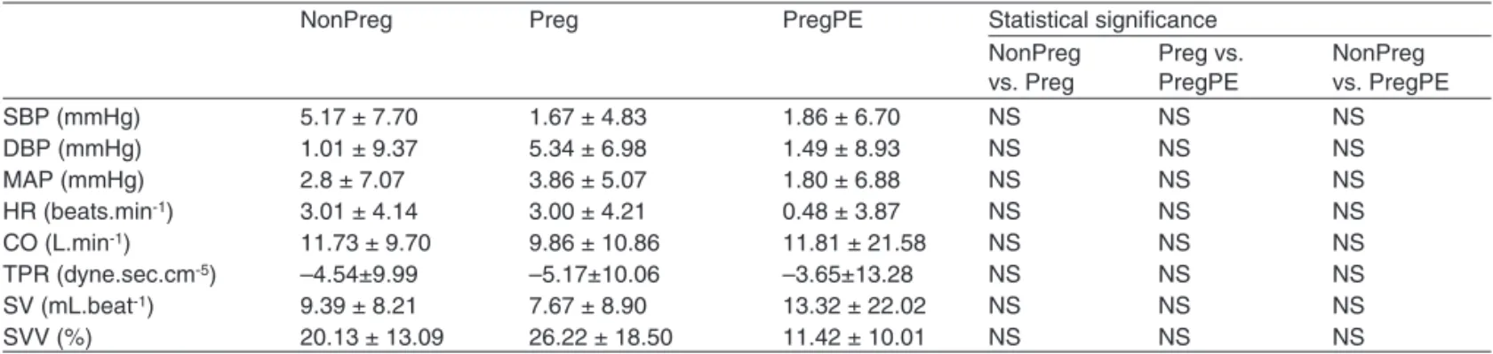
The percent changes in the hemodynamic variables after the PLR tests are summarized in Table III. There were no sig-nificant differences between the three groups.
Table I – Patients’ Characteristics
NonPreg Preg PregPE Statistical significance
NonPreg vs. Preg
Preg vs. PregPE
NonPreg vs. PregPE
Age (years) 30.1 ± 5.6 26.5 ± 5.1 31.6 ± 7.9 NS NS NS
Height (cm) 164.26 ± 6.3 163.98 ± 8.3 164.84 ± 33.9 NS NS NS
Weight (kg) 60.57 ± 10.7 76.8 ± 15.8 85.1 ± 20.8 p = 0.016 NS p = 0.001
BMI 22.27 ± 2.9 29.36 ± 7.0 31.14 ± 7.5 p = 0.009 NS p < 0.001
Gestational age (weeks) 39.89 (36.6-42.1)* 36.98 (35.2-40.2)*
NonPreg: non-pregnant women, Preg: healthy pregnant women, PregPE: mildly preeclamptic pregnant women, BMI: body mass index, NS: not significant; Data are presented as mean ± standard deviation. * Mean and range
Table II – Hemodynamic Data
NonPreg Preg PregPE Statistical significance
NonPreg vs. Preg
Preg vs. PregPE
NonPreg vs. PregPE SBP (mmHg) 101.2 ± 11.9 114.5 ± 12.1 145.5 ± 12.6 NS p < 0.001 p < 0.001
DBP (mmHg) 66.7 ± 10.4 71.7 ± 8.4 94.5 ± 9.1 NS p < 0.001 p < 0.001
MAP (mmHg) 78.1 ± 10.6 85.9 ± 9.3 111.5 ± 9.8 NS p < 0.001 p < 0.001
HR (beats.min-1) 67.9 ± 9.5 78.8 ± 11.6 85.4 ± 8.4 NS NS p = 0.001
CO (L.min-1) 5.6 ± 0.7 5.9 ± 1.1 6.6 ± 0.7 NS NS p = 0.013
TPR (dyne.sec.cm-5) 1,136.9 ± 149.8 1,206.7 ± 254.0 1,369.9 ± 173.5 NS NS p = 0.014
CPO (watts) 1.0 ± 0.2 1.1 ± 0.3 1.6 ± 0.3 NS p < 0.001 p < 0.001
SV (mL.beat-1) 83.3 ± 12.5 76.4 ± 17.0 77.7 ± 7.2 NS NS NS
VET (milliseconds) 265.0 ± 28.8 213.3 ± 19.3 221.6 ± 22.4 p < 0.001 NS p = 0.001 NonPreg: non-pregnant women, Preg: healthy pregnant women, PregPE: mildly preeclamptic pregnant women, SBP: systolic blood pressure, DBP: diastolic blood pressure, MAP: mean arterial pressure, HR: heart rate, CO: cardiac output, TPR: total peripheral resistance (MAP/CO), CPO: cardiac power output, SV: stroke volume, VET: ventricular ejection time, NS: not significant;
Data are presented as mean ± standard deviation.
Table III – Percent Changes of Hemodynamic Variables with a Passive Leg Raising (PLR) Test
NonPreg Preg PregPE Statistical significance
NonPreg vs. Preg
Preg vs. PregPE
NonPreg vs. PregPE
SBP (mmHg) 5.17 ± 7.70 1.67 ± 4.83 1.86 ± 6.70 NS NS NS
DBP (mmHg) 1.01 ± 9.37 5.34 ± 6.98 1.49 ± 8.93 NS NS NS
MAP (mmHg) 2.8 ± 7.07 3.86 ± 5.07 1.80 ± 6.88 NS NS NS
HR (beats.min-1) 3.01 ± 4.14 3.00 ± 4.21 0.48 ± 3.87 NS NS NS
CO (L.min-1) 11.73 ± 9.70 9.86 ± 10.86 11.81 ± 21.58 NS NS NS
TPR (dyne.sec.cm-5) –4.54±9.99 –5.17±10.06 –3.65±13.28 NS NS NS
SV (mL.beat-1) 9.39 ± 8.21 7.67 ± 8.90 13.32 ± 22.02 NS NS NS
SVV (%) 20.13 ± 13.09 26.22 ± 18.50 11.42 ± 10.01 NS NS NS
NonPreg: non-pregnant women, Preg: healthy pregnant women, PregPE: mildly preeclamptic pregnant women, SBP: systolic blood pressure, DBP: diastolic blood pressure, MAP: mean arterial pressure, HR: heart rate, CO: cardiac output, TPR: total peripheral resistance, SV: stroke volume, SVV: stroke volume variation, NS: not significant;
DISCUSSION
The results of our study confirm existing data on the hemo-dynamic features of pregnant and non-pregnant women, and also introduce new hemodynamic concepts that may be of interest to clinicians. Our results suggest that bio-reactance may be one step closer to an operator-independent, consis-tent, simple-to-use, non-invasive monitor that can function as a much needed tool for clarifying the hemodynamics of heal-thy and high risk pregnancies.
Pregnancy induces significant hemodynamic changes secondary to an increase in plasma volume, a decrease in systemic vascular resistance, and an increase in myocardial performance. These changes are necessary to meet the in-creasing metabolic demands of pregnancy. They start early in pregnancy, peak at the end of the second trimester, and gradually trend to non-pregnant levels towards term 2.
Speci-fically, SBP, DBP, MAP and TPR decrease, and SV, HR and CO increase until mid-pregnancy, then the trend is reversed towards term, except for HR, which tends to remain elevated throughout pregnancy. Most of the reversed trends observed in the third trimester are attributed to the compression of the inferior vena cava, especially when the pregnant woman is in the supine position. It has been shown that aortocaval com-pression is present in 40% of supine pregnant women, even when they are tilted between 0°-34°, as well as when they are lying in the semi-recumbent position 22, 23.
Our results comparing healthy pregnant women at term with non-pregnant women are consistent with previous data in the literature. We did not find any significant difference in the hemodynamic variables between them, except for a shor-ter VET in pregnant patients. The left ventricular ejection time (LVET), an index of left ventricular performance,has been shown in previous studies to correlate with ejection fraction and intrinsic cardiac contractility 24-26. This is an interesting
new variable that could be explored in future studies, as it may indicate that myocardial performance is increased in healthy pregnant women at term.
The preeclamptic women, however, showed a very dis-tinct hemodynamic profile when compared both with the non-pregnant and healthy non-pregnant women. When compared to the Preg group, the SBP, DBP, MAP and CPO of the PregPE group were higher. The CPO takes into account both the pressure- and flow-generating capacities of the heart, and provides a more complete representation of the overall car-diac performance. Maximal CPO may be better than maximal CO and left ventricular stroke work index (SWI) independen-tly in representing cardiac pumping capability 26.The TPR
was not statistically different. These results may suggest that PregPE patients show a hyperdynamic state, in the context of a similar TPR.
When compared with the NonPreg group, the PregPE re-sults are even more remarkable. The SBP, DBP, MAP and CPO were higher in the PregPE group, similar to the previous com-parison. However, in the PregPE the HR, CO and TPR were also significantly higher. These results suggest a hyperdynamic state in the PregPE group in the context of an increased TPR.
The apparent discrepancy in findings related to TPR may be a function of the sample size of our study. The trends ob-served in the Preg and PrePE women were in the same di-rection, only more remarkable in the preeclamptic patients. These results also suggest that ideally each woman should serve as her own control for her hemodynamic assessment throughout pregnancy.
The passive leg raise (PLR) test has been suggested to predict fluid responsiveness. PLR induces an abrupt increase in preload due to the auto-transfusion of blood from the ca-pacitance veins of the legs to the intrathoracic compartment, leading to an increase in cardiac output in preload-dependent patients. The PLR test has recently been introduced as an essential part of hemodynamic monitoring, since the effects of auto-transfusion on aortic blood flow or cardiac output enable the assessment of fluid responsiveness 27,28. In this study,
ho-wever, we did not find any significant difference in the percent changes of the hemodynamic parameters with PLR testing among the three groups, indicating similar fluid responsive-ness in all groups.
Recently, both minimally invasive and non-invasive mo-nitoring has received significant attention from investigators and clinicians. A great deal of useful information about the obstetric patient has been gained, but each type of monito-ring has its own limitations. The minimally invasive techniques such as PiCCO, LiDCO, and Vigileo still require an arterial line and other procedures that will always be a limiting factor in obstetrics. Hence, high expectations have been placed on non-invasive monitoring techniques. However, some of these methods, such as transthoracic echo and suprasternal Dop-pler are operator-dependent, which is also a barrier to its im-plementation.
The totally non-invasive, operator-independent techniques of bioimpedance and bio-reactance are, therefore, where efforts can be directed if we want to have a monitor that will enable further understanding about hemodynamic changes in pregnancy, and the potential utility of goal-directed prevention or therapy for some patients.
Transthoracic bioimpedance (intrabeat measurement of chan-ges in transthoracic voltage amplitude in response to an injected high-frequency current) was the first non-invasive method devi-sed for continuous non-invasive monitoring of cardiac output. It has been used to study the hemodynamic patterns in pregnancy and preeclampsia17. Although its clinical use has increased, it is
limited in some clinical settings because of a low signal-to-noise ratio which apparently limits its accuracy in environments where there is ambient electrical noise. Also, the technique is sensitive to the placement of the electrodes on the body, variations in pa-tient body size and other physical factors, such as temperature and humidity, that impact on electrical conductivity between the electrodes and skin 18.
Unlike bioimpedance, bioreactance-based non-invasive CO measurement does not use static impedance, and does not depend on the distance between the electrodes for the cal-culations of SV and CO, thereby significantly increasing the accuracy of the result. Moreover, its readings were shown to correlate well with results obtained from the pulmonary artery catheter thermodilution-derived measurements of cardiac ou-tput 20. In addition, it has also been shown that the NICOM
system has acceptable accuracy, precision and responsive-ness for CO monitoring in patients experiencing a wide range of circulatory situations 21.
In summary, the NICOM was simple to use, and provided a very clear and consistent monitoring signal. It identified distinct hemodynamic profiles in the three studied groups that were consistent with previous data. We concluded that the NICOM
is a promising non-invasive monitoring system for obstetric patients, and further studies are warranted in laboring and critically ill patients, and those undergoing operative delive-ries. We suggest that a monitor such as the NICOM may offer a valuable opportunity to make early diagnoses and provide goal-directed therapy in women with preeclampsia, and other medical conditions that affect women during pregnancy.
ACKNOWLEDGEMENTS
The authors acknowledge Kristi Downey, Perinatal Research Coordinator, Department of Anesthesia and Pain Manage-ment, Mount Sinai Hospital, for facilitating various aspects of this project.
06. Gogarten W – Preeclampsia and anaesthesia. Curr Opin Anaesthe-siol, 2009;22:347-351.
07. von Dadelszen P, Magee LA, Roberts JM – Subclassification of pre--eclamspia. Hypertens Pregnancy, 2003;22:143-148.
08. Huppertz B – Placental origins of preeclampsia: challenging the cur-Huppertz B – Placental origins of preeclampsia: challenging the cur-rent hypothesis. Hypertension, 2008;51:970-975.
09. Valensise H, Vasapollo B, Gagliardi G et al. – Early and late pre-eclampsia: two different maternal hemodynamic states in the latent phase of the disease. Hypertension, 2008;52: 873-880.
10. Benedetti TJ, Cotton DB, Read JC et al. – Hemodynamic observa-tions in severe pre-eclampsia with a flow-directed pulmonary artery catheter. Am J Obstet Gynecol, 1980;136:465-470.
11. Benedetti TJ, Kates R, Williams V – Hemodynamic observations in severe preeclampsia complicated by pulmonary edema. Am J Obstet Gynecol, 1985;152:330-334.
12. Sandham JD, Hull RD, Brant RF et al. – A randomized, controlled trial of the use of pulmonary artery catheters in high-risk surgical patients. N Engl J Med, 2003;348:5-14.
13. Dyer RA, James MF – Maternal hemodynamic monitoring in obstetric anesthesia. Anesthesiology, 2008;109:765-767.
14. Milsom I, Forssman L, Sivertsson R et al. – Measurement of cardiac stroke volume by impedance cardiography in the last trimester of pregnancy. Acta Obstet Gynecol Scand, 1983; 62:473-479.
15. Masaki DI, Greenspoon JS, Ouzounian JG – Measurement of cardiac output in pregnancy by thoracic electrical bioimpedance and thermodilution. A preliminary report. Am J Obstet Gynecol, 1989;161:680-684.
16. Easterling TR, Watts DH, Schmucker BC et al. – Measurement of cardiac output during pregnancy: validation of Doppler technique and clinical observations in preeclampsia. Obstet Gynecol, 1987;69:845-850.
17. San-Frutos LM, Fernández R, Almagro J et al. – Measure of he-Measure of he-modynamic patterns by thoracic electrical bioimpedance in normal pregnancy and in preeclampsia. Eur J Obstet Gynecol Reprod Biol, 2005;121:149-153.
18. Engoren M, Barbee D – Comparison of cardiac output determined by bioimpedance, thermodilution, and the Fick method. Am J Crit Care, 2005;14:40-45.
19. Cheetah Medica – Bio-reactance. [Disponível em http://www.cheetah--medical.com/Bio-reactance].
20. Keren H, Burkhoff D, Squara P – Evaluation of a non-invasive con-tinuous cardiac output monitoring system based on thoracic bio-reac-tance. Am J Physiol Heart Circ Physiol, 2007;293:H583-589. 21. Squara P, Denjean D, Estagnasie P et al. – Non-invasive cardiac
output monitoring (NICOM): a clinical validation. Intensive Care Med, 2007;33:1191-1194.
22. Eckstein KL, Marx GF – Aortocaval compression and uterine displace-ment. Anesthesiology, 1974:40:92-96.
23. Kinsella SM, Whitwam JG, Spencer JAD – Aortic compression by the uterus: identification with the Finapres digital artery pressure instru-ment. Br J Obstet Gynaecol, 1990;97:700-705.
24. Swaminathan M, Phillips-Bute BG, Mathew JP – An assessment of two different methods of left ventricular ejection time mea-surement by transesophageal echocardiography. Anesth Analg, 2003;97:642-647.
25. Aronow WS, Bowter AF, Kaplan MA – External isovolemic contraction times and left ventricular ejection time/external isovolemic contraction time rations at rest and after exercise in coronary heart disease. Cir-culation, 1971;43:59-65.
26. Bromley PD, Hodges LD, Brodie DA – Physiological range of peak cardiac power output in healthy adults. Clin Physiol Funct Imaging, 2006;26:240-246.
27. De Backer D – Can passive leg raising be used to guide fluid adminis-tration? Crit Care, 2006;10:170-171.
28. Jabot J, Teboul JL, Richard C et al. – Passive leg raising for predict-ing fluid responsiveness: importance of the postural change. Intensive Care Med, 2009;35:85-90.
Resumen: Ohashi Y, Ibrahim H, Furtado L, Kingdom J, Carvalho JCA – Evaluación Hemodinámica no invasiva de mujeres no embaraza-REFERÊNCIAS / REFERENCES
01. Monga M – Maternal Cardiovascular, Respiratory and Renal Adapta-tion to Pregnancy, em: Creasy RK, Resnik R, Iams JD et al. – Craesy and Resnik’s Maternal Fetal Medicine: Principles and Practice. 6th Ed,
Philadelphia, Sauders Elsevier, 2009;101-109.
02. Metcalfe J, McAnulty JH, Ueland K – Cardiovascular physiology. Clin Obstet Gynecol, 1981; 24:693-710.
03. Leeman L, Fontaine P – Hypertensive disorders of pregnancy. Am Fam Physician, 2008;78: 93-100.
04. Marik PE – Hypertensive disorders of pregnancy. Postgrad Med, 2009;121:69-76.
das, embarazadas sanas y embarazadas con preeclampsia usando biorreactancia.
Justificativa y objetivos: Comparamos los perfiles hemodinámicos de embarazadas sanas y con preeclampsia ligera a término, como tam-bién los controles sanos de las no embarazadas, usando un nuevo monitor de débito cardíaco no invasivo (NICOM, del inglés), con base en la biorreactancia.
Métodos: Estudiamos embarazadas sanas a término (Embara-zadas, n = 10), embarazadas a término con preeclampsia ligera (EmbarazadasPE, n = 10) y mujeres sanas no embarazadas (No Embarazadas, n = 10). Con las pacientes en posición de semide-cúbito lateral izquierdo, 4 electrodos del NICOM fueron coloca-dos en la pared del tórax. Esa colocación fue secundada por un período de descanso de 15 minutos. Variables hemodinámicas, incluyendo presión arterial sistólica (PAS), diastólica (PAD) y pro-medio (PAM), como también la frecuencia cardíaca (FC), volumen sistólico (VS), resistencia periférica total (RPT), débito cardíaco (DC), potencia cardíaca (PC) y tiempo de eyección ventricular (TEV), fueron monitorizados por 15 minutos.
Resultados: Los grupos Embarazada y No Embarazada, presen-taron perfiles hemodinámicos parecidos, excepto por un TEV más corto en el grupo Embar. (213,3 ± 19,3 ms versus 265,0 ± 28,8 ms, p < 0,001). El grupo Embar.PE presentó PAS, PAD y PAM más eleva-dos, y PC (145,5 ± 12,6 mmHg; 94,5 ± 9,1 mmHg; 111,5 ± 9,8 mmHg; 1,6 ± 0,3 watts), cuando se comparó con los grupos Embar. (114 ± 12,1 mmHg; 71,2 ± 8,4 mmHg; 85,9 ± 9,3 mmHg; 1,1 ± 0,3 watts) y No Embarazadas (101,2 ± 11,9 mmHg; 66,7 ± 10,4 mmHg; 78,1 ± 10,6 mmHg; 1,0 ± 0,2 watts). El grupo Embarazada presentó FC, DC y RPT más altos y TEV más corto (85,4 ± 8,4 latidos.min-1; 6,6 ±
0,7 L.min-1; 1.369,9 ± 173,5 dina.seg.cm5, 221,6 ± 22,4 ms) cuando se le
comparó con el grupo No Embarazadas (67,9 ± 9,5 latidos.min-1; 5,6 ± 0,7 L.min-1; 1.136,9 ± 149,8 dina.seg.cm5, 265,0 ±28,8 ms).