ABSTRACT
Objective: To investigate the applicability of ultrasound imaging of the diaphragm in interstitial lung disease (ILD). Methods: Using ultrasound, we compared ILD patients and healthy volunteers (controls) in terms of diaphragmatic mobility during quiet and deep breathing; diaphragm thickness at functional residual capacity (FRC) and at total lung capacity (TLC); and the thickening fraction (TF, proportional diaphragm thickening from FRC to TLC). We also evaluated correlations between diaphragmatic dysfunction and lung function variables. Results: Between the ILD patients (n = 40) and the controls (n = 16), mean diaphragmatic mobility was comparable during quiet breathing, although it was signiicantly lower in the patients during deep breathing (4.5 ± 1.7 cm vs. 7.6 ± 1.4 cm; p < 0.01). The patients showed greater diaphragm thickness at FRC (p = 0.05), although, due to lower diaphragm thickness at TLC, they also showed a lower TF (p < 0.01). The FVC as a percentage of the predicted value (FVC%) correlated with diaphragmatic mobility (r = 0.73; p < 0.01), and an FVC% cut-off value of < 60% presented high sensitivity (92%) and speciicity (81%) for indentifying decreased diaphragmatic mobility. Conclusions: Using ultrasound, we were able to show that diaphragmatic mobility and the TF were lower in ILD patients than in healthy controls, despite the greater diaphragm thickness at FRC in the former. Diaphragmatic mobility correlated with ILD functional severity, and an FVC% cut-off value of < 60% was found to be highly accurate for indentifying diaphragmatic dysfunction on ultrasound.
Keywords: Diaphragm/ultrasonography; Lung diseases, interstitial; Respiratory muscles; Respiratory function tests.
Identifying decreased diaphragmatic
mobility and diaphragm thickening in
interstitial lung disease: the utility of
ultrasound imaging
Pauliane Vieira Santana1,2, Elena Prina1, André Luis Pereira Albuquerque1, Carlos Roberto Ribeiro Carvalho1, Pedro Caruso1,2
Correspondence to:
Pedro Caruso. Avenida Dr. Enéas de Carvalho Aguiar, 44, 5º andar, bloco 2, sala 1, CEP 05403-900, São Paulo, SP, Brasil. Tel./Fax: 55 11 2661-5990. E-mail: [email protected]
INTRODUCTION
Interstitial lung diseases (ILDs) are a heterogeneous group of pulmonary disorders characterized by breath-lessness and decreases in lung volume, gas exchange, and exercise tolerance, as well as by poorer quality of life and lower survival.(1) Although those characteristics
have been attributed to the parenchymal involvement, that concept was recently challenged because it has also been found that peripheral muscle function is impaired in patients with ILD.(2-4) In addition, diaphragm weakness
and expiratory muscle fatigue after maximal exercise have been detected in ILD patients.(5-9) The main hypotheses
for respiratory muscle dysfunction in ILD are related to
systemic inlammation, disuse, hypoxia, malnutrition,
corticosteroid use, and overload due to the increased elastic recoil of the lung.(6,7,10,11)
Ultrasound imaging of the diaphragm has been broadly applied in some chronic respiratory diseases, such as
COPD, asthma, cystic ibrosis, and diaphragmatic paralysis,
as well as during weaning from mechanical ventilation.
(12-19) In comparison with other imaging methods, this
technique has diverse advantages, such as the absence of radiation, portability, real-time imaging, and the fact
that it is noninvasive. In addition, reduced diaphragmatic mobility and diaphragm thickness, as determined by ultrasound, has proven to be a good predictor of failure to wean from mechanical ventilation(18) and has been
shown to correlate signiicantly with disease severity
in COPD.(15)
We hypothesized that, on ultrasound, ILD patients would present diaphragm dysfunction, characterized by lower diaphragmatic mobility and less diaphragm thickening, when compared with healthy age- and gender-matched controls. We also hypothesized that such diaphragm dysfunction would correlate with the extent
of the parenchymal involvement, as quantiied by FVC,
an index used in order to follow up and determine the prognosis in ILD patients.(20)
METHODS
Patients and controls
We recruited 40 consecutive patients from an ILD outpatient clinic at a tertiary care teaching hospital. The diagnosis of ILD was based on clinical features and
pulmonary function test (PFT) results, as well as on the 1. Divisão de Pneumologia, Instituto do
Coração – InCor – Hospital das Clínicas,
Faculdade de Medicina, Universidade
de São Paulo, São Paulo (SP) Brasil. 2. A.C. Camargo Cancer Center, São Paulo
(SP) Brasil.
Submitted: 21 October 2015. Accepted: 27 January 2016.
Financial support: This study received
inancial support from the Fundação de Amparo à Pesquisa do Estado de São Paulo (FAPESP, São Paulo Research Foundation; Grant no. 2010/08947-9). The
indings of CT scans of the chest, bronchoalveolar
lavage, and (in some cases) lung biopsy. Patients were excluded if they required home oxygen therapy, had an active infection, or had been diagnosed with a neuromuscular disease associated with ILD. We recruited a control group of 16 healthy volunteers who were matched to the patients for age, gender, body mass index, and smoking status. The study was approved by the Research Ethics Committee of the University of São Paulo School of Medicine Hospital das Clínicas (Protocol no. 0835/11), and all participants gave written informed consent.
Measurements
We recorded demographic data, as well as data related to comorbidities, corticosteroid use, immunosuppression
therapy, smoking, and dyspnea, as quantiied with the modiied Medical Research Council (mMRC) scale.(21)
Each of the patients and volunteers underwent PFTs and
ultrasound imaging of the diaphragm on the same day.
PFTs
In all patients and control subjects, we used a calibrated pneumotachograph (Medical Graphics Corporation, St. Paul, MN, USA) to obtain the following
variables: FVC, FEV1, and inspiratory capacity. Using
a body plethysmograph (Elite Dx; Medical Graphics Corporation), we also measured lung volumes: functional
residual capacity (FRC) and total lung capacity (TLC).
Predicted values were those derived for the Brazilian population.(22)
Ultrasound imaging of the diaphragm
In all patients and controls, we performed ultrasound imaging of the diaphragm using a portable ultrasound system (Nanomaxx; Sonosite, Bothell, WA, USA). During the procedure, patients were in a semi-recumbent
position with a peripheral oxygen saturation ≥ 92%, receiving supplementary oxygen if necessary. For the
evaluation of diaphragmatic mobility, we placed a 2-5 MHz convex transducer over the anterior subcostal region between the midclavicular and anterior axillary lines. The transducer was angled medially and anteriorly so that the ultrasound beam would reach the posterior third of the right hemidiaphragm. The ultrasound was used in B-mode to visualize the diaphragm and then in M-mode to measure the amplitude of the cranio-caudal diaphragmatic excursion during quiet breathing and deep breathing.(23,24) We recorded the averaged value
of three consecutive measurements. We also assessed the mobility of the diaphragm during a sniff test in order to exclude the presence of paradoxical movement.
Diaphragm thickness was measured in B-mode with a 6-13 MHz linear transducer placed over the diaphragm apposition zone, near the costophrenic angle, between the right anterior and medial axillary lines. Diaphragm thickness was measured from the
most supericial hyperechoic line (pleural line) to the
deepest hyperechoic line (peritoneal line).(25-27) We
measured the thickness of the diaphragm at FRC
and then at TLC. Again, the averaged value of three consecutive measurements was recorded for each. We
also calculated the thickening fraction (TF, proportional thickening of the diaphragm from FRC to TLC), as deined by the following equation:
TF = [(Tmin − Tmax)/Tmin] × 100
where Tmin is the minimum thickness of the diaphragm
(measured at FRC) and Tmax is the maximum thickness
of the diaphragm (measured at TLC).
Statistical analysis
Categorical data are presented as absolute and relative frequency. Continuous data are presented as
mean ± standard deviation or as median and 25-75%
interquartile range, as appropriate. Categorical variables
were compared using the chi-square test or Fisher’s
exact test. Continuous variables were compared using
the Student’s t-test or Mann-Whitney test, according
to their distribution.
We used a linear model as well as an exponential
model to correlate PFT variables with diaphragmatic mobility and diaphragm thickness. To identify the PFT
variable that presented the strongest correlation with the diaphragmatic mobility or diaphragm thickness, we used a ROC curve, thus also identifying the best
PFT cut-off value for identifying abnormalities in
diaphragmatic mobility or diaphragm thickening.
Diaphragmatic dysfunction was deined as showing
values for diaphragmatic mobility or diaphragm thickness
that were below the 95% conidence interval of the
values obtained for the controls.
To adjust for possible confounders and to evaluate risk factors associated with the occurrence of diaphragmatic dysfunction, we performed a logistic
regression analysis. To avoid overitting of the logistic
regression model, we did not use stepwise procedures and chose the predictor variables that yielded p < 0.20 in a univariate regression analysis and had clinical
relevance. Four independent variables (age, body mass index, the percentage of the predicted FVC, and the percentage of the predicted FEV1) were selected
for the logistic regression model. Values of p ≤ 0.05 were considered statistically signiicant. All statistical
analyses were performed with the IBM SPSS Statistics software package, version 20.0 (IBM Corporation, Armonk, NY, USA).
RESULTS
The controls were well matched to the patients for age, gender, body mass index, and smoking status. As expected, lung volumes were lower in the ILD patients than in the controls (Table 1). The etiologies of the
ILD were as follows: usual interstitial pneumonia (n = 6); nonspeciic interstitial pneumonia (n = 7); ibrotic hypersensitivity pneumonitis (n = 5); giant mixed desquamative interstitial pneumonia (n = 1); associated connective tissue disease (n = 9); sarcoidosis (n = 2); pneumoconiosis (n = 2); and unknown (n = 8). Most of
a signiicant proportion had breathlessness, more than 60% scoring ≥ 2 on the mMRC scale (Table 1).
Diaphragmatic mobility during quiet breathing did
not differ signiicantly between the patients and the
controls. However, during deep breathing, the degree of diaphragmatic mobility was lower in the patients (Table 2). None of the patients presented paradoxical movement of the diaphragm during the sniff test.
The diaphragm thickness at FRC was signiicantly
higher in the patients than in the controls. However,
the diaphragm thickness at TLC was signiicantly lower in the patients, resulting in a lower TF (Table 2).
None of the PFT variables correlated with diaphragm thickness at FRC, diaphragm thickness at TLC, the
TF, or diaphragmatic mobility during quiet breathing
(Table 3). However, diaphragmatic mobility during
deep breathing correlated with all of the PFT variables
(Table 3). The correlations were slightly stronger when
we used exponential itting than when we used linear itting (Figure 1), and the strongest correlation was for FVC as a percentage of the predicted value; patients with lower FVC had lower diaphragmatic mobility during
deep breathing. After we adjusted for confounders (age, body mass index, and the percentage of the predicted
FEV1), the percentage of the predicted FVC was the
only factor associated with decreased diaphragmatic
mobility during deep breathing (p = 0.01), as shown
in Table 4.
Table 1. Characteristics of healthy volunteers (controls) and interstitial lung disease patients.a
Characteristic Controls Patients p
(n = 16) (n = 40)
Age, years 55 ± 11 55 ± 15 0.81
Male gender, n (%) 8 (50) 23 (57) 0.61
BMI, kg/m2 26.8 ± 3.6 25.6 ± 4.5 0.32
Smoking status, n (%) 0.99
Never smoker 12 (75) 30 (75) Former smoker 4 (25) 10 (25)
Current smoker 0 0
FVC, L 3.26 ± 0.73 1.96 ± 0.71 < 0.01 FVC, % of predicted 88 ± 9 57 ± 16 < 0.01 FEV1, L 2.68 ± 0.63 1.67 ± 0.58 < 0.01 FEV1, % of predicted 90 ± 10 62 ± 19 < 0.01 FEV1/FVC ratio 0.82 ± 0.05 0.85 ± 0.06 0.04
TLC, % of predicted 61 ± 12
Corticosteroid use, n (%)
Never - 24 (60)
Current - 16 (40)
Prednisone, < 20 mg/day - 10 (62.5)
Prednisone, ≥ 20 mg/day - 6 (37.5)
mMRC scale score, n (%)
0 (no dyspnea) - 0 (0)
1 (mild dyspnea) - 12 (30)
2 (moderate dyspnea) - 16 (40) 3 (severe dyspnea) - 10 (25) 4 (very severe dyspnea) - 2 (5)
aValues expressed as mean ± SD, except where otherwise indicated. BMI: body mass index; TLC: total lung capacity; and mMRC: modiied Medical Research Council.
Table 2. Diaphragmatic mobility, diaphragm thickness, and the thickening fraction (proportional diaphragm thickening from
functional residual capacity to total lung capacity) in interstitial lung disease patients and healthy volunteers (controls).a
Variable Controls Patients p
(n = 16) (n = 40)
Diaphragmatic mobility
During quiet breathing (cm) 1.78 ± 0.58 1.80 ± 0.67 0.91 During deep breathing (cm) 7.62 ± 1.44 4.46 ± 1.73 < 0.01 Diaphragm thickness
At functional residual capacity (cm)
0.17 ± 0.04 0.19 ± 0.03 0.05
The 95% conidence interval for diaphragmatic mobility
during deep breathing was 4.80-10.44 cm, and values below 4.80 cm during deep breathing were therefore considered indicative of decreased diaphragmatic
mobility. A cut-off value of 60% of the predicted FVC
had the highest accuracy for identifying decreased
diaphragmatic mobility, with a sensitivity of 92%, a speciicity of 81%, a positive predictive value of 88%, and a negative predictive value of 87% (Figure 2).
DISCUSSION
In the present study, we showed that patients with ILD presented decreased diaphragmatic mobility during
deep breathing and a lower TF, in comparison with the
control subjects. We also showed that diaphragmatic dysfunction is associated with the percentage of the
predicted FVC and that a cut-off value of 60% of the predicted FVC has high accuracy for the diagnosis of
diaphragm dysfunction.
The standardization of techniques has made the measurement of diaphragmatic mobility and diaphragm thickness feasible and reproducible, promoting the use of ultrasound imaging of the diaphragm in many
respiratory diseases, such as asthma, cystic ibrosis,
COPD, diaphragmatic paralysis, acute respiratory failure, and mechanical ventilation weaning.(12-14,17,28) To our
knowledge, there has been only one previous study using ultrasound to evaluate the diaphragms of ILD patients and showing that diaphragmatic mobility was similar between ILD patients and healthy controls. (23)
However, the sample in that study was small (18 patients) and was composed exclusively of patients
with idiopathic pulmonary ibrosis. In addition, the
authors did not evaluate diaphragm thickness.(29)
There are several possible causes of diaphragm atrophy in ILD patients, such causes including systemic
inlammation, disuse, hypoxia, malnutrition, and
respiratory myopathy secondary to the use of corti-costeroids. (6,7,10,11) In contrast, the diaphragm overload
resulting from the increased elastic recoil of the lung can, due to a training effect, increase diaphragm muscle mass.(30) The effects of those opposing forces depend
on the extent of the parenchymal involvement, disease duration, use of drugs, hypoxemia level, physical activity level, and individual susceptibility. Previous studies using volitional tests reported unaltered respiratory muscle function in ILD patients.(31,32) However, two
recent studies using non-volitional tests, although not employing ultrasound, demonstrated respiratory muscle dysfunction in ILD patients.(5,9) These discrepant
results regarding the impact of the ILD on inspiratory muscle strength might be due to differences in the severity of ILD among the studies.
In patients with COPD, it has been shown that lung
hyperinlation shifts the diaphragm caudally, imposing
a mechanical disadvantage upon it.(33) In contrast, the
decreased lung volume in ILD patients dislodges the diaphragm cranially, imposing an equivalent mechanical disadvantage, although it does so by shortening the radius of the diaphragm. In addition, the increased elastic recoil of the lung impairs diaphragmatic mobility during inspiration. Therefore, it should not
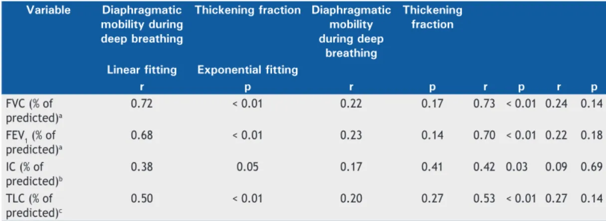
Table 3. Pulmonary function test variables in correlation with diaphragmatic mobility during deep breathing and with
the thickening fraction (proportional diaphragm thickening from functional residual capacity to total lung capacity). Variable Diaphragmatic
mobility during deep breathing
Thickening fraction Diaphragmatic mobility during deep
breathing
Thickening fraction
Linear fitting Exponential fitting
r p r p r p r p
FVC (% of predicted)a
0.72 < 0.01 0.22 0.17 0.73 < 0.01 0.24 0.14
FEV1 (% of predicted)a
0.68 < 0.01 0.23 0.14 0.70 < 0.01 0.22 0.18
IC (% of predicted)b
0.38 0.05 0.17 0.41 0.42 0.03 0.09 0.69
TLC (% of predicted)c
0.50 < 0.01 0.20 0.27 0.53 < 0.01 0.27 0.14
IC: inspiratory capacity; and TLC: total lung capacity. an = 40. bn = 25. cn = 31.
Figure 1. Linear and exponential correlations between
diaphragmatic mobility during deep breathing and FVC as a percentage of the predicted value.
10.0
9.0
8.0
7.0
6.0
5.0
4.0
3.0
2.0
1.0
.0
20 40 60 80 100
Diaphragmatic mobility during deep breathing (cm)
FVC (% of predicted)
be surprising that diaphragmatic mobility is reduced under these conditions of reduced lung volumes. However, the relationship between pulmonary volume and diaphragm excursion is debated and controversial in the literature. Cohen et al.,(34) studying ten normal
subjects, recorded simultaneously the diaphragmatic excursion (using ultrasound in M-mode) and the tidal volume at different inspiratory volumes. The authors
found that, at 15-87% of inspiratory capacity, there
was a linear relationship between diaphragmatic excursion and tidal volume.(34) Houston et al.,(35)
assessing hemidiaphragmatic movement, also found a linear relationship between inspiratory lung volume and diaphragmatic excursion. Another study, designed to validate ultrasound imaging of the diaphragm as an alternative to whole-body plethysmography, showed that the mean diaphragmatic excursion (during quiet breathing and at maximal sniff) correlated poorly with all of the lung volumes measured, although the authors did not investigate excursion during deep breathing.(36)
Fedullo et al.,(37) investigating diaphragmatic motion after
coronary artery bypass surgery, found no relationship between diaphragmatic mobility and vital capacity.
Similar to other studies that evaluated diaphragm thickness using ultrasound in other respiratory diseases, the present study showed that ILD patients presented
a thicker diaphragm at FRC than did healthy controls,
supporting the hypothesis of diaphragm thickening in response to respiratory muscle overload.(13,16) However,
the greater diaphragm thickness at FRC resulted in a lower TF, probably indicating dysfunctional muscular
hypertrophy or “pseudohypertrophy”, as has previously been demonstrated in the diaphragms of young patients with Duchenne muscular dystrophy.(38) In ILD, the
diaphragm appears to thicken over time but is unable to increase during maximal inspiration, unlike the physiological pattern seen in healthy controls.
The results of the present study demonstrate that ultrasound imaging of the diaphragm is a feasible method to evaluate diaphragmatic function. Diaphragmatic mobility is correlated with lung volume, a correlation that has been described in other lung diseases, such as COPD.(39) In clinical practice, ultrasound imaging of
the diaphragm has high sensitivity and speciicity to
identify reduced diaphragmatic mobility in ILD patients
with an FVC < 60% of the predicted value.
We showed that ultrasound imaging of the diaphragm
is associated with PFT variables and that PFT trends
constitute a major prognostic determinant of ILD. Therefore, we believe that ultrasound can be added to the arsenal of methods to follow up patients with ILD.(20) In addition to the chronic course of ILD, the
identiication of diaphragm dysfunction could alert
physicians to the need to avoid or change the use of drugs that induce myopathy, such as corticosteroids.
The main limitation of our study was that we did not measure diaphragm force. However, Ueki et al.(27) found
that a higher thickening ratio was strongly correlated with inspiratory strength in healthy subjects. Another study also showed a strong correlation between maximal
inspiratory pressure and the TF in patients recovering
from diaphragmatic paralysis.(40) In addition, because
our results show not only reduced diaphragmatic
mobility but also a reduced TF, we can conclude that
diaphragmatic dysfunction is common in ILD patients. Another limitation is that, although our study has a
power of 100% to estimate the coeficient of correlation between PFT and diaphragmatic mobility, it has a power of only approximately 40% to estimate that between PFT and diaphragm thickening.
In comparison with healthy controls, patients with ILD
show reduced diaphragmatic mobility and a lower TF.
Table 4. Comparison between interstitial lung disease patients with and without diaphragmatic dysfunction.
Variable Diaphragmatic
dysfunction
p Diaphragmatic
dysfunction
p
No Yes
(n = 16) (n = 24) OR (95% CI)
Age, years 61 ± 10 52 ± 17 0.04 −0.003 (−0.01 to 0.006) 0.82 Male gender, n (%) 11 (48) 12 (52) 0.32
Body mass index, kg/m2 27.4 ± 3.4 24.5 ± 4.9 0.04 −0.01 (−0.04 to 0.01) 0.30 FVC, % of predicted 70 ± 12 49 ± 13 < 0.01 −0.03 (−0.06 to −0.01) 0.04 FEV1, % of predicted 76 ± 14 53 ± 16 < 0.01 0.12 (−0.01 to 0.37) 0.34 Corticosteroid use, n (%) 6 (37) 11 (46) 0.75
aValues expressed as mean ± SD, except where otherwise indicated.
Figure 2. ROC curve of FVC as a percentage of the predicted value (FVC%) and the occurrence of decreased diaphragmatic mobility, showing the area under the curve (AUC).
0.0 0.2 0.4 0.6 0.8 1.0
1.0
0.8
0.6
0.4
0.2
0.0
1 − specificity
Sensitivity
In addition, the cut-off point of an FVC < 60% of the
predicted value showed high accuracy and sensitivity to identify decreased diaphragmatic mobility, as well
being signiicantly associated with impaired lung
function. The use of the results of ultrasound imaging of the diaphragm as a follow-up parameter and its relationship to respiratory muscle strength remain to be investigated.
REFERENCES
1. American Thoracic S, European Respiratory S. American Thoracic Society/European Respiratory Society International Multidisciplinary
Consensus Classiication of the Idiopathic Interstitial Pneumonias.
This joint statement of the American Thoracic Society (ATS), and the European Respiratory Society (ERS) was adopted by the ATS board of directors, June 2001 and by the ERS Executive Committee, June
2001. Am J Respir Crit Care Med. 2002;165(2):277-304. Erratum
in: Am J Respir Crit Care Med. 2002;166(3):426. http://dx.doi.
org/10.1164/ajrccm.165.2.ats01
2. Mendoza L, Gogali A, Shrikrishna D, Cavada G, Kemp SV, Natanek SA, et al. Quadriceps strength and endurance in ibrotic idiopathic interstitial pneumonia. Respirology. 2014;19(1):138-43. http://dx.doi.
org/10.1111/resp.12181
3. Spruit MA, Thomeer MJ, Gosselink R, Troosters T, Kasran A, Debrock AJ, et al. Skeletal muscle weakness in patients with sarcoidosis and its relationship with exercise intolerance and reduced health status.
Thorax. 2005;60(1):32-8. http://dx.doi.org/10.1136/thx.2004.022244 4. Watanabe F, Taniguchi H, Sakamoto K, Kondoh Y, Kimura T, Kataoka
K, et al. Quadriceps weakness contributes to exercise capacity in
nonspeciic interstitial pneumonia. Respir Med. 2013;107(4):622-8.
http://dx.doi.org/10.1016/j.rmed.2012.12.013
5. Elia D, Kelly JL, Martolini D, Renzoni EA, Boutou AK, Chetta A, et al. Respiratory muscle fatigue following exercise in patients with
interstitial lung disease. Respiration. 2013;85(3):220-7. http://dx.doi. org/10.1159/000338787
6. Hansen JE, Wasserman K. Pathophysiology of activity limitation in patients with interstitial lung disease. Chest. 1996;109(6):1566-76. http://dx.doi.org/10.1378/chest.109.6.1566
7. Holland AE. Exercise limitation in interstitial lung disease - mechanisms, signiicance and therapeutic options. Chron Respir Dis. 2010;7(2):101-11. http://dx.doi.org/10.1177/1479972309354689 8. O’Donnell DE, Chau LK, Webb KA. Qualitative aspects of exertional
dyspnea in patients with interstitial lung disease. J Appl Physiol
(1985). 1998;84(6):2000-9.
9. Walterspacher S, Schlager D, Walker DJ, Müller-Quernheim J,
Windisch W, Kabitz HJ. Respiratory muscle function in interstitial lung disease. Eur Respir J. 2013;42(1):211-9. http://dx.doi. org/10.1183/09031936.00109512
10. Dekhuijzen PN, Decramer M. Steroid-induced myopathy and its
signiicance to respiratory disease: a known disease rediscovered. Eur Respir J. 1992;5(8):997-1003.
11. Prasse A, Müller-Quernheim J. Non-invasive biomarkers in
pulmonary ibrosis. Respirology. 2009;14(6):788-95. http://dx.doi. org/10.1111/j.1440-1843.2009.01600.x
12. Baria MR, Shahgholi L, Sorenson EJ, Harper CJ, Lim KG, Strommen
JA, et al. B-mode ultrasound assessment of diaphragm structure and
function in patients with COPD. Chest. 2014;146(3):680-5. http://
dx.doi.org/10.1378/chest.13-2306
13. de Bruin PF, Ueki J, Watson A, Pride NB. Size and strength of the respiratory and quadriceps muscles in patients with chronic asthma.
Eur Respir J. 1997;10(1):59-64. http://dx.doi.org/10.1183/09031936. 97.10010059
14. DiNino E, Gartman EJ, Sethi JM, McCool FD. Diaphragm ultrasound as a predictor of successful extubation from mechanical
ventilation. Thorax. 2014;69(5):423-7. http://dx.doi.org/10.1136/
thoraxjnl-2013-204111
15. Dos Santos Yamaguti WP, Paulin E, Shibao S, Chammas MC, Salge
JM, Ribeiro M, et al. Air trapping: The major factor limiting diaphragm mobility in chronic obstructive pulmonary disease patients. Respirology. 2008;13(1):138-44.
http://dx.doi.org/10.1111/j.1440-1843.2007.01194.x
16. Dufresne V, Knoop C, Van Muylem A, Malfroot A, Lamotte M, Opdekamp C, et al. Effect of systemic inlammation on inspiratory and limb muscle strength and bulk in cystic ibrosis. Am J Respir Crit Care Med. 2009;180(2):153-8. http://dx.doi.org/10.1164/
rccm.200802-232OC
17. Gottesman E, McCool FD. Ultrasound evaluation of the paralyzed
diaphragm. Am J Respir Crit Care Med. 1997;155(5):1570-4. http:// dx.doi.org/10.1164/ajrccm.155.5.9154859
18. Kim WY, Suh HJ, Hong SB, Koh Y, Lim CM. Diaphragm dysfunction assessed by ultrasonography: inluence on weaning from mechanical ventilation. Crit Care Med. 2011;39(12):2627-30. http://dx.doi. org/10.1097/ccm.0b013e3182266408
19. Caruso P, Albuquerque AL, Santana PV, Cardenas LZ, Ferreira JG,
Prina E, et al. Diagnostic methods to assess inspiratory and expiratory
muscle strength. J Bras Pneumol. 2015;41(2):110-23. http://dx.doi. org/10.1590/S1806-37132015000004474
20. Latsi PI, du Bois RM, Nicholson AG, Colby TV, Bisirtzoglou D,
Nikolakopoulou A, et al. Fibrotic idiopathic interstitial pneumonia: the prognostic value of longitudinal functional trends. Am J Respir
Crit Care Med. 2003;168(5):531-7. http://dx.doi.org/10.1164/ rccm.200210-1245OC
21. Mahler DA, Weinberg DH, Wells CK, Feinstein AR. The measurement
of dyspnea. Contents, interobserver agreement, and physiologic
correlates of two new clinical indexes. Chest. 1984;85(6):751-8. http://dx.doi.org/10.1378/chest.85.6.751
22. Pereira CA, Sato T, Rodrigues SC. New reference values for forced
spirometry in white adults in Brazil. J Bras Pneumol. 2007;33(4):397-406. http://dx.doi.org/10.1590/S1806-37132007000400008 23. Boussuges A, Gole Y, Blanc P. Diaphragmatic motion studied by
m-mode ultrasonography: methods, reproducibility, and normal
values. Chest. 2009;135(2):391-400. http://dx.doi.org/10.1378/ chest.08-1541
24. Testa A, Soldati G, Giannuzzi R, Berardi S, Portale G, Gentiloni Silveri N. Ultrasound M-mode assessment of diaphragmatic kinetics by anterior transverse scanning in healthy subjects.
Ultrasound Med Biol. 2011;37(1):44-52. http://dx.doi.org/10.1016/j.
ultrasmedbio.2010.10.004
25. Boon AJ, Harper CJ, Ghahfarokhi LS, Strommen JA, Watson
JC, Sorenson EJ. Two-dimensional ultrasound imaging of the diaphragm: quantitative values in normal subjects. Muscle Nerve.
2013;47(6):884-9. http://dx.doi.org/10.1002/mus.23702
26. Cohn D, Benditt JO, Eveloff S, McCool FD. Diaphragm thickening
during inspiration. J Appl Physiol (1985). 1997;83(1):291-6.
27. Ueki J, De Bruin PF, Pride NB. In vivo assessment of diaphragm contraction by ultrasound in normal subjects. Thorax.
1995;50(11):1157-61. http://dx.doi.org/10.1136/thx.50.11.1157 28. Pinet C, Cassart M, Scillia P, Lamotte M, Knoop C, Casimir G, et al.
Function and bulk of respiratory and limb muscles in patients with
cystic ibrosis. Am J Respir Crit Care Med. 2003;168(8):989-94. http://dx.doi.org/10.1164/rccm.200303-398OC
29. He L, Zhang W, Zhang J, Cao L, Gong L, Ma J, et al. Diaphragmatic
motion studied by M-mode ultrasonography in combined pulmonary
ibrosis and emphysema. Lung. 2014;192(4):553-61. http://dx.doi. org/10.1007/s00408-014-9594-5
30. de Troyer A, Yernault JC. Inspiratory muscle force in normal subjects and patients with interstitial lung disease. Thorax. 1980;35(2):92-100. http://dx.doi.org/10.1136/thx.35.2.92
31. Garcia-Rio F, Pino JM, Ruiz A, Diaz S, Prados C, Villamor J.
Accuracy of noninvasive estimates of respiratory muscle effort during spontaneous breathing in restrictive diseases. J Appl
Physiol (1985). 2003;95(4):1542-9. http://dx.doi.org/10.1152/
japplphysiol.01010.2002
32. Nishimura Y, Hida W, Taguchi O, Sakurai M, Ichinose M, Inoue H, et
al. Respiratory muscle strength and gas exchange in neuromuscular diseases: comparison with chronic pulmonary emphysema and
idiopathic pulmonary ibrosis. Tohoku J Exp Med. 1989;159(1):57-68. http://dx.doi.org/10.1620/tjem.159.57
33. De Troyer A. Effect of hyperinlation on the diaphragm. Eur Respir J. 1997;10(3):708-13.
34. Cohen E, Mier A, Heywood P, Murphy K, Boultbee J, Guz A.
Excursion-volume relation of the right hemidiaphragm measured
35. Houston JG, Angus RM, Cowan MD, McMillan NC, Thomson NC.
Ultrasound assessment of normal hemidiaphragmatic movement:
relation to inspiratory volume. Thorax. 1994;49(5):500-3. http://dx.doi. org/10.1136/thx.49.5.500
36. Scott S, Fuld JP, Carter R, McEntegart M, MacFarlane NG. Diaphragm ultrasonography as an alternative to whole-body plethysmography in
pulmonary function testing. J Ultrasound Med. 2006;25(2):225-32. 37. Fedullo AJ, Lerner RM, Gibson J, Shayne DS. Sonographic
measurement of diaphragmatic motion after coronary artery bypass
surgery. Chest. 1992;102(6):1683-6. http://dx.doi.org/10.1378/
chest.102.6.1683
38. De Bruin PF, Ueki J, Bush A, Khan Y, Watson A, Pride NB. Diaphragm
thickness and inspiratory strength in patients with Duchenne
muscular dystrophy. Thorax. 1997;52(5):472-5. http://dx.doi. org/10.1136/thx.52.5.472
39. Levine S, Nguyen T, Kaiser LR, Rubinstein NA, Maislin G, Gregory
C, et al. Human diaphragm remodeling associated with chronic
obstructive pulmonary disease: clinical implications. Am J Respir Crit Care Med. 2003;168(6):706-13. http://dx.doi.org/10.1164/
rccm.200209-1070OC
40. Summerhill EM, El-Sameed YA, Glidden TJ, McCool FD. Monitoring