Received from Hospital Biocor, Belo Horizonte, MG, Brazil.
1. MSc from Universidade Federal de Minas Gerais (UFMG); Anesthesiologist of Hospital Biocor
2. R3 in Anesthesiology of Hospital Júlia Kubistchek Submitted on September 8, 2010.
Approved on February 21, 2011. Correspondence to:
Dra. Michelle Nacur Lorentz Rua da Paisagem, 290, 6° andar Nova Lima
34000000 – Belo Horizonte, MG, Brazil E-mail: [email protected]
REVIEW ARTICLE
Cardiac Dysrhythmias and Anesthesia
Michelle Nacur Lorentz, TSA
1, Bruna Silviano Brandão Vianna
2Summary: Lorentz MN, Vianna BSB – Cardiac Dysrhythmias and Anesthesia.
Background and objectives: Cardiac dysrhythmias are relatively common in the perioperative period and should be adequately diagnosed and treated by the anesthesiologist whenever indicated. The objective of this article was to review the most relevant aspects of cardiac dysrhythmias, as well as establishing the cause-effect relationship between drugs used in the perioperative period and dysrhythmias.
Contents: The mechanisms of dysrhythmias, drugs that can potentially cause dysrhythmias, besides diagnosis and treatment in the perioperative period are presented.
Conclusions: Perioperative dysrhythmias oftentimes do not require treatment and in others the treatment can generate iatrogenicity. Therefore, the knowledge of cardiac dysrhythmias and triggering factors allows a better approach of the perioperative period by the anesthesiologist avoiding wrong or unnecessary treatment.
Keywords: Anesthesia; Arrhythmias, Cardiac.
©2011 Elsevier Editora Ltda. All rights reserved.
INTRODUCTION
Cardiac dysrhythmias and anesthesia
Dysrhythmias represent an important cause of perioperative complications because during this period there are several clini cal situations that may trigger changes in cardiac rhythm 1. These rhythm changes may be due to a primary etiology or to reversible causes that should be corrected. The prevalence of cardiac dysrhythmias varies according to the literature, type of surgical procedure, and the patient. In a multicenter study with 17,201 patients undergoing general anesthesia, dysrhythmias (tachycardia, bradycardia, or other dysrhythmias) were obser-ved in 70.2%, of which only 1.6% required treatment 2. A lar-ge number of patients undergoing non-cardiac surlar-geries may develop dysrhythmias 3,4 and the incidence of atrial fibrillation (AF) is low after exploratory thoracotomy, but in elderly pa-tients undergoing lobectomy, pneumectomy, and esophago-gastrectomy the incidence rises to 12% to 33% 4.
Note that antidysrhythmic drugs can also cause dys-rhythmias and oftentimes the anesthesiologist in an attempt
to treat perioperative dysrhythmia can cause iatrogenesis, and as such the knowledge of the physiology of the cardiac rhythm, anesthetic pharmacology, and risk-benefits of antidys-rhythmic drugs is mandatory. Most perioperative dysrhythmias are benign, without significant hemodynamic consequences. Symptomatic patients, whose dysrhythmias can evolve to life-threatening malignant dysrhythmias should be treated with antidysrhythmic drugs or electrotherapy 5.
Etiology
In children the main causes of dysrhythmias include congenital cardiopathies, cardiomyopathies, and myocardial inflamma-tory disorders 6. However, there are patients with premature ventricular contractions (PVCs) whose hearts are structurally normal. The incidence of benign PVCs has a biphasic distri-bution with a peak of approximately 15% in the first weeks of life decreasing to < 5% before adolescence, with a gradual in-crease in adolescents 6. In the pediatric population with struc-turally normal myocardium sustained ventricular PVCs are re-latively rare. Benign dysrhythmias usually have sinus rhythm, repolarization, normal ventricular function, and, in general, patients do not have a significant family history of mortality.
is greater 4. Development of supraventricular tachycardia (SVT) or non-sustained ventricular tachycardia (VT) can be secondary to hypoxemia, hypercarbia, acidosis, hypotension, electrolyte imbalance, mechanical irritation, pulmonary artery catheter, thoracic tube, hypothermia, micro- or macroshocks, adrenergic stimulation (e.g., superficial anesthesia), use of dysrhythmogenic drugs, and myocardial ischemia. Periope-rative dysrhythmias are usually reversible and before being treated the most common causes should be excluded.
Mechanisms of dysrhythmias
Dysrhythmias are secondary to changes in cardiac ion chan-nels (sodium, calcium, and potassium chanchan-nels) and adrener-gic receptors are the targets. To better understand the mecha-nism of dysrhythmias and antidysrhythmic agents, one should remember that the action potential is divided in five phases (0 to 4) 7. The initial period of the action potential corresponds to phase 0 and it initiates the conduction in the cardiac tissue. In atria and ventricles the impulse originates in the sodium current. In the sinoatrial (SA) and atrioventricular (AV) nodes phase 0 is produced by calcium current. Phases 1, 2, and 3 represent repolarization; the plateau (maintained by calcium current) is phase 2, and its end (phase 3) is maintained by potassium current. During phase 4, nodal cells undergo spon-taneous depolarization while atrial and ventricular tissues are hyperpolarized.
Dysrhythmias may be due to changes in the formation of the electric impulse (automaticity) or in conduction. Abnormal impulse generation can occur in the sinus node or in ectopic foci. Automaticity refers to abnormal atrial or ventricular depo-larization during the repodepo-larization (phases 2 or 3) or resting (phase 4) period of the action potential 6. Some molecular substrates, such as prolongation of the QT interval and low potassium (K+) concentrations can trigger automaticity. Muta-tions in ion channels responsible for repolarization, and that can prolong it, make cardiac cells more sensitive to dysrhyth-mias 8. Factors that increase automaticity include increased activity of the sympathetic nervous system, hypokalemia, hypomagnesemia, catecholamines, digoxin, hypoxemia, and atrial and ventricular dilation 9. Besides the abnormal gene-ration of impulses, abnormal conduction (reentry) is also res-ponsible for dysrhythmias.
Three factors must be present for reentry to occur: 1) pre-sence of two conduction pathways; 2) unidirectional blockade of one of the pathways prevents progression of the impulse, but is allows retrograde conduction; and 3) reduced impul-se velocity in one of the pathways giving time for the other pathway to depolarize. Reentry is the mechanism of several supraventricular and ventricular dysrhythmias implying the presence of a pathologic circuit of electrical impulse around a functional or anatomic loop, which is seen in Wolff-Parkin-son-White syndrome (WPW). Ischemia also predisposes the development of reentry tachycardia. Drugs that terminate re-entry do this by two mechanisms: suppression of the current responsible for phase 0 of the action potential that prolongs
or blocks conduction in the reentry pathway, interrupting the dysrhythmia. Drugs that prolong the action potential (with K+-channel blocking properties) prolong cellular and reentry circuit refractory period, blocking propagation of impulses through the circuit. Reentry is responsible for 90% of SVTs in children. The main mechanism of monomorphic VT is also reentry around the infracted myocardium.
Risk factors for dysrhythmias
Risk factors for the development of dysrhythmias can be clas-sified as modifiable and non-modifiable. The non-modifiable risk factors include dilated cardiac diseases, ischemic car-diomyopathy, autonomic changes of the conduction system, polymorphism of ion channels, or congenital long-QT syndro-me (LQTS). Among modifiable factors there are electrolyte changes: K+ changes can generate increases in automaticity and abnormal formation of impulses. Changes in serum K+ le-vels are closely related with the development of dysrhythmias, and abrupt changes are less tolerable than chronic changes. The relationship between preoperative K+ and perioperati-ve adperioperati-verse eperioperati-vents was demonstrated by Wahr et al.10, and serum K+ levels below 3.5 mEq.L-1 can predict the develop-ment of perioperative dysrhythmias.
Magnesium is important for several physiologic functions activating ATPase and promoting transport of cations, such as calcium and potassium 11. Severe hypomagnesemia in-creases automaticity and predisposes to the development of
torsades de pointes (TdP). Although magnesium deficiency
can contribute for several dysrhythmias, especially after car-diac surgeries 12,13, and magnesium seems effective in de-creasing catecholamine-induced dysrhythmias 14,15, TdP is the only dysrhythmia in which magnesium has been proven to be effective 16. Hypomagnesemia usually is concomitant with hypokalemia and hypocalcemia, making it difficult to ade-quately replace K+ or calcium without replacing magnesium. Hypermagnesemia can cause bradycardia, first degree atrio-ventricular block, and increase the QT interval.
Differential diagnosis of dysrhythmias
Supraventricular tachycardia can be defined as sustained, non-sinus related, acceleration of the cardiac rhythm, origi-nating above the AV node. On the other hand, autonomic ta-chycardia is rare and can be defined as a tata-chycardia initiated and sustained by an ectopic focus. Automatic atrial tachycar-dia is a type of automatic tachycartachycar-dia that involves primarily the atrial tissue. The continuous type is usually symptomatic and often results from dilated cardiomyopathy. The repetitive type is frequently interrupted by periods of sinus rhythm, is less severe, and only becomes symptomatic during periods of very fast heart rate (HR).
re-gular tachycardia represent a diagnostic and treatment chall-enge. History, factors like history of AMI, physical exam, and electrocardiographic (ECG) findings, such as AV dissociation are useful to distinguish the origin of the tachycardia, although none of these factors is sensitive and specific. Adenosine is useful to classify SVTs because tachycardia caused by atrial reentry such as AF or flutter have transient response of de-creased frequency after adenosine, but it does not terminate the dysrhythmia; on the other hand, SVTs due to reentry in the AV node ceases after adenosine. Ventricular dysrhythmias do not respond to adenosine because these dysrhythmias ori-ginate in distal tissues of the conduction system. This also allows the use of adenosine to distinguish between wide-com-plex SVT and VT, since adenosine causes rapid AV block, with a half-life of 9 seconds, and terminates most SVTs due to reentry; on the other hand, it causes transient AV block and is incapable of terminate most VT, but since adenosine has a brief effect, in case of VT it does not cause major problems.
However, the safety of adenosine has been questioned and, according to the 2005 guidelines of the American Heart Association 17, it should not be used for differential diagno-sis between wide-complex SVT and VT due to its vasodilator properties, bronchospasm, paradoxal increase in conduction in the accessory pathway, persistent bradycardia or asystole, and degeneration to ventricular fibrillation (VF). In contrast, in a study by Marril et al. 18, in which consecutive patients were treated with adenosine between 1991 and 2006, the authors observed that adenosine is useful and safe to distinguish bet-ween VT and SVT in patients with stable and regular tachy-cardia. Besides, it has been postulated that in these cases adenosine would be useful to prevent treatment of VT with a long-acting AV node blocker, which would be risky since it causes vasodilation and longer-lasting preload reduction.
Non-sustained ventricular dysrhythmias can be divided according to their morphology (monomorphic or polymorphic) and duration (sustained or non-sustained). The main mecha-nism of monomorphic VT is the formation of a reentrant pa-thway around a tissue scar of a healed myocardial infarction. In monomorphic VT the amplitude of the QRS complex is constant, while in polymorphic VT the morphology of the QRS changes continuously. Non-sustained ventricular tachycardia (NSVT) is defined as three or more premature ventricular con-tractions with a frequency higher than 100 beats per minute (BPM) lasting 30 seconds or less without hemodynamic com-promise. These dysrhythmias are routinely seen in the ab-sence of cardiac disease and might not require drug therapy in the perioperative period. Non-sustained ventricular tachy-cardia is seen in approximately 50% of patients during or after large size cardiac or vascular surgeries and they do not affect late mortality in patients without ventricular dysfunction.
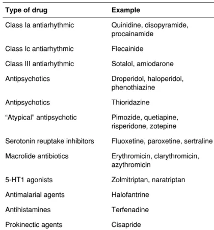
Drugs that prolong the QT interval
Normal QT interval measures up to 450 msecs. Approxima-tely 60 medications increase the QT interval and can cause TdP. Type Ia (for example, quinidina) and type III
antiarhyth-mics can cause TdP. Type Ib and Ic antiarhythantiarhyth-mics rarely cause TdP 19-25. Despite its ability to prolong the QT interval amiodarone only causes TdP in 1% of patients when com-pared to other type III drugs that can cause TdP in 2% to 4% of patients. Drugs that precipitate TdP delay repolarization, allowing formation of several reentry sites (Table I). Risk fac-tors for induction of TdP include bradycardia, recent cardio-version of AF, heart failure, hypomagnesemia, hypokalemia, digoxin, rapid infusion of drugs that increase the QT interval, long-QT, female gender, LQTS, and ion channel polymor-phism 26,27. In patients receiving drugs that increase the QT interval, QT interval monitoring is indicated for prophylaxis of TdP 26,27. To correct the QT interval, which is influenced by HR and gender, and to find the corrected QT (QTc), Fridericia (QTc = QT/3RR) or Bazett (QTc = QT/RR) formulas are used. Bazett correction is used more often, but it can generate hy-percorrection in short RR intervals and hyhy-percorrection in long RR intervals 28.
Prodysrhythmia
A term used to describe antiarhythmic drug-induced dysrhyth-mia 29. Vaughan Williams classification 30 divides antiarhyth-mic drugs into four classes according to their effects on ion channels. Singh 25 reviewed the drugs used to treat dysrhyth-mias and found that the use of class Ia drugs are declining; class Ib drugs such as lidocaine are still widely used in the pe-rioperative period; while class Ic drugs should not be used in patients with structural cardiomyopathies due to the high risk
Table I – Drugs that Affect Repolarization, Prolong the QT Interval, with Documented Cases of Torsades de Pointes
Type of drug Example
Class Ia antiarhythmic Quinidine, disopyramide, procainamide
Class Ic antiarhythmic Flecainide
Class III antiarhythmic Sotalol, amiodarone
Antipsychotics Droperidol, haloperidol, phenothiazine
Antipsychotics Thioridazine
“Atypical” antipsychotic Pimozide, quetiapine, risperidone, zotepine
Serotonin reuptake inhibitors Fluoxetine, paroxetine, sertraline
Macrolide antibiotics Erythromicin, clarythromicin, azythromicin
5-HT1 agonists Zolmitriptan, naratriptan
Antimalarial agents Halofantrine
Antihistamines Terfenadine
of prodysrhythmia. In patients after myocardial infarction slow conduction due to class Ic antiarhythmics leads to an increa-se in reentry allowing the development of VT and, therefore, class Ic drugs should not be used in patients with ischemia. Class Ia drugs can also slow down conduction and prolong repolarization, and may cause TdP. Procainamide, sotalol, and bipyridyl can cause TdP. Some antiarhythmic drugs such as verapamil and amiodarone can occasionally lead to TdP, although amiodarone prolongs the QT interval by more than 500 msecs and, despite this, the development of TdP is rare.
Class Ib drugs (lidocaine and mexiletine) are more selecti-ve to abnormal or damaged myocytes and are not associated with prodysrhythmia. Class III agents (amiodarone, sotalol, and dofetilide) block potassium channels prolonging repolari-zation, and have the potential to induce TdP. Although this is observed with sotalol and dofetilide, it rarely occurs with amio-darone, which is considered safe and neutral in patients after myocardial infarction. Amiodarone also has class II (blocking adrenergic receptors) and class IV (calcium blockers) proper-ties and it can cause bradycardia. Beta blockers (class II) may be used to treat SVTs, as well as calcium channel blockers (class IV), and they do not have a great risk of prodysrhythmic phenomena. Class III drugs such as amiodarone and sota-lol prolong the action potential and, currently, are replacing class I drugs due to the inherent risk of these drugs for cau-sing dysrhythmias. In reality, all antiarhythmic drugs have the potential to generate dysrhythmias and for this reason care should be taken when using them, especially in patients with cardiac abnormalities and electrolytic changes 31,32.
Dysrhythmia related to drugs used in the perioperative period
Propofol, nitrous oxide (N2O), and sevoflurane have little arr-hythmogenic potential and very few side effects 31. Halothane is not a good choice in the presence of dysrhythmias. Isoflura-ne causes ventricular dysrhythmias in 2.5% of patients, while desflurane increases the HR. Sevoflurane, halothane, and isoflurane can delay ventricular repolarization and prolong the QT interval. Propofol does not change the HR, has little effect on cardiac conduction, and, whether it is associated with a negative inotropism, this is due to a reduction in sympathetic tonus and increase in parasympathetic sensitivity. Ketamine causes node dysrhythmia and decreases contractility, but the HR can increase.
Opioids except meperidine reduce the HR through a cen-tral mechanism that reduces sympathetic tonus and increa-ses vagal tonus 31. Fentanyl has a direct effect on SA node. Midazolam has a biphasic effect on HR, since it affects the sympathetic and parasympathetic nervous system and its effects on dysrhythmias are not clear. Bronchodilators stimu-late adrenergic receptors, increasing the risk of cardiovascu-lar events; in patients with baseline tachycardia these drugs have the potential to exacerbate dysrhythmias. Although le-valbuterol has been developed to avoid the cardiovascular effects of albuterol, tachydysrhythmias still occur in 2.7% of
patients. Drugs that induce bradydysrhythmias include beta-blockers, calcium channel beta-blockers, amiodarone, clonidine, and dexmedetomidine. Bradycardia is seen in 5% of patients with dexmedetomidine and this agent should be avoided in patients with heart block.
Droperidol has been associated with ventricular dysr-hythmias 32 and in 2001 it was placed on the Food and Drug Administration’s (FDA) black list, as it is associated with pro-longed QT interval and malignant dysrhythmias such as TdP. The relative risk of dysrhythmias with droperidol compared to other antiemetic drugs or placebo is not clear; therefore, recommendation for droperidol requires 12-lead ECG befo-re administration and continuous ECG monitoring for 2 to 3 hours after administration. If the QTc is prolonged in ba-seline ECG, droperidol administration is not recommended. Extreme caution is recommended when using droperidol in patients with risk factors for prolonged QT interval, such as congestive heart failure, bradycardia, use of diuretics, ven-tricular hypertrophy, hypokalemia, hypomagnesemia, or use of drugs that prolog the QT interval. Droperidol should be ini-tiated in low doses and adjusted until the desired results are obtained.
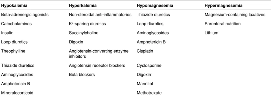
Inotropics, such as dobutamine and milrinone, may preci-pitate arrhythmias and ventricular fibrillation. Dobutamine is directly dysrhythmogenic, causing dose-dependent tachycar-dia; doses higher than 5 µg.kg-1.min-1 are prone to cause dys-rhythmias and generate little benefit in oxygen transportation. Milrinone increases the inotropism without activating adrener-gic receptors, but it also has a dysrhythmic potential. Digoxin increases intracellular calcium in cardiac myocytes, slows AV node conduction, and can increase automaticity, triggering any type of dysrhythmia, the most common being premature ventricular contraction, heart blocks of any degree (although rarely Mobitz type II), paroxysmal atrial tachycardia with heart block, accelerated junctional rhythm, and bidirectional VT. Risk factors for digoxin intoxication include renal dysfunction and electrolyte imbalance, such as hypokalemia, hypomag-nesemia, and hypercalcemia. Drugs that cause electrolyte imbalances have the potential to cause dysrhythmias and are on Table II.
Treatment of dysrhythmias
Table II – Drugs that Cause Electrolyte Abnormalities
Hypokalemia Hyperkalemia Hypomagnesemia Hypermagnesemia
Beta-adrenergic agonists Non-steroidal anti-inflammatories Thiazide diuretics Magnesium-containing laxatives
Catecholamines K+-sparing diuretics Loop diuretics Parenteral nutrition
Insulin Succinylcholine Aminoglycosides Lithium
Loop diuretics Digoxin Amphotericin B
Theophylline Angiotensin-converting enzyme inhibitors
Cisplatin
Thiazide diuretics Angiotensin receptor blockers Cyclosporine
Aminoglycosides Beta blockers Digoxin
Amphotericin B Mannitol
Mineralocorticoid Methotrexate
Table III – Electrophysiological Changes of Anesthetics
Drug Action Effects
Inhalational Antagonize calcium and increase depolarization in Purkinje fibers
Junctional rhythm, atrioventricular asynchrony
Propofol Stimulate muscarinic receptors Bradycardia
Succynilcholine Activate muscarinic or nicotinic receptors Tachycardia or bradycardia, it can lead to asystole
Pancuronim Increase catecholamines and automatism Tachycardia
Vecuronium Decrease automatism by sympathetic blockade Bradycardia and junctional rhythm
Local Anesthetics Block calcium channels Widening of the QRS, Tachycardia, and VF
Opioids Decrease frequency of the SA node Bradycardia
Prolong AV conduction
Ketamine Increase SA node frequency by sympathetic activation
Tachycardia
Clonidine and Dexmedetomidine Sympathetic blockade Bradycardia
Table IV – Interaction between Anesthetics and Antiarhythmics
Adenosine Vasodilation with isoflurane and neuraxial block, Bronchoconstriction with neostigmine,
Asystole with neostigmine, dexmedetomidine and opioids, Antagonism with aminophylline
Amiodarone Myocardial depression and vasodilation with inhalational agents
Digoxin Bradycardia is potentiated by halothane and succinylcholine,
Care should be taken when administering calcium and using diuretics (hypokalemia)
Beta blocker Myocardial depression is potentiated by halothane, Bronchoconstriction with neostigmine and atracurium
Quinidine Prolongs the effects of neuromuscular blockers (NMB)
Procainamide Antagonizes neostigmine,
Ventricular dysrhythmias when combined with phenotiazides
Calcium channel blocker Bradycardia and myocardial depression with halogenated agents and dantrolene, Potentiates NMB
Magnesium Prolongs the action of NMB
should be used with caution, and correction of electrolyte im-balances and prevention of bradycardia are required in the management of dysrhythmias 33.
Sinus tachycardia
Tachycardia increases myocardial oxygen consumption ge-nerating ischemic episodes and increasing mortality, therefo-re, it should be treated. Beta blockers can be used for this purpose and during anesthesia drugs with short half-life and continuous infusion such as esmolol should be used.
Recently, Landiolol has been introduced into clinical prac-tice in some countries. It is a beta-blocker whose cardioselec-tive properties are greater than the esmolol and has a shorter half-life (2 to 4 minutes), as it is rapidly hydrolyzed by plas-ma esterases. Harasawa et al. 34 administered Landiolol to treat tachycardia during anesthesia and to obtain protection against myocardial ischemia to evaluate its dose-dependent bolus effects in doses of 0.1, 0.2, or 0.3 mg.kg-1. They did not observe hypotension, bradycardia, or ischemic changes on ECG with 0.1 mg.kg-1, and for this reason the authors su-ggested that this would be the optimal dose to reduce the effects of tachycardia. On the other hand, higher doses such as 0.3 mg.kg-1 caused a reduction in HR and blood pressure and they can be used in patients with tachycardia and hyper-tension in response to surgical stress 34. In a case report by Chrysostomou et al. 35 of dexmedetomidine 0.3 mg.kg-1 used to treat persistent sinus tachycardia resistant to treatment with esmolol the authors found no side effects, therefore, they su-ggested that this drug could be useful in patients with bron-chospasm and tachycardia.
Supraventricular tachycardia (SVT)
Perioperative SVTs should be initially considered as a sign of potentially life-threatening underlying clinical condition. The-refore, the initial conduct is to look for an underlying cause, usually procedure-related, and possible repercussions. The-se conditions are often reversible and antiarhythmics drugs should be considered only after these etiologies have been ruled out. Another fundamental step in the management of dysrhythmias in general is recognizing symptoms of dysrhyth-mia-related hemodynamic instability such as hypotension, changes in the level of consciousness, thoracic pain, or any other sign of poor tissue perfusion. In such cases, electrical cardioversion is the initial conduct.
Remember that perioperative cardioversion may not have maximum effectiveness or even may not be able to maintain an organized rhythm for adequate time, taking into conside-ration that cardioversion itself does not reverse the baseline cause of dysrhythmias. But cardioversion is indicated in any case of tachydysrhythmia with hemodynamic repercussions and in the perioperative period it can be useful in that, during a small period of sinus rhythm, there might be time to initiate measures to reverse the baseline cause 7. A study of patients with SVTs (especially AF) undergoing cardiac surgery with
extracorporeal circulation demonstrated that electrical low-energy cardioversion was effective in 80% of cases, but the recurrence rate after 1 minute was greater than 50% 36. It has been recommended, when deciding for perioperative electi-ve electrical cardioelecti-version that initially an attack dose of an antiarhythmic agent should be administered to minimize the recurrence of SVT after the shock. Adenosine is the drug of choice to treat SVTs involving the SA or AV node (6 mg IV rapid flush, which can be repeated as 12 mg bolus), important especially in cases of node reentry, which is not the main me-chanisms of perioperative SVTs.
Most patients with perioperative SVTs maintain hemody-namic stability and, therefore, they do not need immediate electrical cardioversion. For this reason, heart rate control is the most important conduct in the treatment of this disorder; for this end, AV node blockers, such as beta blockers and calcium channel blockers (class II or IV), are used. Among IV beta-blockers, esmolol, due to its pharmacologic characteris-tics (short half-life and easy titration) is the drug of choice in the perioperative period. Verapamil and diltiazem also promo-te fast ventricular rapromo-te control in SVTs. Intravenous diltiazem has less negative inotropic effects when compared with vera-pamil, being preferable in patients with heart failure. In these patients, diltiazem, digitalis, and amiodarone are the recom-mended drugs to control heart rate.
A prospective randomized study compared the efficacy of diltiazem versus amiodarone to control HR in patients with atrial tachycardia and HR > 120 bpm. Diltiazem showed better HR control than amiodarone; however, it was associated with a higher rate of hypotension 37. In patients with history of SVT due to the presence of an anomalous pathway (WPW) the use of AV node blockers is contraindicated due to the in creased risk of developing malignant ventricular dysrhythmias. These rugs accentuate the refractory period of the anomalous pa-thway. In this case, it is possible to use procainamide and amiodarone 7. Chemical cardioversion of these perioperative dysrhythmias has little significance, being reserved for the cases of failure to control heart rate or absence of reversion and hemodynamic instability with electrical cardioversion. The efficacy of antiarhythmics in cardioversion is questioned in several studies, as several patients showed reversion of the rhythm with the use of placebo in randomized studies. A randomized clinical assay demonstrated 60% conversion in 24 hours in the placebo group compared with 68% in the amiodarone group 38.
Even knowing that with high doses of amiodarone the rate of chemical conversion is considerable, the potential for postoperative side effects should be considered to establish the risk-benefit ratio. Drugs like procainamide and amioda-rone may be useful to treat any of those dysrhythmias, but they may be ineffective in some cases, in addition to having adverse effects, especially procainamide currently used in ex-ceptional cases.
AF and they can represent important adjuvant therapy in the prevention of this dysrhythmia 39. Amiodarone can also be effective after cardiac surgeries, although a study questioned its prophylactic efficacy post-thoracotomy. In case of AF, stra-tegies to reduce HR should be adopted in the first 24 hours as more than 85% of the episodes will resolve within this period. Class Ic or III antiarhythmics can be used.
Regarding prophylaxis of AF, a perioperative cardiopa-thy very common in cardiac surgeries, Beaulieu et al. 40 in a randomized prospective study of 120 patients who received amiodarone or placebo for prevention of AF in this type of surgery concluded that amiodarone was not effective to pre-vent AF.
VENTRICULAR TACHYCARDIA (VT)
Non-sustained ventricular tachycardia (NSVT)
It is a common perioperative occurrence and in the absence of cardiac disease it does not require treatment. In patients with cardiopathies this non-sustained rhythm can predict ma-lignant ventricular dysrhythmias. The main strategy in NSVT would be prevention, instituting immediate treatment when possible risk factors arise 41. A study of patients undergoing cardiac surgery demonstrated that correction of magnesium after ECC reduced the incidence of non-sustained VT 12.
Sustained ventricular tachycardia
It is divided into two categories: monomorphic or polymorphic. Lidocaine, procainamide, or amiodarone are used in mono-morphic VT 42. The management in polymorphic ventricular tachycardia is based on the prior presence of a prolonged QT interval and consequent development of TdP. In such cases, the treatment of dysrhythmia consists of reversing the prolon-ged QT interval and especially discontinuation of drugs that can cause increased QT.
The approach of TdP may include cardioversion, although magnesium is the treatment of choice (2 g administered IV slowly). Because TdP is usually recurrent, efforts should be made to increase HR between 150 and 120 bpm through pa-cemaker or inotropic drugs. Drugs that prolong repolarization time such as procainamide are contraindicated during TdP. Discontinuation of drugs that prolong the QT interval and co-rrection of electrolyte is usually necessary. In refractory high risk patients, one should consider placing a pacemaker and defibrillator. In cases in which there is doubt if polymorphic VT is the result of prolonged QT interval, it is recommended using a channel blocking drug and replace magnesium empirically 7. Among the drugs that prolong the QT interval, the incidence of
TdP with amiodarone is smaller; therefore, amiodarone IV is a good choice as an alternative therapy for refractory polymor-phic VT of unknown etiology.
Unstable VT, pulseless VT, VF
The main maneuvers for patients with perioperative unstable VT, pulseless VT, or VF are not pharmacological. They are immediate defibrillation, cardiopulmonary resuscitation (CPR) in case of cardiorespiratory arrest, and correction of reversible causes.
CONCLUSION