Rev Bras Anestesiol SCIENTIFIC ARTICLE 2011; 61: 6: 713-719
Revista Brasileira de Anestesiologia 713
Vol. 61, No 6, November-December, 2011
Received from Faculdade de Ciências Médicas da Santa Casa de São Paulo (FCM-SCSP); Irmandade da Santa Casa de Misericórdia de São Paulo (ISCMSP), Brazil.
1. MSc in Medicine from Faculdade de Ciências Médicas da Santa Casa de São Paulo (FCM-SCSP); Assistant Physician of Irmandade da Santa Casa de Misericórdia de São Paulo (ISCMSP); Co-responsible for the CET-SCSP
2. Associate Professor of FCM-SCSP; Director of the Anesthesiology Department and Serv-ice of ISCMSP
3. Professor of FCM-SCSP; Chief of Thoracic Surgery Department of ISCMSP
Submitted on November 11, 2010. Approved on April 4, 2011.
Correspondence to:
Dra. Ligia Andrade da S. Telles Mathias Alameda Campinas 139/41 01404000 – São Paulo, SP, Brazil E-mail: [email protected] SCIENTIFIC ARTICLE
The Influence of Posture on Spirometric Values in Grade III
Obese Patients
Ayrton Bentes Teixeira, TSA
1, Ligia Andrade da S. Telles Mathias, TSA
2, Roberto Saad Junior
3Summary: Teixeira AB, Mathias LAST, Saad Junior R – The Influence of Posture on Spirometric Values in Grade III Obese Patients.
Background and objectives: The change from the sitting position to supine position, general anesthesia, and surgical procedure reduce lung volumes and this effect can be greater in obese patients. The objective of the present study was to evaluate the influence of the sitting position, 30° dorsal inclination, and horizontal dorsal decubitus on spirometry of grade III obese patients.
Methods: Twenty-six adult patients in the preoperatory period were selected according to the following criteria: BMI > 40 kg.m-2, age between18
years and 60 years, and female gender. Variables analyzed included: age, weight, height, BMI, percentage of predictive values of FVC, FEV1, and
VEF1/FVC in the sitting position (90o), 30o dorsal elevation, and horizontal dorsal decubitus (0o). ANOVA, followed or not by Tukey test were used
to compare mean predicted values on the different positions, considering significant a p value lower than 0.05.
Results: Percentage values of FVC, FEV1, and FEV1/FVC ratio regarding predicted values in the sitting position (90o), 30o dorsal elevation,
and horizontal dorsal decubitus (0o), and p value of the corresponding statistical analysis were, respectively: FVC = 92.8%, 88.2%, and 86.5%,
p = 0.301 (ANOVA); FEV1: 93.1%, 83.8%, and 83.3%, p = 0.023 (ANOVA), p = 0.038 (Tukey test – 90o x 0o); FEV1/FVC: 100,8%, 95.5%, and
96.8%, p = 0.035 (ANOVA), p = 0.035 (Tukey test – 90o x 30o).
Conclusions: Changes in position produced changes in spirometry results of patients with grade III obesity.
Keywords: Spirometry; Morbid Obesity; Preoperative Care.
©2011 Elsevier Editora Ltda. All rights reserved.
INTRODUCTION
Obesity causes changes in the respiratory system; among them, changes in respiratory mechanics, muscular contraction and strength, pulmonary gas exchange, respiration control, lung function tests, and exercise capacity 1. Total resistance
of the respiratory system increases when obese patients change from the sitting position to the supine position 2.
Spirometry is more commonly performed on the sitting position, although the supine position is also accepted. The most common abnormality in spirometry of obese patients is the reduction in expiratory reserve volume (ERV) and re-sidual functional capacity (RFC). Vital capacity (VC) and total
lung capacity (TLC) show discrete variations, even in differ-ent obese populations and in grade III obesity 3,4. Differences
in position can significantly change the values of lung func-tion tests. Gudmundsson et al. 5 demonstrated that in obese
individuals the forced vital capacity (FVC) is greater when it is measured with the patient in the supine compared to the sitting position. Forced expiratory volume in the first second (FEV1) did not show differences between the sitting and
su-pine positions.
General anesthesia and surgical procedures reduce lung volumes, and this effect can be greater in obese patients 3,6-8.
In normal individuals, the site of surgery affects the respiratory function, which is more frequently impaired after abdominal procedures in relation to non-abdominal procedures8.
The objective of this study was to determine whether the change from the sitting position (90o) to 30o dorsal
eleva-tion and horizontal dorsal decubitus (0o) causes spirometric
changes in grade III obese patients.
METHODS
After approval of the Ethics on Research Committee of the Ir-mandade da Santa Casa de Misericórdia de São Paulo (ISC-MSP) and signing of the informed consent, adult patients from the Morbid Obesity outpatient clinic were selected for this trans-versal study. Inclusion criteria were: BMI > 40 kg.m-2; age > 18
TEIXEIRA, MATHIAS, SAAD JUNIOR
714 Revista Brasileira de Anestesiologia
Vol. 61, No 6, November-December, 2011 considered exclusion criteria: pregnancy; smokers; refusal to
participate in the study; users of drugs that cause central ner-vous system depression; inability to perform the spirometry for lack of understanding; prior or current lung disease; and hear-ing disease that prevented verbal communication.
The size of the study population was calculated to identify a 30% difference among variables according to the analysis pow-er based on the following parametpow-ers: type I pow-error (α = 0.05) and type II error (β = 0.8). For this, 24 patients would be nec-essary and, assuming the possibility of losses, we decided to include 26 patients.
In the Morbid Obesity Outpatient Clinic, the spirometry was explained to the patient, the spirometer was shown to the pa-tient, and the position of the mouth piece was demonstrated. Then, the test was performed, according to the criteria of the
American Thoracic Society (ATS) 9. Measurements were
per-formed, first, in the sitting position (90o), followed by the 30o
dorsal inclination, with at least three, but no more than eight, measurements. After these two steps, patients were trans-ferred to another room where they underwent pre-anesthetic evaluation performed by another physician who was not linked to the investigation. After the pre-anesthetic evaluation, the patient returned to the exam room and the horizontal dorsal decubitus (0o) spirometry was performed.
A portable spirometer with a SpiroCard® flow sensor was
used for analysis and plotting the volume-time and flow-vol-ume charts, according to the ATS spirometric criteria.
Variables investigated included: age, weight, height,
BMI, FVC, FEV1, FEV1/FVC ratio in the sitting position
(90o), 30o dorsal elevation, and horizontal dorsal decubitus
(0o) as percentage of predicted values according to Pereira
et al. 10
The Kolmogorov-Smirnov test evaluated all variables in this study regarding the normalcy of distribution. Comparison of predicted values was done by analysis of variance (ANOVA), and the Tukey test was used to compare the different posi-tions, considering significant whenever p < 0.05.
RESULTS
Twenty-six female patients participated in this study. Mean values and standard deviation of anthropometric data were: age (years), 42.07 ± 10.79; weight (kg), 123.51 ± 17.43; height (m), 1.59 ± 0.05; and BMI (kg.m-2), 48.51 ± 6.19.
All variable were within normal distribution according to the Kolmogorov-Smirnov test (p > 0.05).
Table I shows the results of mean percentage of predicted values for FVC, FEV1, and FEV1/FVC, and the value of p on
analysis of variance on all three positions.
Tables II and III show the results of the Tukey test for FEV1
and the FEV1/FVC ratio, respectively, among the different
po-sitions.
Table I – Mean Percentages of the Predicted Values of FVC, FEV1, and FEV1/FVC in the Sitting Position (90o), 30o Dorsal Elevation, and
Horizontal Dorsal Decubitus (0o), and Analysis of Variance (ANOVA)
Sitting Position 30° Dorsal Elevation HDD p (ANOVA)
FVC 92.82 88.21 86.46 0.301
FEV1 93.08 83.78 83.29 0.023*
FEV1/FVC 100.76 95.50 96.81 0.035*
FVC: forced vital capacity; FEV1: forced expiratory volume in the first second; FEV1/FVC ratio: forced expiratory volume in the first second/forced vital capacity ratio;
HDD: horizontal dorsal decubitus; ANOVA: analysis of variance; * = p < 0.05.
Table II – Comparison of the FEV1 in the Different Positions by the Tukey Test
Sitting Position 30o Dorsal Elevation HDD
Sitting Position - 0.051 0.038*
30o Dorsal Elevation - - 0.992
FEV1: forced expiratory volume in the first second; HDD: horizontal dorsal decubitus; * = p < 0.05.
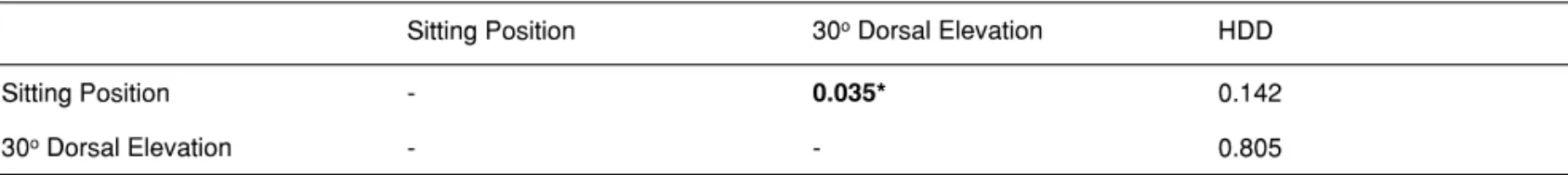
Table III – Comparison of the FEV1/FVC in the Different Positions by the Tukey Test
Sitting Position 30o Dorsal Elevation HDD
Sitting Position - 0.035* 0.142
30o Dorsal Elevation - - 0.805
THE INFLUENCE OF POSTURE ON SPIROMETRIC VALUES IN GRADE III OBESE PATIENTS
Revista Brasileira de Anestesiologia 715
Vol. 61, No 6, November-December, 2011
DISCUSSION
The prevalence of grade III obesity among women is greater than in men and, in our service, it is not different. Due to diffi-culties of having homogenous groups of obese patients of both genders and the fact that lung volumes and the FEV1/FVC
ra-tio are different in both genders, we decided to include only females. Elderly patients and/or smokers were also excluded, since both conditions change the results of spirometry.
The correlation of varying the decubitus and lung function in patients with grade III obesity in the preoperative period was evaluated by different authors using other variables not used in the present study 8,11,12. We did not find in the
litera-ture studies comparing predicted FVC, FEV1, and FEV1/FVC
ratio on all three positions in the preoperative period of pa-tients with grade III obesity.
Several studies have included preoperative FVC of obese patients only in the sitting position 7,10-14. Some of them
pre-sented absolute values of spirometric variables 8,11-14.
Com-parison of those results with the present study was not pos-sible, since the normalcy pattern of spirometric values varies according to the anthropometric characteristics of weight, height, gender, and race, and has specific formulas deter-mined by different authors. We used the reference values de-fined by Pereira et al. 10 in our study.
The values observed in the present study on the sitting position (90o) were similar to those observed by Rasslam15
in a study that evaluated, in patients of both genders, the ef-fects of grades I and II obesity on spirometry. Mean FVC of 101.0% was observed in female patients with mean BMI of 34.2 kg.m-2.
On the study of Sarikaya et al. 14, comparing spirometries
of non-obese patients and grades I, II, and III obese patients in the sitting position, higher values in the group of grade III obesity (BMI > 40 kg.m-2; 86% females) were observed, with
mean FVC of 108.26%. Domingos-Benício et al. 16 compared
the spirometry of eutrophic and obese (grades I, II, and III) non-smoker volunteers of both genders on orthostatic, sitting, and supine positions. The numeric values were not included in the publication, only histograms, showing that the mean FVC value of grade III obese patients in the sitting position is be-tween 90% and 95%. In their results, as well as in ours, there is a reduction of spirometry results in the supine position.
The mean FEV1 value in the sitting position observed in the
present study, 93.1%, was similar to that observed by other authors in studies with comparable methods: between 90.0% and 96.0% 14,16-18.
Razi and Moosavi 17, in a study with patients of both
gen-ders, observed that, in a group of non-asthmatic patients in the sitting position, their FEV1 = 101.0% 17. However, the
mean BMI of 36.69 kg.m-2 of that study was smaller than that
of our study, justifying higher FEV1 values.
The mean value of the FEV1/FVC ratio in the sitting
po-sition observed in the present study was 100.76%, higher than the values observed in some studies with comparable methods (76.5% and 86.0%) 14, but similar to that observed
by Rasslam (100.0%) 15.
Studies with male patients, smokers and non-smokers,
showed lower FEV1/FVC ratio: 81.6% and 82.5%, as well as
in studies with patients of both genders: between 80% and 86.4% 9,19,20.
The results regarding the horizontal position confirm the results of Domingos-Benício et al. 16, the only one found in the
literature that used a similar method and evaluated patients in horizontal dorsal decubitus. They reported a statistically sig-nificant reduction in FVC, FEV1, and FEV1/FVC ratio in dorsal
decubitus in relation to the sitting position, which was not ob-served in the present study.
The present study demonstrated that, in adult female pa-tients with grade III obesity, non-smokers, without lung dis-ease, 30o dorsal inclination and horizontal dorsal decubitus
(0o) cause changes in spirometry. This has practical
implica-tions, since the beds in the recovery unit at ISCMSP reach, the most, an inclination of 30o. Note that patients with grade III
obesity have difficulties in maintaining the semi-sitting position in the postoperative period. Even at 30o inclination, very often
they slide in the bed, resulting in a lower inclination, which potentially hinders their respiratory pattern.
One can infer that male and elderly patients also suffer in-fluence of the decubitus; however, similar studies with those groups should be performed.