ABSTRACT
HPV16 in oropharynx as compared to oral cavity
Hideo SHIGEISHI10DVDUX68*,<$0$2, Kouji OHTA1, Mohammad Zeshaan RAHMAN1, Masaaki TAKECHI1
1- Hiroshima University, Institute of Biomedical and Health Sciences, Department of Oral and Maxillofacial Surgery, Hiroshima, Japan. 2- Hiroshima University, Institute of Biomedical and Health Sciences, Department of Public Oral Health, Hiroshima, Japan.
Corresponding address: Hideo Shigeishi - Department of Oral and Maxillofacial Surgery, Division of Cervico-Gnathostomatology - Institute of Biomedical and Health Sciences - Hiroshima University 1-2-3 Kasumi - Minami-ku - Hiroshima - 734-8553 - Japan - Phone: +81-82-257-5673 - FAX: +81-82-257-5671 - e-mail: [email protected]
6XEPLWWHG-DQXDU\0RGL¿FDWLRQ$SULO$FFHSWHG$SULO
O
bjective: The objective of this study was to clarify differences regarding HPV16 infectionMaterial and Methods: The subjects were 94 healthy asymptomatic individuals (41 males, 53 females; mean age 58.6 years, range 16-97 years) who visited the Department of Oral and Maxillofacial Reconstructive Surgery of the Hiroshima University Hospital from 2014 to 2015. Oral epithelial cells were collected from oral rinse and pharynx gargle samples and placed in saline. The human endogenous retrovirus gene ERV3-1 was used as a reference to estimate the number of human cells in each sample. DNA samples were extracted from
primer. Similarly, we analyzed the HPV16 viral copy number in HPV16-positive cases using
cases was higher in the gargle (28.7%) as compared to the rinse (16.0%) samples. In the
(7.5%) (P=0.021). Importantly, in older subjects (aged 60-89 years), gargle samples
(P=0.034). The average number of viral copies was approximately 8 times higher in the gargle than in the oral rinse samples (0.16±0.27 vs. 1.35±1.26 copy numbers per cell),
is more susceptible to HPV16 infection as compared to the oral cavity, while HPV16 gene
Ke yw or ds:
I N TROD UCTI ON
The human papilloma virus (HPV) consists of a circle of double-stranded DNA and belongs to the family of papillomaviridae, with infection generally occurring in the skin and mucosa7,8. HPV infection
remains in basal cells, though is discharged outside the body upon differentiation of the infected cells into keratinocytes8. The virus, therefore, does not
enter the bloodstream and is able to evade immune surveillance. The HPV genome consists of six early open reading frames (ORFs) (E1, E2, E4, E5, E6, E7), two late ORFs (L1, L2), and a non-coding long control region (LCR)7,8.According to the International
Agency forResearch on Cancer (IARC), 12 HPV types
(HPV16, 18, 31, 33, 35, 39, 45, 51, 52, 56, 58, 59)
3,of which
in the early 1980s and considered to be the most important risk factor for development of cervical cancer30. In addition, persistent infection with oral
HPV16 is also thought to drive the development of oropharyngeal cancer12.
We previously reported that most precancerous lesions caused by HPV16 infection of the oral
associated with malignant transformation of precursor lesions24. However, few epidemiologic
healthy Japanese individuals and that has not been fully elucidated. In this study, we investigated oral rinse and gargle samples obtained from healthy individuals to clarify differences of HPV16 infection
oropharynx.
M ATERI AL AN D M ETH OD S
Su bj e ct s
The subjects of this study were 94 healthy asymptomatic individuals (41 males, 53 females; mean age 58.6 years, range 16-97 years), who visited the Department of Oral and Maxillofacial Reconstructive Surgery of the Hiroshima University Hospital from 2014 to 2015. None had evidence of oral cancer or pre-malignant lesions. The study design was approved by the Ethics Committee of the Hiroshima University and all participants signed an informed consent form.
Or a l r inse a nd ga r gle sa m ple pr oce ssing a nd D N A e x t r a ct ion
Oral rinse samples were used to evaluate HPV16 infection in the oral cavity while gargle samples were used to the evaluation of the oropharynx. Exfoliated epithelial cells were collected in samples obtained by a 30-second oral rinse and a separate 30-second gargle9
their mouth with 10 mL of saline for 30 seconds and then spit into a sterile 50-mL falcon tube. Next, they gargled 10 mL of saline for another 30 seconds and spit that sample into a different 50-mL falcon tube. Immediately after collection, all samples were centrifuged at 3000x g for 10 minutes at 4°C, then the supernatant was decanted, the pellet re-suspended in 10 ml of phosphate-buffered saline (PBS), and centrifugation repeated. Thereafter, the pellets were stored at -80°C until further processing.
Wizard®
Qu a n t it a t ion of h u m a n ce ll n u m be r s
DNA amounts extracted from the oral rinse samples were not equivalent to the amount of human DNA, due to the quantity of non-human derived DNA. Therefore, we employed the human endogenous retrovirus group 3 member 1 (ERV3-1) gene to quantitate human cells using a real-time PCR assay, according to a previously reported method9.
A 1.7-kb Hind III:Pst I fragment within the ERV3-1 genome was prepared and then cloned into a 2.7-kb pUC57 vector. Serial 10-fold dilutions of the pUC57 vector were made with copy numbersranging from 100 to 109. Calculation of number of copies was
performed based on the DNA molecular weight of the plasmid, according to a previously reported method29. Next, we performed real-time PCR assays
primer. Quantitation of DNA levels was done using a CFX connect real-time PCR detection system (BioRad, Hercules, CA, USA) and SYBR Green PCR Master Mix (TOYOBO, Tokyo, Japan), with a reaction mixture containing 1.0 μl of DNA, 12.5 μl of SYBR Green Mix, and 10 μmol of each pair of oligonucleotide primers. The primer sequences were as follows: ERV3-1; 5’- CATGGGAAGCAAGGGAACTAATG-3’ (sense) and 5’- CCCAGCGAGCAATACAGAATTT -3’ (antisense)9
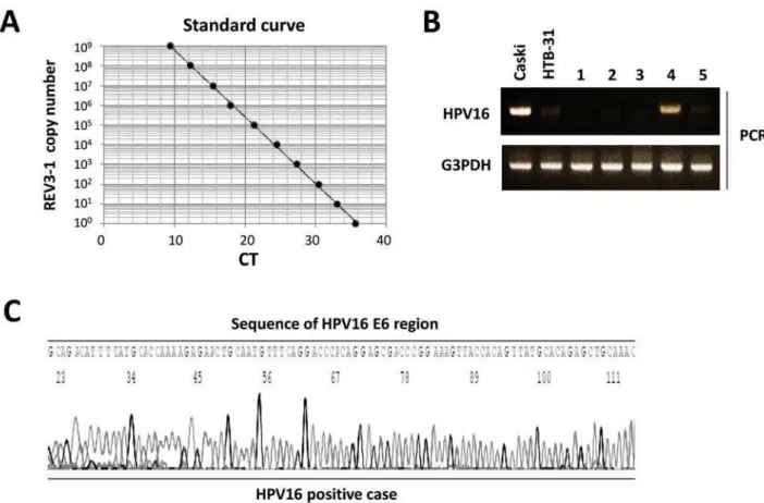
initial melting at 95°C for 5 minutes, then 40 cycles of denaturing at 95°C for 30 seconds, annealing at 55°C for 30 seconds, and extension at 72°C for 1 minute. A standard curve indicating CT value versus ERV3-1 copy number was obtained (Figure 1A). The number of cells, determined by use of the ERV3-1 diploid genome, was used to estimate the number of human cells in each sample.
H PV1 6 D N A de t e ct ion by PCR
The HPV16 sequence was examined by PCR
open reading frame using a PCR HPV Detection Set
fragment with a length of 140 base pairs. Samples
an oral cellular sample and those without G3PDH were excluded from analysis. A positive control containing HPV16 DNA extracted from Caski cells and a negative control containing DNA
were used for the PCR reactions. We prepared
DNA samples extracted from approximately
10,000 human cells in each PCR mixture. Each
10x PCR
buffer (TOYOBO), dNTPs, Taq DNA polymerase,
and HPV16 primers. The primer sequences for
HPV1 6 were 5’- AAGGGCGTAACCGAAATCGGT-3’
(sense) and 5’-GTTTGCAGCTCTGTGCATA-3’
(antisense), and those for G3 PD H were 5’- A C C A C A G TC C ATG C C ATC A C- 3 ’ ( s e n s e ) a n d 5’-TCCACCACCCTGTTGCTGTA-3’ (antisense). The PCR program included initial melting at 95°C for 5 minutes, followed by 28 cycles at 95°C for 30
product was electrophoresed on 3% agarose gels
HPV16 DNA sequence, we performed sequencing for HPV16.
PCR product was analyzed by electrophoresis using 2% agarose gels. For the sequencing reaction, a BigDye Terminator v3.1 cycle sequencing kit (Applied Biosystems, Foster City, CA, USA) was
H PV1 6 vir a l copy qu a n t it a t ion
Real-time PCR assays were performed to determine the viral load of HPV16 DNA in the samples. We prepared a 200-bp fragment within the HPV16 E6 genome, which was then cloned into a 2.7-kb pUC57 vector. Serial 10-fold dilutions of
the vector were made with copy numbersranging
from 100 to 109. Serial 10-fold dilutions were used to generate a standard curve, which indicated the CT value versus the copy number of HPV16 E6. We then prepared DNA samples extracted from approximately 10,000 human cells from each PCR
mixture. Real-time PCR was performed using a
(Takara). The PCR program was as follows: initial melting at 95°C for 5 minutes, followed by 40 cycles at 95°C for 30 seconds, 57°C for 2 minutes, and
72°C for 30 seconds. A standard curve was used
to enumerate the copy number of HPV16 in each sample.
St a t ist ica l a n a lysis
Fisher’s exact test was used to examine the correlation between oral HPV16 infection and clinical
parameters. Student’s t-test was used to evaluate significant differences regarding HPV16 copy
numbers.P values less than 0.05 were regarded as
RESULTS
H PV1 6 pr e va le n ce in or a l r in se a n d ph a r yn x ga r gle sa m ple s
HPV16 prevalence was examined in a total of 94 oral rinse and gargle samples. The product of HPV16 DNA was detected in Caski cells (HPV16 positive),
but not in HTB-31 cells (HPV negative), by PCR
analysis (Figure 1B). Next, the PCR products were
(Figure 1C). We found HPV16-positive samples in 15 (16.0%) oral rinse samples and 27 (28.7%) gargle samples, showing a higher prevalence in the gargle samples. There were only 8 subjects (8.5%) with both HPV16-positive oral rinse and gargle samples, whereas 19 (20.2%) were HPV16 positive in the gargle sample alone.
Figure 1- Detection of HPV16 by PCR. (A) Standard curve indicating CT value vs. ERV3-1 copy number. A standard curve was calculated by plotting obtained CT values against serial 10-fold dilutions produced with ERV3-1 copy numbers ranging from 100 to 109%$JDURVHJHOHOHFWURSKRUHVLVRIDPSOL¿HG3&5SURGXFWVIURPFHOOOLQHVDQGRUDOVDPSOHV7HQ
Cor r e la t ion be t w e e n H PV 1 6 in fe ct ion a n d clin ica l fa ct or s
Males showed a higher rate of HPV16 prevalence than females (26.8% vs. 7.5%) in the oral rinse samples (Table 1). Of the 11 oral HPV16-positive men, 4 were denture users and 4 were young
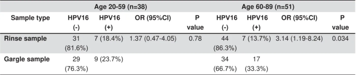
(18-HPV16 infection and male gender found (P=0.021). As for age, subjects aged 20-29 years old showed a high level of HPV16 prevalence (26.8%) in the oral rinse samples. On the other hand, the 30-39, 60-69, and 80-89 year-old groups exhibited high HPV16 prevalence (37.5%, 44.4%, and 37.5%, respectively) in the gargle samples. We also performed statistical analysis to compare young (20-59 years old) with older (60-89 years old) subjects (Table 2). HPV16 prevalence was higher
in the gargle samples as compared to the oral rinse samples in both of those groups. However, the odds ratio for a HPV16-positive gargle sample was higher in the older group (odds ratio, 3.14 vs. 1.37). Furthermore, the gargle samples showed
the oral rinse samples (13.7%) in that older group (P=0.034). In addition, subjects who used dentures showed an increased percentage of HPV16 infection in both oral rinse and gargle samples as compared to non-denture users (oral rinse, 20.7% vs. 13.4%; gargle, 34.5% vs. 25.3%). However, we found no
and denture use in either the oral rinse or gargle samples.
Rinse sample Gargle sample
Clinical characteristics +39 +39 P value +39 +39 P value Gender
Male (41) 0.021 0.36
Female (53)
Age in years
10-19 (3) 0.49 0.61
20-29 (14)
30-39 (8)
40-49 (7)
50-59 (9)
60-69 (18)
70-79 (17)
80-89 (16)
Denture use
(-) (65) 0.54 0.46
(+) (29)
Table 1- Correlation between oral HPV16 infection and clinical parameters
)LVKHU¶VH[DFWWHVWZDVXVHGIRUVWDWLVWLFDODQDO\VLV3YDOXHVOHVVWKDQZHUHUHJDUGHGWREHVWDWLVWLFDOO\VLJQL¿FDQW
Age 20-59 (n=38) Age 60-89 (n=51)
Sample type +39 (-)
+39
OR (95%CI) P value
+39 (-)
+39
OR (95%CI) P value
Rinse sample 31
1.37 (0.47-4.05) 0.78 44
3.14 (1.19-8.24) 0.034
Gargle sample 29
34
17
Table 2- Correlation between oral HPV16 infection and sample type among young adults and older people
Qu a n t it a t ion of H PV 1 6 vir a l copy n u m be r u sin g r e a l- t im e PCR
In HPV16-positive cases, we analyzed the HPV16 viral copy number using real-time PCR to examine
Serial 10-fold dilutions of a plasmid vector containing HPV16 E6 DNA were
used to generate a standard curve (Figure 2A), then
we evaluated the number of HPV16 DNA copies as copy number per human cell. The average number of viral copies was approximately 8 times higher in the gargle samples than in the oral rinse samples (0.16±0.27 vs. 1.35±1.26 copy numbers per cell) (Figure 2B). We found a significant difference regarding HPV16 E6 DNA copy number between the gargle and oral rinse samples (P<0.001). These
is more commonly found in the oropharynx as compared to the oral cavity.
D I SCUSSI ON
Oral HPV infection is considered to be transmitted through sexual behavior, thus marital status, number of sex partners, oral sex, and anal sex are thought to be related variables. Oral HPV prevalence is significantly higher in individuals who have participated in oral sex as compared to those who have not, indicating that as a risk factor for oral HPV infection11. As for other risk factors, smoking
and poor oral hygiene related to such variables as periodontitis, gingivitis, and denture use have been found to be associated with oral HPV infection12,15,18.
As for smoking, the chemical substances contained in tobacco may be involved and suppress
immune mechanisms in the oral cavity11, while
the relationship of denture use to HPV infection suggests that oral mucosal injury caused by mechanical stimulation from denture wearing and poor oral hygiene due to unclean dentures may have an association23. Thus, HPV infection may be
associated with oral environmental changes due to local effects, such as smoking and denture use. Importantly, our results showed that being male
Although the reasons remain unclear, smoking, denture use, sexual activity, and other factors may be related to the high rate of oral infection seen in men.
In the subjects of this study, the prevalence rate of HPV16 was higher in the gargle samples as compared to the oral rinse samples. Gargle samples from the older groups also showed
oral rinse samples from those subjects, indicating an important association between age and HPV16 infection in the oropharyngeal region, and that age distribution is likely to be a cause of differences between oral rinse and gargle samples. In addition,
in the gargle samples. Despite cross-mixing of the samples between the oral cavity and oropharyngeal region, we considered that the collected oral rinse and gargle samples were associated with HPV DNA present primarily in the oral cavity and oropharyngeal region, respectively. Thus, our observations suggest that the oropharynx is more susceptible to HPV16 infection and HPV16 gene
Figure 2-4XDQWL¿FDWLRQRI+39YLUDOFRS\QXPEHUE\UHDOWLPH3&5$6WDQGDUGFXUYHLQGLFDWLQJ&7YDOXHYV+39 copy number. A standard curve was calculated by plotting obtained CT values against serial 10-fold dilutions produced with HPV16 copy numbers ranging from 100 to 109%7KHDYHUDJHQXPEHURIYLUDOFRSLHVZDVVLJQL¿FDQWO\KLJKHULQWKHJDUJOH
compared to the oral cavity.
In a previous study of head and neck cancer cases, the positive rate of HPV was 10% in patients with oral cavity cancer, 83.2% in those with oropharyngeal cancer, and 44.4% in those with nasopharynx cancer, demonstrating the highest prevalence in oropharyngeal cancer cases, while approximately 90% of the HPV-positive oropharyngeal cancer patients also showed HPV16 infection27. Furthermore, Wang, et al.28 (2012)
reported that male oropharyngeal cancer patients showed a higher rate of HPV16 infection than female patients. Our results also showed that the gargle specimens from males had a higher rate of HPV16 infection as compared to those from females. Another study also reported that the HPV16 genome copy number was much higher in oropharyngeal cancer than oral cancer specimens22. Together, these
results support the notion that the oropharynx is a more common site for HPV16 infection as compared to other head and neck regions. In addition, the variability of HPV16 infection rate among different sites (i.e., oral, oropharyngeal, laryngeal) suggests the biological characteristics of HPV-related cancer.
Findings for the prevalence of oral HPV vary substantially among reported studies, which has been attributed to differences in study populations, as well as approaches for specimen collection and detection methods1,2,4-6,13,14,16,17,19-21,25,26. In recent
studies of oral HPV infection, the methods most frequently used for oral sample collection have included oral swabbing, oral rinsing, and gargling with mouthwash or saline,with rinsing with mouthwash or saline thought to be highly effective. PCR assays targeting the HPV L1 region have also been commonly performed in many studies, while direct HPV genotyping without HPV detection was utilized in some1,2,4-6,13,14,16,17,19-21,25,26. In those, HPV
genotypes were determined by PCR using
type-hybridization frequently using the LINEAR ARRAY HPV Genotyping Test , as it is useful for detection of combined HPV infection, though less sensitive and positive rates may vary with different cut-off values10. Thus, we used a combination of PCR with
detection to accurately determine HPV16 infection status in the present subjects.
Taken together, our observations indicate that HPV16 infection has a higher rate of prevalence in gargle samples. Thus, HPV16 is considered to be an important risk factor for development of head and neck cancer, especially oropharyngeal cancer. Future studies are needed to examine the risks of smoking, oral hygiene, and sexual behavior in relation to HPV infection, and will likely lead to a decrease in oral HPV infection rates, while
elucidation of the molecular biology of HPV may reduce the occurrence of HPV-related head and neck cancer.
CON CLUSI ON
Our results indicate that the oropharynx is more susceptible to HPV16 infection as compared to the
more commonly found in the oropharynx.
ACKN OW LED GEM EN TS
This work was supported by a Grant-in-aid for
Japanese Ministry of Education, Culture, Sports, and Technology.
REFEREN CES
1- Antonsson A, Cornford M, Perry S, Davis M, Dunne MP, Whiteman DC. Prevalence and risk factors for oral HPV infection in young Australians. PLoS One. 2014;9:e91761.
2- Beachler DC, Sugar EA, Margolick JB, Weber KM, Strickler HD, Wiley DJ, et al. Risk factors for acquisition and clearance of oral human papillomavirus infection among infected and HIV-uninfected adults. Am J Epidemiol. 2015;181:40-53.
3- Bouvard V, Baan R, Straif K, Grosse Y, Secretan B, El Ghissassi F, et al. A review of human carcinogens-Part B: biological agents. Lancet Oncol. 2009;10:321-2.
4- Cook RL, Thompson EL, Kelso NE, Friary J, Hosford J, Barkley P, et al. Sexual behaviors and other risk factors for oral human papillomavirus infections in young women. Sex Transm Dis. 2014;41:486-92.
5- Dahlstrom KR, Burchell AN, Ramanakumar AV, Rodrigues A, Tellier PP, Hanley J, et al. Sexual transmission of oral human papillomavirus infection among men. Cancer Epidemiol Biomarkers Prev. 2014;23:2959-64.
6- Dalla Torre D, Burtscher D, Edlinger M, Sölder E, Widschwendter A, Rasse M, et al. Comparison of the prevalence of human papilloma
lesions and healthy oral mucosa by brush smear detection. Oral Surg Oral Med Oral Pathol Oral Radiol. 2015;119:333-9. 7- De Villiers EM, Fauquert C, Bernard HU, zur Hausen H.
8- Doorbar J. Molecular biology of human papillomavirus infection and cervical cancer. Clin Sci. 2006;110:525-41.
9- D'Souza G, Sugar E, Ruby W, Gravitt P, Gillison M. Analysis of
in oral rinse samples by PCR. J Clin Microbiol. 2005;43:5526-35. 10- Dunn ST, Allen RA, Wang S, Walker J, Schiffman M. DNA extraction: an understudied and important aspect of HPV genotyping using PCR-based methods. J Virol Methods. 2007;143:45-54.
11- Gillison ML, Broutian T, Pickard RK, Tong ZY, Xiao W, Kahle L, et al. Prevalence of oral HPV infection in the United States, 2009-2010. JAMA. 2012;307:693-703.
13- Grün N, Ährlund-Richter A, Franzén J, Mirzaie L, Marions L, Ramqvist T, et al. Oral human papillomavirus (HPV) prevalence in youth and cervical HPV prevalence in women attending a youth clinic in Sweden, a follow up-study 2013-2014 after gradual introduction of public HPV vaccination. Infect Dis (Lond). 2015;47:57-61.
14- Hang D, Liu F, Liu M, He Z, Sun M, Liu Y, et al. Oral human papillomavirus infection and its risk factors among 5,410 healthy adults in China, 2009-2011. Cancer Epidemiol Biomarkers Prev. 2014;23:2101-10.
15- Hormia M, Willberg J, Ruokonen H, Syrjänen S. Marginal periodontium as a potential reservoir of human papillomavirus in oral mucosa. J Periodontol. 2005;76:358-63.
16- Kero K, Rautava J, Syrjänen K, Willberg J, Grenman S, Syrjänen S. Smoking increases oral HPV persistence among men: 7-year follow-up study. Eur J Clin Microbiol Infect Dis. 2014;33:123-33.
17- King EM, Gilson R, Beddows S, Soldan K, Panwar K, Young C, et al. Oral human papillomavirus (HPV) infection in men who have sex with men: prevalence and lack of anogenital concordance. Sex Transm Infect. 2015;91:284-6.
18- Kreimer AR, Pierce Campbell CM, Lin HY, Fulp W, Papenfuss MR, Abrahamsen M, et al. Incidence and clearance of oral human papillomavirus infection in men: the HIM cohort study. Lancet. 2013;382:877-87.
19- Lima MD, Braz-Silva PH, Pereira SM, Riera C, Coelho AC, Gallottini M. Oral and cervical HPV infection in HIV-positive and HIV-negative women attending a sexual health clinic in São Paulo, Brazil. Int J Gynaecol Obstet. 2014;126:33-6.
20- Mbulawa ZZ, Johnson LF, Marais DJ, Coetzee D, Williamson AL. Risk factors for oral human papillomavirus in heterosexual couples in an African setting. J Infect. 2014;68:185-9.
21- Meyer MF, Huebbers CU, Siefer OG, Vent J, Engbert I, Eslick GD, et al. Prevalence and risk factors for oral human papillomavirus infection in 129 women screened for cervical HPV infection. Oral Oncol. 2014;50:27-31.
22- Ndiaye C, Mena M, Alemany L, Arbyn M, Castellsagué X, Laporte L, et al. HPV DNA, E6/E7 mRNA, and p16INK4a detection in head and neck cancers: a systematic review and meta-analysis. Lancet Oncol. 2014;15:1319-31.
23- Nishimura Y, Maeda H, Hattori M, Azumaya F, Muramatsu I, Kameyama Y, et al. Human papillomavirus infection in the oral cavity of denture wearers. Nihon Hotetsu Shika Gakkai Zasshi. 2004;48:713-22.
24- Sugiyama M, Bhawal UK, Dohmen T, Ono S, Miyauchi M, Ishikawa T. Detection of human papillomavirus-16 and HPV-18 DNA in normal, dysplastic, and malignant oral epithelium. Oral Surg Oral Med Oral Pathol Oral Radiol Endod. 2003;95:594-600.
Prevalence of human papillomaviruses in the healthy oral mucosa of women with high-grade squamous intra-epithelial lesion and of their partners as compared to healthy controls. J Oral Pathol Med. 2015;44:722-7.
26- Vidotti LR, Vidal FC, Monteiro SC, Nunes JD, Salgado JV, Brito LM, et al. Association between oral DNA-HPV and genital DNA-HPV. J Oral Pathol Med. 2014;43:289-92.
27- Walline HM, Komarck C, McHugh JB, Byrd SA, Spector ME, Hauff SJ, et al. High-risk human papillomavirus detection in oropharyngeal, nasopharyngeal, and oral cavity cancers: comparison of multiple methods. JAMA Otolaryngol Head Neck Surg. 2013;139:1320-7.