108
ISSN: 1646-0499
Revista da Faculdade de Ciências da Saúde, nº 7, p. 108-121 (2010) Submetido: 20 Mai. 2010/Aceite: 12 Ago. 2010
HEAD AND NECK CANCER: CONTRIBUTION FOR
THE ANALYSIS OF THE REHABILITATION NEEDS
Daniela Oliveira Vieira
Speech Therapist
Master student in Oncology
Institute of Biomedical Sciences Abel Salazar University of Porto, Porto, Portugal
Eurico Monteiro
Director
Otorhinolaryngology Department
Institute of Oncology of Porto Francisco Gentil, Porto, Portugal Assistant Professor
School of Health Sciences
109
ABSTRACT
With this study, we aim to understand the relevance of the Speech Therapist´s intervention on a Cancer Department, through the analysis of the correlated surgical cases effectuated in the Otorhinolaryngology Department of the Portuguese Cancer Institute of Porto Francisco Gentil – EPE, between January 2005 and December 2008. All surgeries relevant to the Spe-ech Therapist’s intervention were analyzed according to the number of sessions needed for an individualized rehabilitation and, afterwards, for the entire group.
KEYWORDS
Head and Neck Neoplasms, Rehabilitation, Speech Therapy.
RESUMO
Com este estudo pretende-se dar a compreender a pertinência da intervenção dos Terapeu-tas da Fala num serviço oncológico, tendo por base a análise dos casos cirúrgicos tratados no Serviço de Otorrinolaringologia do Instituto Português de Oncologia do Porto Francisco Gentil – EPE, entre Janeiro de 2005 e Dezembro de 2008. Foram seleccionados todos os pro-cedimentos cirúrgicos com relevância para a intervenção dos Terapeutas da Fala e agrupa-dos de acordo com o número de sessões necessárias para uma reabilitação individualizada e calculado posteriormente o impacto para todo o grupo.
PALAVRAS-CHAVE
110
1.
INTRODUCTION
Head and neck tumours are the 6th most common neoplasm, with approximately 640000 new cases per year with a mortality of 5% (IARC-WHO GLOBOCAN Database 2002). Surgical treat-ment of these tumours affects structures involved in breathing, voice, speech and swallowing, apart from aesthetic sequels. This study aims to analyze the surgeries performed in patients treated for cancer in the Otorhinolaryngology (ENT) Department of the Portuguese Institute of Oncology of Porto Francisco Gentil – EPE (IPOPFG-EPE), between January 2005 and December 2008 with relevance for Speech Therapists, analyzing the impact of intervention areas.
This analysis will only address aspects concerning surgical options, once the removed struc-tures, the mobility of remaining ones, and the type of reconstruction, allows us to foresee the implications in voice, speech and swallowing. In this way, it is possible to establish a more precise rehabilitation protocol (Crevièr-Buchman, Brihaye and Tessier).
Another aim of this study is to explore the costs of the rehabilitation, starting with the aver-age time spent in it, because, in the case of Speech Therapists intervention, these studies are almost non-existent, not knowing what the real costs of rehabilitation and its relevance.
2.
MATERIAL AND METHOD
Concerning the importance of the inclusion of Speech Therapists in multidisciplinary teams involved in the rehabilitation of patients with head and neck cancer, was carried out a col-lection of surgical acts effectuated by the ENT Department, during four years (2005 to 2008), with impact on Speech therapist’s intervention areas. We also compare endoscopic techni-ques, assisted by CO2 laser with conventional surgical techniques, seeking to understand its growing application from 2005.
Based on topographic localizations of tumours were structured surgical techniques, for tu-mours of: 1) oral cavity; 2) maxilla/mandible; 3) other locations/undefined lesions of the oral cavity and maxilla/mandible; 4) pharynx; 5) larynx; 6) pharyngolaryngeal region; and 7) others.
3.
OUTCOMES
3.1.
CHARACTERIZATION OF THE SAMPLE
111
Year/Gender 2005 2006 2007 2008 Total % Mean age
Male 138 151 168 192 649 87,82 58
Female 18 22 19 31 90 12,18 60
Total 156 173 187 223 739 100
TABLE 1 - Characterization of the sample by gender.
3.2.
SURGICAL TECHNIQUES
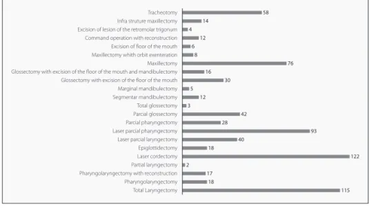
The tables and graphs below highlight the distribution of surgical techniques. The five most commonly performed procedures were laser cordectomy (122), total laryngectomy (115), laser partial pharyngectomy (93), maxilectomy (76) and tracheotomy (58). (Graphic 1) Be-tween 2005 and 2008 the topographical region more frequently addressed was the larynx (297), followed by pharynx (121), maxilla/mandible (101) and oral cavity (99). (Tabela 2)
GRAPHIC 1 - Absolute frequency of surgical techniques.
Topographic regions N
Oral cavity 99
Maxilla/mandible 101
Oral cavity & maxilla/mandible 28
Pharynx 121
Larynx 297
Pharyngolaryngeal region 35
Tracheotomy 58
Total 739
TABLE 2 - Number of surgeries performed by topographic region.
112
with excision of the floor of the mouth (30%), infra structure maxillectomy (14%), excision of the floor of the mouth (6%), resection of lesion of the retromolar trigonum (4%) and total glossectomy (3%).Surgeries performed in the “oral cavity” N %
2005 2006 2007 2008 Total
Partial glossectomy 8 18 7 9 42 43
Total glossectomy 1 1 1 0 3 3
Excision of the floor of the mouth 1 1 2 2 6 6 Infrastructure maxillectomy 8 4 1 1 14 14 Resection of the retromolar trigonum 2 1 1 0 4 4 Glossectomy with excision of the floor of the mouth 7 5 10 8 30 30
Total 27 30 22 20 99 100
TABLE 3 - Absolute frequency of surgeries performed in the “oral cavity”.
In the region of “maxilla/mandible”, the most frequent surgical techniques were Maxillec-tomy (75%), followed by marginal mandibulecMaxillec-tomy (12%), maxillecMaxillec-tomy with orbit exente-ration (8%) and segmentar mandibulectomy (5%), as shown in table 4.
Surgery performed on “Maxilla/Mandible N %
2005 2006 2007 2008 Total
Maxillectomy with orbit exenteration 2 0 3 3 8 8
Maxillectomy 7 17 17 35 76 75
Marginal mandibulectomy 0 0 3 2 5 12 Segmentar mandibulectomy 4 0 2 6 12 5
Total 13 17 25 46 101 100
TABLE 4 - Distribution of absolute and relative frequencies of surgeries performed in the “maxilla/mandible”.
In some situations, it was necessary to remove rather than an anatomical region for a com-plete ablation of tumours mass, having these surgeries grouped in surgical acts like glosso-pelvectomy with mandibulectomy.
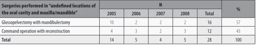
In other locations undefined of the oral cavity and maxilla/mandible, as can be seen in table 5, the surgeries most frequently performed were the glossopelvectomy with mandibulec-tomy (57%) and command operation with reconstruction (43%).
Surgeries performed in “undefined locations of the oral cavity and maxilla/mandible”
N
%
2005 2006 2007 2008 Total
Glossopelvectomy with mandibulectomy 10 2 2 2 16 57 Command operation with reconstruction 4 3 2 3 12 43
Total 14 5 4 5 28 100
113
In the pharynx, according to the typescript in table 6, the surgical procedures more frequen-tly performed were laser partial pharyngectomy (77%), partial pharyngectomy (23%). We should however consider that 99% of surgeries performed in this region were made through endoscopic/transoral procedure instead of external ones (23% - partial pharyngectomy).
Surgeries performed on “pharynx” N %
2005 2006 2007 2008 Total
Laser partial pharyngectomy 14 20 37 22 93 77 Partial pharyngectomy 3 1 11 13 28 23
Total 17 21 48 35 121 100
TABLE 6 - Absolute and relative frequencies of surgeries performed in “pharynx”.
In what larynx is concerned, as shown in table 7 the most common surgery was laser cor-dectomy (41%), followed by total laryngectomy (39%), laser partial laryngectomy (14%) and laser epiglottidectomy (6%). The other surgeries do not have representation (fronto-lateral laryngectomy and near-total Laryngectomy). In this region most of the interventions were also made by endoscopic procedures (61% - laser parcial laryngectomy, laser epiglottidec-tomy and laser cordecepiglottidec-tomy) and only 39% by external approach (fronto-lateral laryngec-tomy, near-total laryngectomy and total laryngectomy).
Topographic Surgeries performed in the region of “larynx”
N
%
2005 2006 2007 2008 Total
Laser partial laryngectomy 5 11 11 13 40 14 Laser epiglottidectomy 1 10 5 2 18 6
Laser cordectomy 18 33 26 45 122 41
Fronto-lateral laryngectomy 1 0 0 0 1 0
Near-total laryngectomy 0 1 0 0 1 0
Total laryngectomy 35 34 20 26 115 39
Total 60 89 62 86 297 100
TABLE 7 - Absolute and relative frequencies of surgeries performed in “larynx”.
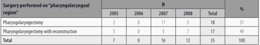
In the pharyngolaryngeal region, the most frequently procedures performed were pharyn-golaryngectomy (51%) and the pharynpharyn-golaryngectomy with reconstruction (49%). (Table 8)
Surgery performed on “pharyngolaryngeal region”
N
%
2005 2006 2007 2008 Total
Pharyngolaryngectomy 2 0 11 5 18 51
Pharyngolaryngectomy with reconstruction 5 0 5 7 17 49
Total 7 0 16 12 35 100
TABLE 8 - Distribution of absolute and relative frequencies of surgeries performed in the “pharyngolaryngeal region”.
114
3.2.1. LASER SURGERYAs can be seen from table 9, the most frequently performed endoscopic surgeries occur-red in two anatomical regions: pharynx and larynx. Regarding the evolution of this type of surgery, it was observed a gradual increase in the endoscopic laser procedures, from 2005 until 2008 (table 9). However, we can see that overall, the conventional techniques (63%) are superior in number to the endoscopic one (37%). (Table 10)
Oral cavity Maxilla/ Mandible
Oral Cavity & Maxilla/ Mandible
Pharynx Larynx
Pharyngo- Laryngeal region
Tracheo-tomy Total
2005 Conventional Surgery 27 13 14 3 35 7 18 117
Laser Surgery 0 0 0 14 25 0 0 39
2006 Conventional Surgery 30 17 5 1 35 0 11 99
Laser Surgery 0 0 0 20 54 0 0 74
2007 Conventional Surgery 22 25 4 11 20 16 10 108
Laser Surgery 0 0 0 37 42 0 0 79
2008 Conventional Surgery 20 46 5 13 26 12 19 141
Laser Surgery 0 0 0 22 60 0 0 82
Total 99 101 28 121 297 35 58 739
TABLE 9 - Absolute frequency of surgeries performed in accordance with topographical region and surgical option.
Type of surgery %
Conventional 63
Laser 37
Total 100
TABLE 10 - Relative frequency of the type of surgery.
The table 11 show the variation within the endoscopic approaches between 2005 and 2008. We see an increase in laser partial pharyngectomy, laser partial laryngectomy and laser cor-dectomy procedures and the remaining (laser epiglotticor-dectomy) also ranged during the same period. However, it has been increasingly used the endoscopic approach.
Surgery 2005 2006 2007 2008 Total
Pharynx Laser partial pharyngectomy 14 20 37 22 92
Larynx
Laser partial laryngectomy 5 11 11 13 40
Laser epiglottidectomy 1 10 5 2 18
Laser cordectomy 18 33 26 45 122
TOTAL 38 74 79 82 273
% 14% 27% 29% 30% 100%
115
4.
AREAS OF INTERVENTION OF THE SPEECH THERAPIST IN HEAD
AND NECK CANCER
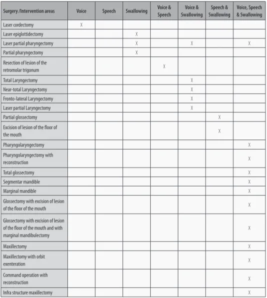
The various regions of the head and neck area are clearly involved in breathing, mastication, swallowing, voice and speech. Thus, partial or total removal of some of these structures will have side effects in some of these functions, alone or in association. (Boyle and Kraus; Tonini). Being Speech Therapists the professionals that assess and treat these disorders, their inclu-sion in multidisciplinary teams will be beneficial for a fast and effective rehabilitation, balan-cing the structures functioning as well as improving the psychosocial and emotional state of patients and caregivers (Gulfier). The knowledge of these possible surgical techniques in this functional complex enables us to predict the after-effects post-surgical in contexts of voice, speech and swallowing. (Crevièr-Buchamn, Brihaye and Tessier).
4.1.
ILLUSTRATION OF POSSIBLE AREAS OF INTERVENTION BASED
ON SURGERIES PERFORMED FROM ENT DEPARTMENT AT IPOPFG – EPE
116
Surgery /Intervention areas Voice Speech Swallowing Voice & Speech
Voice & Swallowing
Speech & Swallowing
Voice, Speech & Swallowing Laser cordectomy X
Laser epiglottidectomy X
Laser partial pharyngectomy X X X
Partial pharyngectomy X Resection of lesion of the
retromolar trigonum X
Total Laryngectomy X
Near-total Laryngectomy X
Fronto-lateral Laryngectomy X Laser partial Laryngectomy X
Partial glossectomy X
Excision of lesion of the floor of
the mouth X
Pharyngolaryngectomy X
Pharyngolaryngectomy with
reconstruction X
Total glossectomy X
Segmentar mandible X
Marginal mandible X
Glossectomy with excision of lesion
of the floor of the mouth X
Glossectomy with excision of lesion of the floor of the mouth and with marginal mandibulectomy
X
Maxillectomy X
Maxillectomy with orbit
exenteration X
Command operation with
reconstruction X
Infra structure maxillectomy X
TABLE 12 - Areas of intervention of the Speech Therapist, according to the surgical technique. (Behlau et al.; Camargo cit. in Mello; Carvalho, “Influência”; Carvalho,A atuação; Cichero; Crary and Groher; Crevièr-Buchman, Brihaye and Tes-èr-Buchman, Brihaye and Tes--Buchman, Brihaye and Tes-sier; Dwivedi et al.; Figueiredo et al.; Fouquet, Amaral and Vicente; Furia cit. in Carvalho, A atuação; Gielow; Gielow cit. in Carvalho, A atuação; Le Huche and Allali; Matos; Mello; Murray; Perkins, Hancock and Ward; Sanchez; Seif; Steffen and Feijó cit. in Mello; Vale et al.; Vicente; Zago and Sawada cit. in Carvalho,A atuação).
117
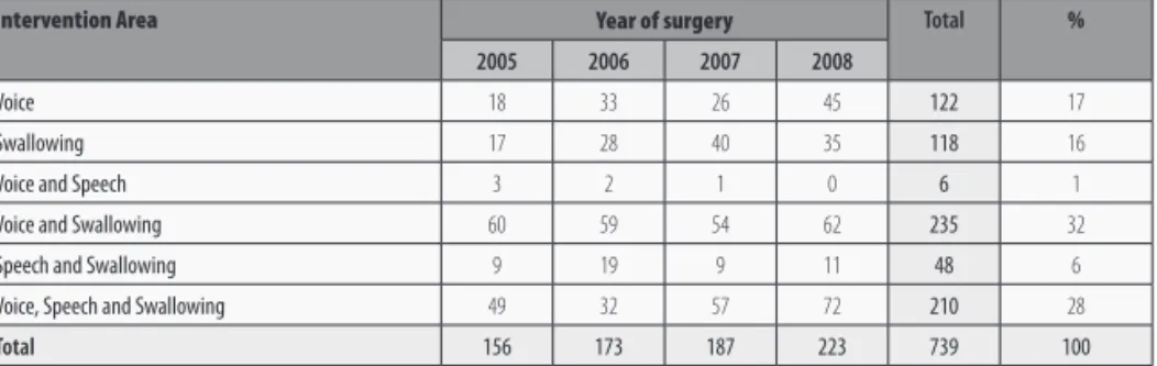
Intervention Area Year of surgery Total %
2005 2006 2007 2008
Voice 18 33 26 45 122 17
Swallowing 17 28 40 35 118 16
Voice and Speech 3 2 1 0 6 1
Voice and Swallowing 60 59 54 62 235 32 Speech and Swallowing 9 19 9 11 48 6 Voice, Speech and Swallowing 49 32 57 72 210 28
Total 156 173 187 223 739 100
TABLE 13 - Absolute Frequency of areas of intervention of Speech therapists.
According to the analysis, we can conclude that 82% of surgeries have direct implications in swallowing, 78% in voice and 35% in speech. These sequels can be multiple and associated.
5.
TEMPORAL ANALYSIS OF REHABILITATION PROCESS
Currently, the investigations that assess the costs of the intervention are more frequent, con-tributing to a more rational and meticulous rehabilitation. Head and neck cancer treatment involved different medical resources, high direct financial costs (services/institutions, me-dications, equipments) and indirect (working time spent by professionals, caregivers, other family resources and social support for patients). For these reasons, it is urgent to analyze carefully the different financial costs involved in this process (Terrell and Wilkins).
In this specific kind of rehabilitation, the frequency of Speech Therapy sessions should be whenever possible daily, while the patient is in the hospital and weekly after hospital dischar-ge. Each session should last about 30 minutes, but this could vary according to the needs of the patient, the Speech Therapist’s approach and time available. In the case of total laringec-tomies, the ideal intervention should be 3 to 4 weekly sessions (Lazarus, Ward and Yiu).
To accomplish this study, it was used as a measure the mean time of a Speech Therapy ses-sion, despite being a relative question, because it depends on physical and psychological factors as well as the commitment and motivation of the patients and professionals expe-rience, enable us to extrapolate some results. For this effect it was considered a 8-working hour, with weekly 30-minute sessions in outpatients. So, during a day, with a fully comple-ted schedule without breaks, a Therapist can perform 16 sessions. Consequently, during the time-span of this study (2005 to 2008), a Speech Therapist would make 14672 sessions, that multiplied by 30 minutes would give 7336 hours of work (table 14).
Year No sessions
2005 3664
2006 3648
2007 3680
2008 3680
Total 14672
118
Through the analysis of table 15 and based on the complexity of interventions in this area, we can ascertain that the pharyngolaryngeal topographical region is the one that requires more time of rehabilitation, followed by other locations/undefined lesions of the oral cavity and maxilla/mandible. This happens because the surgeries performed in these regions are more invasive and extensive.Another aspect to consider is the total number of sessions needed (19800) for the entire sample (739 surgeries). From the comparison between the table 14 and 15 we can see that 26% of the surgeries (192) would not have an appropriate rehabilitation. Taking into consi-deration this results, we can consider that to meet the requirements of this population, it would be needed at least two Speech Therapists to be part of the team in this Department, so that all patients could have access to a well-timed rehabilitation.
Topographical Region No total sessions No middle sessions No minimum sessions No maximum sessions
Oral cavity 1678 19,95 4 144
Maxilla/Mandible 2868 28,40 5 144
Oral cavity & Maxilla/Mandible 4032 144 144 144
Pharynx 2484 20,53 4 24
Larynx 3698 12,45 3 24
Pharyngolaryngeal region 5040 144 144 144
TABLE 15 - Distribution of the total number, mean, minimum, and maximum sessions per topographical region. (Behlau et al.; Berlin cit. in Mathieson; Berlin et al. cit. in Martin; Carvalho, “Influência”; Crevièr-Buchman, Brihaye and Tessier; Figueiredo et al.; Furia, Carrara-de Angelis e Mourão; Goode cit. in Mathieson; Laccoureye et al. cit. in Yeager and Grillone; Le Huche and Allali; Logemann; Mekaru et al.; Murray; Perkins, Hancock and Ward; Pou; Santos; Singer; Thomas and Keith; van As-Brooks, Finizia, and Ward; Yeager and Grillone).
6.
CONCLUSION
With this study, we aim to understand the reality of an Oncology Department, in what con-cern the necessity of Speech Therapists to include in the multidisciplinary teams. Cancer patients from an ENT Department need the intervention of these professionals and the amount of work is growing due to the high number of new cases.
119
7.
BIBLIOGRAPHY
Behlau, Mara et al. “Disfonias por câncer de cabeça e pescoço.” Voz - o livro do especialista. Ed. Mara Behlau. Vol. II. Rio de Janeiro: Editora Revinter, 2005. 213-85.
Boyle, Jay O., and Dennis H. Kraus. “Functional rehabilitation.” Essentials of Head and Neck
On-cology. Ed. Lanny G.Close, David L. Larson, and Jatin P. Shah. New York: Thieme Medical
Pub-lishers, 1998. 369-78.
Camargo, Zuleica A. “Reabilitação fonoaudiológica em câncer de laringe.” Fundamentos em
fonoaudiologia - tratando os distúrbios da voz. Ed. Sílvia M. Pinho. 2.ª Ed. (1.ª Ed. 1998). Rio de
Janeiro: Editora Guanabara Koogan, 2003. 101-16.
Carvalho, Viviane A. “Influência das próteses obturadoras e rebaixadoras de palato na terapia fonoaudiológica.” Fonoaudiologia em cancerologia. Ed. Ana P. Barros et al. São Paulo: Funda- São Paulo: Funda-: Funda-ção Oncocentro de São Paulo - Comité de Fonoaudiologia em Cancerologia, 2000. 93-98. Internet. 6 Jul. 2009 <http://bvsms.saude.gov.br/bvs/publicacoes/cd06_03.pdf>.
Carvalho, Melissa A. A atuação fonoaudiológica em pacientes laringectomizados totais. Mo-nografia. Centro de Especialização em Fonoaudiologia Clínica, 2001. Internet. 24 Jul. 2007 <www.cefac.br/library/teses/7e74eb76845190acc90347584b51ae6b.pdf>.
Cichero, Julie. “Conditions Commonly Associated with Dysphagia.” Dysphagia - Foundation, Theory
and Practice. Ed. Julie Cichero, and Bruce Murdoch. Chichester: John Wiley and Sons, 2006. 237-98.
Crary, Michael A., and Michael E. Groher. Introduction to Adult Swallowing Disorders. Maryland Heights, MO: Butterworth-Heinemann, 2003.
Crevièr-Buchamn, Lise, Sylvie Brihaye, and Christophe Tessier. La déglutition, après chirurgie
partielle du larynx. Marseille: Solal Éditeurs, 1998.
Dwivedi, Raghav C. et al.“Evaluation of Speech Outcomes Following Treatment of Oral and Oropharyngeal Cancers.” Cancer Treatment Reviews 35.5 (2009): 417-24.
Figueiredo, Elaine S. et al. “Aspectos fonoaudiológicos no pós-operatório da laringectomia near-total.” Fonoaudiologia em cancerologia. Ed. Ana P. Barros et al. São Paulo: Fundação On- São Paulo: Fundação On-: Fundação On-cocentro de São Paulo - Comité de Fonoaudiologia em Cancerologia, 2000. 207-13. Internet. 6 Jul. 2009 <http://bvsms.saude.gov.br/bvs/publicacoes/cd06_03.pdf>.
Fouquet, Marina L., Teresa C. Amaral, and Laélia C. Vicente. “Inteligibilidade de fala em pa-cientes com ressecção de tumor de cavidade de boca e/ou orofaringe.” Fonoaudiologia em
cancerologia. Ed. Ana P. Barros et al. São Paulo: Fundação Oncocentro de São Paulo - Comité
de Fonoaudiologia em Cancerologia, 2000. 195-204. Internet. 6 Set. 2007 <www.univag.com. br/biblioteca/Enfermagem/Cancer/Fonoaudiologia%20em%20cancerologia/cd06_03.pdf>.
Furia, Cristina L., Elisabete Carrara-de-Angelis, e Lúcia F. Mourão. “Inteligibilidade de fala nas dissecções de cavidade oral.” Fonoaudiologia em cancerologia. Ed. Ana P. Barros, et al. São Paulo:
120
Gielow, Ingrid. “Reabilitação fonoaudiológica da disfagia em pós-operatório de cirurgia de cabeça e pescoço.” Disfagias orofaríngeas. Ed. Ana M. Furkim, e Celia S. Santini (1.ª reimp. 2001). São Paulo: Pró-fono, 1999. 203-27.Gulfier, Blacy C. “Atuação fonoaudiológica em câncer de cabeça e pescoço - um estudo de caso.” Fonoaudiologia e lusofonia - I Simposium Luso-Brasileiro de Terapia da Fala. Ed. A. P. de Almeida. Porto: Edições U Fernando Pessoa, 2005. 251-54.
International Agency for Research on Cancer and World Health Organization. Cancer
Inci-dence, Mortality and Prevalence Worldwide, Estimates for the Year 2002, 2005. Internet. 15 Fev.
2010. <http://www.dep.iarc.fr/>.
Lazarus, Cathy L., Elizabeth C. Ward, and Edwin M. Yiu. “Speech and Swallowing Following Oral, Oropharyngeal, and Nasopharyngeal Cancers.” Head and Neck Cancer - Treatment,
Reha-bilitation, and Outcomes. Ed. Elizabeth C. Ward, and Corina J. van As-Brooks. San Diego: Plural
Publishing, 2007. 103-22.
Le Huche, François, and André Allali. La voz - patología vocal de origen orgánico. Vol. 3. Barce-Vol. 3. Barce-lona: Masson, 2004.
Logemann, Jeri A. Evaluation and Treatment of Swallowing Disorders. 2ª Ed. (1ª Ed. 1983). Austin: Pro-Ed, 1998.
Martin, Daniel. “Rehabilitación poslaringectomía.” La voz patológica. Ed. María C. Jackson--Menaldi. Buenos Aires: Editora Médica Panamericana, 2002. 99-117.
Mathieson, Lesley. Greene & Mathieson’s - The Voice and its Disorders. 6ª Ed. (1ª Ed. 1957). London: Whurr Publishers, 2005.
Matos, Belmiro J. “Laringectomias parciais.” Barros, Ana P. B. et al. (Org.). Fonoaudiologia em
Cancerologia. Ed. M. C. Jackson-Menaldi. São Paulo: Fundação Oncocentro de São Paulo -
Co-mité de Fonoaudiologia em Cancerologia, 2000. 38-47. Internet. 6 Jul. 2009 <http://bvsms. saude.gov.br/bvs/publicacoes/cd06_03.pdf>.
Mekaru, Daniela T. et al. “Laringectomizados totais: aspectos da reabilitação fonoaudiológi-“Laringectomizados totais: aspectos da reabilitação fonoaudiológi-ca.” Fonoaudiologia em cancerologia. Ed. Ana P. Barros et al. São Paulo: Fundação Oncocentro de São Paulo - Comité de Fonoaudiologia em Cancerologia, 2000. 226-36. Internet. 6 Jul. 2009 <http://bvsms.saude.gov.br/bvs/publicacoes/cd06_03.pdf>.
Mello, Jamara N. A atuação fonoaudiológica em laringectomias parciais. Monografia. Centro de Especialização em Fonoaudiologia Clínica, 2001. Internet. 24 Jul. 2009 <www.cefac.br/ library/teses/ b9268a083cbef4d6d0a75 ec4ff86 4a6a.pdf>.
Murray, Joseph. Manual of Dysphagia Assessment in Adults. San Diego: Singular Publishing Group, 1999.
Perkins, Kylie, Kelli L. Hancock, and Elizabeth C. Ward. “Speech and Swallowing Following Larynge-al And HypopharyngeLarynge-al Cancer.” Head and Neck Cancer - Treatment, Rehabilitation, And Outcomes.
121
Pou, Anna M. “Tracheoesophageal Voice Restoration with Total Laryngectomy.” The
Otolaryn-gologic Clinics of North America 37.3 (2004): 531-45.
Sanchez, Renata F. “Reabilitação fonoaudiológica após as laringectomias parciais.” Fonoaudiolo-gia em CanceroloFonoaudiolo-gia. Ed. Ana P. Barros, et al. São Paulo: Fundação Oncocentro de São Paulo - Co- São Paulo: Fundação Oncocentro de São Paulo - Co-: Fundação Oncocentro de São Paulo - Co-mité de Fonoaudiologia em Cancerologia, 2000. 78-82. Internet. 6 Set. 2007 <www.univag.com. br/biblioteca/Enfermagem/Cancer/Fonoaudiologia%20em%20cancerologia/cd06_03.pdf>.
Santos, Luiz R. “Laringectomia quase-total.” Fonoaudiologia em cancerologia. Ed. Ana P. Barros, et al. São Paulo: Fundação Oncocentro de São Paulo - Comité de Fonoaudiologia em Can- São Paulo: Fundação Oncocentro de São Paulo - Comité de Fonoaudiologia em Can-: Fundação Oncocentro de São Paulo - Comité de Fonoaudiologia em Can-cerologia, 2000. 48-52. Internet. 6 Jul. 2009 <http://bvsms.saude.gov.br/bvs/publicacoes/ cd06_03.pdf>.
Seif, Célia S. “Fonoaudiologia em cabeça e pescoço.” Diagnóstico e tratamento dos tumores de
cabeça e pescoço. Ed. Mauro M. Barbosa, Geraldo M. Sá, e Roberto A.Lima, São Paulo: Editora
Atheneu, 2001. 209-15.
Singer, Mark I. “The Development of Successful Tracheoesophageal Voice Restoration.” The
Otolaryngologic Clinics of North America 37.3 (2004): 507-17.
Terrell, Jeffrey E., and Edwin G. Wilkins. “Outcomes and Cost-Effectiveness of Contemporary Head and Neck Cancer Treatment.” Essentials of Head and Neck Oncology. Ed. Lanny G. Close, David L. Larson, and Jatin P. Shah. New York: Thieme Medical Publishers, 1998. 425-32.
Thomas, Jack E., and Robert L. Keith. Looking Forward… The Speech and Swallowing Guide-book for People with Cancer of the Larynx or Tongue. 4ª Ed. New York: Thieme Medical Publi-ª Ed. New York: Thieme Medical Publi-New York: Thieme Medical Publi-shers, 2005.
Tonini, Monique D. “Câncer de cabeça e pescoço: trabalho com voz, disfagia ou fala?”
Motri-cidade orofacial - como atuam os especialistas.Ed.Comité de Motricidade Orofacial da
Socie-dade Brasileira de Fonoaudiologia. São Paulo: Pulso Editorial, 2004. 259-62.
Vale, Luciana P. et al. “Laringectomia parcial supracricoídea: avaliação perceptiva auditiva e acústica da voz.” Fonoaudiologia em cancerologia. Ed. Ana P. Barros et al. São Paulo: Fundação Oncocentro de São Paulo - Comité de Fonoaudiologia em Cancerologia, 2000. 246-52. Inter-net. 6 Jul. 2009 <http://bvsms.saude.gov.br/bvs/publicacoes/cd06_03.pdf>.
van As-Brooks, Corina J., Caterina A. Finizia, and Elizabeth C. Ward. “Rehabilitation of Olfaction and Taste Following Total Laryngectomy.” Head and Neck Cancer - Treatment, Rehabilitation,
and Outcomes. Ed. Elizabeth C. Ward, and Corina J. van As-Brooks. San Diego: Plural
Publish-ing, 2007. 325-46.
Vicente, Laélia C. “Desafios e perspectivas no tratamento do câncer da boca e orofaringe: rea-bilitação fonoaudiológica das disfagias.” Fonoaudiologia em Cancerologia. Ed. Ana P. Barros et al. São Paulo: Fundação Oncocentro de São Paulo - Comité de Fonoaudiologia em Cancerologia, 2000. 152-158. Internet. 6 Jul. 2009 <http://bvsms.saude.gov.br/bvs/publicacoes/cd06_03.pdf>.