RESEARCH
Marcelo Buzzi
I, Franciele de Freitas
II, Marcos de Barros Winter
III I Rede Sarah de Hospitais de Reabilitação. Brasília, Distrito Federal, Brazil.II Phytoplenus Bioativos Sociedade Anônima. Pinhais, Paraná, Brazil.
III Hospital da Santa Casa de Misericórdia de Curitiba, Dermatology Department. Curitiba, Paraná, Brazil.
How to cite this article:
Buzzi M, Freitas F, Winter MB. Pressure ulcer healing with Plenusdermax® Calendula officinalis L. extract.
Rev Bras Enferm [Internet]. 2016;69(2):230-6. DOI: http://dx.doi.org/10.1590/0034-7167.2016690207i
Submission: 01-11-2015 Approval: 09-29-2015
ABSTRACT
Objective: to evaluate the therapeutic benefi ts of the bioactive extracts of Plenusdermax®Calendula offi cinalis for pressure
ulcer healing. Method: an observational cohort study, including 41 patients with a diagnosis of pressure ulcer that was stable in size for more than three months. Patients were evaluated every two weeks, over 30 weeks, for: reduction of the wound area, infection control, types of tissue and exudate, and ulcer microbiology. Results: the proportions of patients who were completely healed after 15 and 30 weeks of treatment were 63% and 88%, respectively, and the mean healing time was 12.5±7.8 weeks. No adverse events were observed during treatment. Conclusion: the results of the study indicate that bioactive C. offi cinalis Plenusdermax® is a safe treatment that promotes healing of pressure ulcers.
Key words: Pressure Ulcer; Wound Healing; Infl ammation; Anti-infl ammatory; Calendula Offi cinalis.
RESUMO
Objetivo: Avaliar os benefícios terapêuticos do extrato de bioativos de Calendula offi cinalis Plenusdermax® na cicatrização de
úlceras de pressão. Métodos: estudo observacional de coorte realizado com quarenta e um pacientes com diagnóstico de úlcera por pressão com tamanho da ferida estável por mais de três meses. Os pacientes foram avaliados quinzenalmente durante 30 semanas, em relação a redução da área da lesão, controle de infecção, tipos de tecido e exsudato e microbiologia das úlceras.
Resultados: a proporção de pacientes que cicatrizaram completamente após 15 e 30 semanas de tratamento foi 63% e 88%, respectivamente, sendo que a média de tempo de cicatrização foi de 12,5 ± 7,8 semanas. Não foram observados eventos adversos durante o tratamento. Conclusão: os resultados do estudo indicam que Plenusdermax® de bioativos de C. offi cinalis é
um tratamento seguro que promove a cicatrização de úlceras de pressão.
Descritores: Úlcera por Pressão; Cicatrização; Infl amação, Antiinfl amatório; Calendula Offi cinalis.
RESUMEN
Objetivo: evaluar los benefi cios terapéuticos del extracto bioactivo de Calendula offi cinalis Plenusdermax® en la cicatrización
de úlceras por presión. Método: estudio observacional de cohorte con cuarenta y un pacientes con diagnóstico de úlceras por presión con un tamaño de herida estable durante más de tres meses. Se evaluó a los pacientes durante 30 semanas cada dos semanas, incluyendo reducción del área de lesión, control de infecciones, tipos de tejidos y secreciones y microbiología de las úlceras. Resultados: la proporción de pacientes con cicatrización completa después de 15 y 30 semanas de tratamiento fue de 63% y 88%, respectivamente, y el promedio de todos los tiempos de cicatrización fue de 12.5±7.8 semanas. No se observaron eventos adversos durante el tratamiento. Conclusión: los resultados del estudio indican que Plenusdermax® con bioactivos de C. offi cinalis es un tratamiento seguro que promueve la cicatrización de úlceras por presión.
Palabras clave: Úlcera por Presión; Cicatrización de Heridas; Infl amación; Anti-infl amatoria; Calendula Offi cinalis.
Pressure ulcer healing with Plenusdermax
®
Calendula offi cinalis
L. extract
Cicatrização de úlceras por pressão com extrato Plenusdermax
®de Calendula offi cinalis L.
Cicatrización de úlceras por presión con extracto de Plenusdermax
®Calendula offi cinalis L.
Marcelo Buzzi E-mail: [email protected]
in the treatment of pressure ulcers, specifically in the process-es of healing, control of microbial colonization, tissue evolu-tion of the wounds, pain management, and also in nursing interventions such as debridement and dressing changes of patients who are bedridden at home or those who are wheel-chair users.
METHOD
Ethical aspects
This study was approved by the Institutional Committee of Ethics on Research at PUC-PR and was registered in Plata-forma Brasil with the National Commission for Research Eth-ics. Informed consent was obtained from all patients before screening.
Study design, setting and period
The efficacy of Plenusdermax®C. officinalis hydroglycolic
extract in the healing of pressure ulcers was clinically evalu-ated in a prospective, observational study over a 30-week pe-riod. The study was performed between May of 2012 and De-cember of 2013, and the bedridden and wheelchair patients with pressure ulcers were first seen at the outpatient clinic of the Dermatology Department of Hospital da Santa Casa de Mi-sericórdia, Curitiba, Catholic University of Paraná (PUC-PR).
Inclusion and exclusion criteria
Patients with pressure ulcers in the metropolitan area of Curitiba were selected, according to the inclusion and exclu-sion criteria shown in Box 1.
Study protocol
The nursing team monitored the patients every two weeks for 30 weeks, or until complete healing of the ulcer occurred. Healing was confirmed one week after complete healing and was monitored for another two weeks. At base-line, a complete medical history and an assessment of the current condition of the patients were recorded. Sociodemo-graphic data, medical history, clinical evaluation of wounds, and cleaning procedures were collected and assessed. The initial patient conditions were evaluated in terms of the du-ration of the ulcer, glycemic control, current activity level, nutritional status, and pain score according to the numerical pain scale(9). Patients were scored according to the Pressure
Ulcer Scale for Healing (PUSH) tool (NPUAP) to estimate the severity and staging of the pressure ulcers(4). Blood tests
results were included: glucose level, serum albumin, blood count and erythrocyte sedimentation rate, bacterioscopy and culture of the ulcer bed.
The microbiological flora was evaluated based on bio-gram/antibiogram of swab collection from the wound bed. The wounds were photographed and the area determined by computerized planimetry, using ImageJ® software (National
In-stitutes of Health, Bethesda, MD, USA), and an area known as standard(10). The clinical appearance of the wounds was
char-acterized according to the presence of granulation tissue, fi-brin, exudate, unpleasant odor, epithelialization and necrosis.
INTRODUCTION
Pressure ulcers, also known as skin ulcers, decubitus ulcers and bedsores, are complex, chronic wounds. They are a fre-quent cause of morbidity in patients in hospitals and nursing homes, requiring expensive and time-consuming treatments(1).
The healing process of chronic wounds, such as pressure ul-cers, is more complex than that of acute wounds, which leads to difficulties in treatment management. Chronic wounds such as pressure ulcers might never heal if not properly treated(2).
Pressure ulcers are areas of localized skin injuries and under-lying tissues that usually develop over bony prominences. They occur as a result of continuous pressure on the skin, soft tissue, muscle and bone, leading to localized ischemia, fol-lowed by a cascade of processes that result in necrosis(3). The
body areas commonly prone to pressure ulcers include the heels, hips, elbows, shoulders, back of the head, knees, thighs and toes(3). Ulcer severity is assessed in several ways, but the
pressure ulcer staging system in the United States (United States National Pressure Ulcer Advisory Panel - NPUAP) is the most widely used across the health care community. The NPUAP system includes a four-stage classification, represent-ing progressive severity from intact skin with non-blanchable redness of an area in Stage I, to full thickness tissue loss with exposed bone, tendon or muscle in Stage IV(2-4).
Local treatment of pressure ulcers is based on the use of dressings that protect the wound and provide a favorable environment for healing to occur. International guidelines for the treatment of pressure ulcers include essential proce-dures, such as: pressure relief therapy on the injured area, through consistent repositioning of the patient in bed; nutri-tional diet that is rich in proteins, carbohydrates, vitamins, minerals and important microelements to support formation of granulation tissue; infection control with adequate asepsis and antibiotic therapy; wound bed preparation of injury bed with debridement of necrosis and devitalized tissue; special-ized dressings for the preservation of the wound bed; plastic surgery; and, use of topical therapies that are adjuvant to stimulate tissue repair(2-4).
Several topical products have been developed to reduce the healing time and pain, absorb exudate and blood, and promote rapid healing of the wound(5-6). The topical adjuvant
therapies based on natural products and plants for chronic ulcers have been widely used to reduce healing time, infec-tion, inflammation and edema(7). Calendula officinalis flower
extracts are used in many topical preparations, such as an-ti-inflammatory agents, healing agents of skin and mucous membrane injuries, and for the treatment of herpes, solar erythemas, burns, and dermatitis(8). The treatment of
pres-sure ulcers in hospitals and home care remains challenging for the nursing team, which motivates research for new, more effective products, despite the various adjuvant therapies cur-rently available, such as antimicrobials, advanced dressings, prepared from natural products, growth factors, among others.
ulcers, the product was applied in excess to reach inside the cavity. In the case of Stage II pressure ulcers, the bubbles were not burst and Plenusdermax® was applied twice a day in the
wound region, leaving the product to dry for five minutes. In Stage III pressure ulcers, the product was applied twice a day in the wound region and allowed to dry for five minutes. After application and drying of the product, the wound area was occluded with gauze and affixed with sterile micropore. As preventive measures, the incident pressure was reduced and intensive decubitus change was performed (2/2 h to 1/1h). In tunneled ulcers, no gauze was packed into the tunnels, only the orifice was occluded. Exudate control was performed by observing the amount drained, and adjustments were made with more frequent changes of sterile gauze dressings.
In all visits, photographs of the ulcers were obtained by the nursing team for the planimetry, to estimate the wound areas and to evaluate their clinical aspects. Quantitative parameters were evaluated every two weeks from the beginning until the completion of the study (30 weeks). Complete wound closure was defined as complete ulcer epithelialization without any drainage. Patients who experienced an allergic reaction to Ple-nusdermax®, or a substantial bacterial infection, were treated by
the medical director investigator. Patients who suffered allergic reactions were removed from the clinical trial, whereas antibi-otic therapy was administered in cases of infection. Patients who had fever and other infectious complications resulting from their wounds during the treatment period were excluded.
Statistical analysis
Quantitative variables (mean, median, minimum, maximum, and standard deviation) were used for descriptive statistics. The qualitative variables were described as frequencies and per-centages. The wound contraction (mm2/week) per week (WoC)
was calculated as the initial wound area (iWA) minus the final wound area (fWA), divided by the number of weeks:
The healing rate by week (WHR%) was calculated as follows:
The healing time was evaluated based on the calculation of the cumulative frequency plot. The Mann-Whitney’s U test for matched pairs was used to evaluate the differences in the measured variables at the beginning and at the end of the treatment period. Results with a p<0.05 were considered statistically significant. Data were analyzed using the Statisti-cal Package for the Social Sciences (SPSS) statistiStatisti-cal software, version 20.0 (IBM Corp. Armonk, NY).
RESULTS
The patient demographics and ulcer characteristics at base-line and after 30 weeks of treatment with Plenusdermax® are
Box 1 – Inclusion and exclusion criteria for people with pressure ulcer
Inclusion criteria Exclusion criteria
• Age between 18 and 90 years.
• No previous history of allergy to any plant of the
Asteraceae family
• Pressure ulcer (target ulcer) determined by clinical as-sessment, present for at least five weeks from the date of the screening visit, with a surface area between 1-30 cm2 measured by compu-terized planimetry, in the sacral or trochanteric region, classified as stage II to III, according to NPUAP PUSH scale.
• Non-diabetic patients (type 1 or type 2), as diagnosed by a physician, with adequate glycemic control during selection. Blood glucose, glycosylated hemoglobin A1c, serum albumin, blood count, erythrocyte sedimen-tation rate, bacterioscopy, swab culture and antibio-gram of the wound bed, measured at the screening visit, to help the researcher in selection.
• Willingness to be visited by the nursing team during the study period.
• Individuals or caregivers who can follow strict protocol recommendations required during the study period.
• Stage IV pressure ulcers or necrotic tissue that could not be adequately debrided by the nursing team.
• Pressure ulcers with clinical evidence of apparent infec-tion, e.g., ulcers surrounded by an advanced, hard red edge, hot or tender with very purulent exudate.
• Individuals with an infected target ulcer, including (but not limited to) cellulitis, osteomyelitis, or gangrene, infections of deeper tissues. • Clinically significant medical
conditions that can impair wound healing (e.g., kidney, liver, anemia, severe malnu-trition and immunocompro-mising).
• Pregnant women of chil-dbearing age who do not use an approved method of contraception.
• Individuals who are receiving or have received corticoste-roids, immunosuppressants, radiation or chemotherapy systemically (oral/intravenous) within four weeks of entering the study.
• History of Plenusdermax® use in the target ulcer less than 12 weeks before the screening visit.
• History of drug or alcohol abuse in the previous year, or those who currently use illicit drugs.
• Individuals with any other condition which, in the opinion of the investigator, would render the patient ineligible for the study.
Treatment with Plenusdermax®
The healing product based on Phytoplenus Plenusdermax®
(Phytoplenus Bioativos S.A., Pinhais, PR, Brazil), with a liquid presentation and topical spray application, was used twice dai-ly after cleaning the wound with sterile saline. The study par-ticipants, patients and their caregivers, were informed verbally and in writing, about the dosage, conditions for drug storage, as well as guidelines for use, care for and change of dressings, along with the informed consent. The spray was thoroughly applied to the wound, leaving the wound bed and wound edges moistened with the product. In the case of tunneled
WoCs
=
(
iWA – fWA
)
weeks
WHR%s=
weeks
(iWA – fWA)
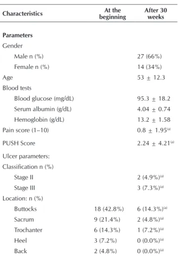
shown in Table 1. Of the 41 patients treated with Plenusder-max®, 14 (34%) were female and 27 (66%) were male. Pa-tients had a mean PUSH score of 9.63±2.41, which rose to 2.24±4.21 at the end of the 30-week treatment. The neuro-pathic pain scale showed a mild mean pain score (1.6) due to the high number of spinal cord injury patients. The glucose, serum albumin and hemoglobin levels were normal in all pa-tients during the study. There were no significant differences in blood parameters between patients who achieved com-plete wound closure within 30 weeks and those who did not (p=0.503, Student’s t-test).
Most patients had Stage III pressure ulcers (70.7%), typical-ly in the buttocks (43%), sacral (21%) and trochanteric (14%) regions. All pressure ulcers and wounds were characterized as chronic, because the patients developed them on average 41.4±33.6 weeks before the start of the study. The mean wound area at baseline was 3.74±2.34cm2 (range: 1.1 to
11.0 cm2). The biogram/antibiogram swab tests collected from
the wound beds and within the lesions showed that 43.9% (18/41) of the ulcers were clinically colonized. Staphylococ-cus aureus and Escherichia coli were the predominantly pres-ent pathogens (15%, 6/41 each), followed by Pseudomonas aeruginosa (7%, 3/41) and Klebsiella pneumoniae (5%, 2/41).
Table 1 – Demographic characteristics of patients with pres-sure ulcers at the beginning and after 30 weeks of treatment
Characteristics At the
beginning
After 30 weeks
Parameters
Gender
Male n (%) 27 (66%)
Female n (%) 14 (34%)
Age 53 ± 12.3
Blood tests
Blood glucose (mg/dL) 95.3 ± 18.2 Serum albumin (g/dL) 4.04 ± 0.74 Hemoglobin (g/dL) 13.2 ± 1.58 Pain score (1–10) 0.8 ± 1.95(a)
PUSH Score 2.24 ± 4.21(a)
Ulcer parameters: Classification n (%)
Stage II 2 (4.9%)(a)
Stage III 3 (7.3%)(a)
Location: n (%)
Buttocks 18 (42.8%) 6 (14.3%)(a)
Sacrum 9 (21.4%) 2 (4.8%)(a)
Trochanter 6 (14.3%) 1 (7.2%)(a)
Heel 3 (7.2%) 0 (0.0%)(a)
Back 2 (4.8%) 0 (0.0%)(a)
Thigh 2 (4.8%) 0 (0.0%)(a)
Leg 1 (2.4%) 0 (0.0%)(a)
Toes 1 (2.4%) 0 (0.0%)(a)
Duration (weeks) 41.4 ± 36.6
-Ulcer baseline area (cm2) 3.74 ± 2.34 0.54 ± 1.66(a)
Aspect of the wound bed
% Epithelium 5.1% 87.8%(a)
% Granulation 68.4% 21.3%(a)
% Moderate exudate 78.1% 12.2%(a)
% Fibrin 26.8% 7.3%(a)
% Necrosis 12.2% 0.0%(a)
% Unpleasant odor 36.6% 2.4%(a)
Ulcer microbiology
Staphylococcus aureus 14.8% 0.0%(a)
Pseudomonas aeruginosa 7.3 % 2.4%(a)
Klebsiella pneumoniae 4.9 % 0.0%(a)
Escherichia coli 14.8% 4.8%(a)
Others 12.2% 2.4%(a)
Notes: (a)Significant difference (Mann-Whitney’s U Non-parametric Test, p
<0.05); Values are mean ± standard deviation, unless otherwise stated.
Healing time and reduction of wound area
The time analysis indicated a linear increase in the propor-tion of patients who achieved complete healing after two to 30 weeks of treatment (Figure 1). Thus, a minimum eight-week pe-riod of treatment was necessary for complete ulcer healing, the mean healing time of about 12.5±7.8 weeks. After 15 weeks of treatment, 63% of ulcers were completely healed, with notable improvements in appearance, compared to baseline. After 30 weeks, the proportion of completely healed wounds was 88%. No adverse events were observed during treatment.
Figure 1 – Proportion of healed pressure ulcers during the 30-week treatmentwith Plenusdermax®
Based on the fact that pressure ulcers with larger areas take longer to heal than smaller ulcers, and due to the large varia-tion (range 1.1 to 11.0 cm2) in the initial area of the ulcers, it
Table 1 (concluded)
To be continued
0,0 10,0 20,0 30,0 40,0 50,0 60,0 70,0 80,0 90,0 100,0
1 2 3 4 5 6 7 8 9 10 11 12 13 14 15 16 17 18 19 20 21 22 23 24 25 26 27 28 29 30 31
Length of Treatment (weeks)
was necessary to separate the sample into two groups, based on the initial ulcer area in order to more accurately assess the healing rates. The first group included those with smaller ulcers ranging from 1.0 to 3.9 cm2; the second included those
with larger ulcers, ranging from 4.0 to 11.0 cm2 (Table 2).
Regarding the duration of ulcers before the study, there was no significant difference between the group of smaller ulcers and the group of larger ulcers (p=0.850, Mann-Whitney’s U test) (Table 2). As predicted, the group of smaller ulcers had a mean ulcer area at baseline (2.23 cm2) that was significantly
lower (p≤0.001) than the group of larger ulcers (7.71cm2).
At the end of 30 weeks, all ulcers smaller than 4.0 cm2 were
completely healed, whereas 58% of larger ulcers were com-pletely healed (Table 2).
Despite the mean healing time of smaller ulcers being lower than that of larger ulcers during the 30-week period of the study, these values are not statistically different (p=0.857, Mann-Whitney’s U test). Although not statistically significant (p=0.465), the wound contraction rate per week (WoC - mm2/s) was 52% higher in the larger ulcers than in the smaller
ones, indicating, in absolute terms, a greater contraction ratio in the larger wounds. The percentage healing rate by week (WHR%), which considers the initial wound area in smaller ulcers (14.3%) was significantly two times faster than in the larger ulcers (6.6%). Collectively, these data suggest that the healing benefits of Plenusdermax® is not dependent on the
initial size of the pressure ulcer.
Inflammation and microbiology of the pressure ulcers
After 30 weeks of treatment with Plenusdermax®, the
to-tal number of colonized pressure ulcers was significantly re-duced from 18 (44%) to 5 (12.1%) (Mann-Whitney’s U test; p=0.011; Table 1). It is also important to mention that Plenus-dermax® was equally effective against all pathogens identified
in the wounds. Therefore, the number of wounds that had an
unpleasant odor at baseline (36.6%) was significantly lower after two weeks, and was reduced to 2.4% after treatment. These data are consistent with the dramatic reduction in in-flammatory cells and fibrin induced by Plenusdermax®.
Equal-ly interesting is the complete disappearance of necrotic tissue after treatment. Taken together, these data demonstrate that Plenusdermax® promotes closure of chronic injury through
strong antimicrobial and anti-inflammatory properties that is able to prevent tissue damage.
DISCUSSION
In this observational cohort study, the beneficial effects of the bioactive Plenusdermax®C. officinalis spray was tested in
healing pressure ulcers in bedridden patients and wheelchair users who used the product regularly at home, with instruc-tions by the nursing team which is a reality in most treatments. The treatment administered to patients followed interna-tional guidelines, including repositioning and control of tis-sue overload, colonization and infection control of ulcers as well as cleaning procedures and care as adjunctive therapy, i.e., Plenusdermax®. However, evidence indicates that in
Bra-zil, the international guidelines for the treatment of pressure ulcers are not regularly followed. An observational study in a general hospital found a low level of adherence to the use of guidelines for the treatment of pressure ulcers, and, in the case of Stage II ulcers, nursing interventions consisted pre-dominantly of cleansing with saline (57%), using vegetable oils (40%) and decubitus change maneuvers, whereas in Stage III ulcers, cleaning interventions with saline (84%) and soapy water (19%), use of debridants such as papain (32 %) and collagenase (14%), and use of antimicrobials, such as silver sulfadiazine (10%) predominated(11). Another observational
cohort study showed that bedridden patients with pressure ul-cers treated at home had a high risk of aggravating the healing process, had an overall worse condition which resulted in a
Table 2 – Healing parameters of patients with pressure ulcer treated with Plenusdermax® for 30 weeks
Groups according to ulcer size (cm²) All Smaller 1.0 – 3.9 Bigger 4.0 – 10.0 p value*
Number of patients 41 29 12
Duration (weeks) 41.4 ± 33.6 42.3 ± 36.2 39.3 ± 22.6 0.850
Initial area (cm²) 3.74 ± 2.82 2.23 ± 0.81 7.71 ± 2.65 ≤ 0.001
Final area (cm²) 0.55 ± 1.66 0.00 ± 0.00 2.03 ± 2.65 ≤ 0.001
Mean time until healing (weeks) 12.0 ± 7.85 11.7 ± 7.64 12.9 ± 9.45 0.857
% healing (30 weeks) 87.8 100.0 58.3
Wound contraction index per week (WoC - mm²/week) 36.1 ± 32.6 30.2 ± 24.3 45.8 ± 41.8 0.465
Percentage wound rate (WHR%) 12.4 ± 11.1 14.3 ± 11.7 6.6 ± 5.3 0.027
three times higher morbidity when compared to other types of injuries(11). The same study also showed that domiciled
pa-tients with increased risk of developing pressure ulcers were those that had limited repositioning capability, did not feel the need to reposition, had fecal incontinence, dementia and/or difficulty feeding themselves(12).
The results obtained with the use of the bioactive extract Plenusdermax® as an adjuvant topical therapy demonstrated
that, despite the adversity of home treatment, the ulcer treat-ment protocol is limited. Eighty-eight percent of patients who had stable ulcers for more than three months achieved complete healing over the 30-week period of the study; the mean healing time was approximately 12 weeks. A percentage of 58% of pa-tients had more difficulty achieving complete healing, probably because they had larger ulcers, some with areas up to 11cm2.
Nevertheless, the mean time to healing in these patients was not statistically different from those with smaller ulcers.
The proportion of patients who achieved complete healing with this treatment during the study period corroborated effec-tive treatments for pressure ulcers, since studies showed that high rates of Stage III ulcer healing can reach 59% at 24 weeks, with treatment of up to one year being necessary in some patients(13).
A significant reduction in odor, edema, erythema and bacte-rial count was observed in the studied patients. This reduction is due to the anti-inflammatory and antibacterial properties of the bioactive extract of Plenusdermax®C. officinalis, since
bacterioscopy swabs collected from the wound bed showed significant reduction of colonization by Staphylococcus au-reus and Escherichia coli after using Plenusdermax®. Calen-dula officinalis extracts have proven to have bactericidal and fungicidal effects against pathogenic microorganisms isolated from hospital patients(14). The formation of bacterial biofilms
in the wound bed of the lesions is extremely harmful to the tis-sue and a major contributor to the establishment of a chronic and degrading condition of the ulcers. This situation is further aggravated by the emergence of multidrug-resistant bacteria in response to the abuse of antibiotics, intensifying the search for new strategies to combat bacterial colonization(15). In many
pressure ulcer healing therapies, aggressive oxidizing antisep-tics are still used very frequently, such as hydrogen peroxide, hypochlorite, acetic acid, chlorhexidine, povidone/iodine, ce-trimide, among others, which have antibacterial properties, but are very toxic to healthy granulation tissue(2).
The use of natural extracts for the treatment of chronic skin injuries has always been an alternative adjuvant therapy in
the healing area. With the advent of recent and better tech-nologies for obtaining natural bioactives, new therapies have contributed to tissue repair in chronic injuries, preparing the wound bed for better healing(16-18).
Plenusdermax® extract showed an increase in
anti-inflam-matory, anti-edematous, anti-erythematous and healing activi-ties that corroborate previous clinical and experimental stud-ies using C. officinalis phyto-preparations(19-24). The beneficial
effects of Plenusdermax® in healing of pressure ulcers are due
to bioactive constituents such as triterpenoids monoesters, tri-terpene alcohols, triterpenic oligoglycosides and flavonoids, which may work synergistically to promote wound healing.
The therapy using the bioactive Plenusdermax®C. officina-lis spray offered advantages when compared to other adjuvant therapies for the treatment of chronic injuries. The bioactive C. officinalis extract is an aqueous preparation that leaves no solid residue adhering to the wound bed, has ease of application, fa-cilitates the cleaning process and dressing changes, and helps the debridement process. Therefore, it minimizes friction and prevents small traumas on the emerging granulation tissue.
Although the present study is an uncontrolled observational cohort, which has limitations in determining the guaranteed clinical efficacy of randomized controlled clinical trials, the results showed that the use of the bioactive Plenusdermax® C. officinalis spray was a powerful adjuvant in the treatment of pressure ulcers, as it aids nursing interventions such as dressing changes and care of ulcers. Accordingly, the use of the spray helps clean the wound bed, aids in debridement and stimula-tion of granulastimula-tion tissue growth, providing a better healing.
CONCLUSION
The results of this study indicate that the bioactive extract of Plenusdermax® C. officinalis is a safe and promising therapy
for the treatment of pressure ulcers in bedridden and wheel-chair users treated at home. The use of Plenusdermax® spray
as an adjunctive topical therapy resulted in complete healing of pressure ulcers in 88% of patients during the 30-week pe-riod, in addition to helping with wound debridement, dress-ing change and promotion of significant reduction in bacte-rial colonization. However, randomized controlled tbacte-rials on a larger scale with a longer follow-up are required to demon-strate clinical efficacy, to establish optimal treatment guide-lines using Plenusdermax®, and also to allow for these results
to be validated in a broad population.
REFERENCES
1. Cooper L, Vellodi C, Stansby G, Avital L. The prevention and management of pressure ulcers: summary of updated NICE guidance. J. Wound Care [Internet]. 2015[cited 2014 Feb 09];24(4):179-81. Available from: http://www.magonline library.com/doi/full/10.12968/jowc.2015.24.4.179 2. Whitney J, Phillips L, Aslam R, Barbul A, Gottrup F, Gould
L, et al. Guidelines for the treatment of pressure ulcers.
Wound Repair Regen [Internet]. 2006[cited 2014 Feb 09];14:663-79. Available from: http://onlinelibrary.wiley. com/doi/10.1111/j.1524-475X.2006.00175.x/epdf 3. National Pressure Ulcer Advisory Panel, European
Media: Osborne Park, Western Australia; 2014.
4. Santos V, Azevedo M, Silva T, Carvalho V, Carvalho V. [Crosscultural adaptation of the pressure ulcer scale for healing to the portuguese language]. Rev Latino-Am En-fermagem [Internet]. 2005[cited 2014 Feb 09];13(3):305-13. Available from: http://www.scielo.br/pdf/rlae/v13n3/ v13n3a04.pdf Portuguese.
5. Reddy M, Gill SS, Kalkar SR, Wu W, Anderson PJ, Rochon PA. Treatment of pressure ulcers: a systematic review. JAMA [Internet]. 2008[cited 2014 Feb 09];300(22):2647-62. Available from: http://jama.jamanetwork.com/article. aspx?articleid=183029
6. Banks V. Pressure sores: topical treatment and the healing process. J Wound Care. 1998 May;7(5):265-6.
7. Avijgan M. Phytotherapy: an alternative treatment for non-healing ulcers. J Wound Care [Internet]. 2004[cited 2014 Feb 09];13(4):157-8. Available from: http://www.ncbi. nlm.nih.gov/pubmed/15114830
8. Arora D, Rani A, Sharma A. A review on phytochemis-try and ethnopharmacological aspects of genus Calen-dula. Pharmacogn Rev [Internet]. 2013[cited 2014 Feb 09];7(14):179-87. Available from: http://www.phcogrev. com/text.asp?2013/7/14/179/120520
9. Hawker GA, Mian S, Kendzerska T, Franch M. Measures of adult pain. Arthritis Care Res [Internet]. 2011[cited 2014 Feb 09];63(s11):s240-52. Available from: http://online library.wiley.com/doi/10.1002/acr.20543/epdf
10. Caetano KS, Frade MAC, Minatel DG, Santana LA, En-wemeka CS. Phototherapy improves healing of chronic venous ulcers. Photomed Laser Surg [Internet]. 2009[cited 2014 Feb 09];27(1):111-8. Available from: http://online. liebertpub.com/doi/abs/10.1089/pho.2008.2398 11. Rangel EML, CaliriI MHL. Uso das diretrizes para
trata-mento da úlcera por pressão por enfermeiros de um hos-pital geral. Rev Eletr Enf [Internet]. 2009[cited 2014 Feb 09];11(1):70-7. Available from: http://www.fen.ufg.br/re-vista/v11/n1/v11n1a09.htm
12. Smith DM. Pressure ulcers in the nursing home. Ann Inter Med [Internet]. 1995[cited 2014 Feb 09];123(6):433-8. Avail-able from: http://annals.org/article.aspx?articleid=708996 13. Thomas DR. Prevention and treatment of pressure ulcers:
what works? what doesn’t? Cleve Clin J Med [Internet]. 2001[cited 2014 Feb 09];68(8):704-7. Available from: http://www.ccjm.org/cgi/doi/10.3949/ccjm.68.8.704 14. Efstratiou E, Hussain AI, Nigam PS, Moore JE, Ayub MA,
Rao JR. Antimicrobial activity of Calendula officinalis petal extracts against fungi, as well as negative and Gram-positive clinical pathogens. Complement Ther Clin Pract [Internet]. 2012[cited 2014 Mar 14];18(3):173-6. Available from: http://www.ncbi.nlm.nih.gov/pubmed/22789794 15. Ammons M. Anti-biofilm strategies and the need for
inno-vations in wound care. Recent Pat Antiinfect Drug Discov [Internet]. 2010[cited 2014 Feb 09];5(1):10-7. Available from: http://www.eurekaselect.com/85459/article
16. Dorai A. Wound care with traditional, complementary and alternative medicine. Indian J Plast Surg [Internet]. 2012[cit-ed 2014 Feb 09];45(2):418-24. Available from: http://www. ncbi.nlm.nih.gov/pmc/articles/PMC3495394/.
17. Preethi KC, Kuttan R. Wound healing activity of flower ex-tract of Calendula officinalis. J Basic Clin Physiol Pharma-col [Internet]. 2009[cited 2014 Feb 09];20:73-9. Available from: http://www.ncbi.nlm.nih.gov/pubmed/19601397 18. Hamburger M, Adler S, Baumann D, Förg A, Weinreich
B. Preparative purification of the major anti-inflamma-tory triterpenoid esters from Marigold (Calendula offi-cinalis). Fitoterapia [Internet]. 2003 Jun [cited 2014 Feb 09];74(4):328-38. Available from: http://www.ncbi.nlm. nih.gov/pubmed/12781802
19. Della Loggia R, Tubaro A, Sosa S, Becker H, Saar S, Isaac O. The Role of Triterpenoids in the Topical Anti-Inflamma-tory Activity of Calendula officinalis Flowers. Planta Med [Internet]. 1994[cited 2014 Feb 09];60(6):516-20. Available from: http://www.ncbi.nlm.nih.gov/pubmed/9254116 20. Zitterl-Eglseer K, Sosa S, Jurenitsch J, Schubert-Zsilavecz M,
Della Loggia R, Tubaro A, et al. Anti-oedematous activities of the main triterpendiol esters of marigold (Calendula of-ficinalis L.). J Ethnopharmacol [Internet]. 1997[cited 2014 Feb 09];57(2):139-44. Available from: http://www.ncbi. nlm.nih.gov/pubmed/9254116
21. Babaee N, Moslemi D, Khalilpour M, Vejdani F, Moghad-amnia Y, Bijani A, et al. Antioxidant capacity of Calendula officinalis flowers extract and prevention of radiation induced oropharyngeal mucositis in patients with head and neck can-cers: a randomized controlled clinical study. Daru [Internet]. 2013[cited 2014 Feb 09];21(1):18. Available from: http://daru jps.biomedcentral.com/articles/10.1186/2008-2231-21-18 22. Duran V, Matic M, Jovanovć M, Mimica N, Gajinov Z, Poljacki
M, et al. Results of the clinical examination of an ointment with marigold (Calendula officinalis) extract in the treatment of venous leg ulcers. Int J Tissue React [Internet]. 2005[cited 2014 Feb 09];27(3)(March):101-6. Available from: https:// www.researchgate.net/publication/7399603_Results_of_th e_clinical_examination_of_an_ointment_with_marigold_Cal endula_Officinalis_extract_in_the_treatment_of_venous_ leg_ulcers
23. Lavagna SM, Secci D, Chimenti P, Bonsignore L, Ottavi-ani a, Bizzarri B. Efficacy of Hypericum and Calendula oils in the epithelial reconstruction of surgical wounds in childbirth with caesarean section. Farmaco [Internet]. 2001[cited 2014 Feb 09];56(5-7):451-3. Available from: http://www.ncbi.nlm.nih.gov/pubmed/11482776