Cop
yright
© ABE&M t
odos os dir
eit
os r
eser
vados
.
Correspondence to: Denise Prado Momesso Rua Eduardo Guinle, 20/ 904 22260-090 − Rio de Janeiro, RJ, Brazil
dmomesso@terra.com.br
Received on Apr/21/2010 Accepted on Feb/22/2011
Body composition, metabolic
syndrome and insulin resistance
in type 1 diabetes mellitus
Composição corporal, síndrome metabólica e resistência insulínica no diabetes melito tipo 1
Denise Prado Momesso1, Isabela Bussade1,2, Giovanna A. Balarini Lima1, Leniane Pereira Coelho Fonseca1, Luis Augusto Tavares Russo3, Rosane Kupfer1
ABSTRACT
Objective: Our aim was to determine the relationship between body fat composition, metabo-lic syndrome (MS), and insulin resistance in type 1 diabetes (DM1). Subjects and methods:
Forty-five DM1 women (36 ± 9 years; body mass index 24.6 ± 4.4 kg/m2) had body composition and insulin resistance determined by dual-energy X-ray absorptiometry and estimated glucose disposal ratio (eGDR), respectively. Twenty patients (45%) had MS according to World Health Organization (WHO) criteria.Results: Women with DM1 and MS had increased central fat and lower eGDR than women without MS (41.9 ± 2.0 vs. 33.7 ± 1.8%; p = 0.004 and 4.99 ± 0.40 vs.
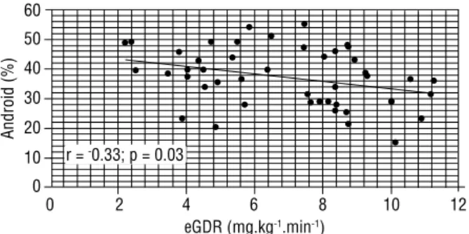
8.37 ± 0.39; p < 0.0001, respectively). Total body fat and peripheric fat were similar between the groups. Central fat negatively correlated with eGDR (r = -0.33; p = 0.03). Conclusion: Central fat deposition in young non-obese DM1 women was related to MS and insulin resistance. Thus, body fat composition analysis might be important to identify DM1 patients with increased me-tabolic risk. Arq Bras Endocrinol Metab. 2011;55(3):189-93
Keywords
Type 1 diabetes; body composition; metabolic syndrome; insulin resistance
RESUMO
Objetivo: Avaliar a relação entre composição corporal, síndrome metabólica (SM) e resistência insulínica (RI) no diabetes tipo 1 (DM1). Sujeitos e métodos: Quarenta e cinco mulheres com DM1 (36 ± 9 anos; índice de massa corporal 24,6 ± 4,4 kg/m2) foram submetidas à análise de compo-sição corporal e RI por meio de densitometria por dupla emissão de raios-X e taxa de disponibi-lização de glicose estimada (eGDR), respectivamente. Vinte mulheres (45%) apresentavam SM, conforme critérios da Organização Mundial da Saúde (OMS). Resultados: Mulheres com SM apresentaram maior gordura central e menor eGDR do que as sem SM (41,9 ± 2,0 vs. 33,7±1,8%;
p = 0,004 e 4,99 ± 0,40 vs. 8,37 ± 0,39; p < 0,0001). A gordura corporal total e a gordura periférica
não diferiram entre os grupos. A gordura central foi inversamente correlacionada com eGDR (r = -0,33; p = 0,03). Conclusão: Deposição de gordura central em mulheres jovens não obesas com DM1 esteve associada com SM e RI. Avaliação da composição corporal pode ser importan-te na identificação de pacienimportan-tes com risco metabólico elevado. Arq Bras Endocrinol Metab. 2011;55(3):189-93
Descritores
Diabetes tipo 1; composição corporal; síndrome metabólica; resistência insulínica
1 Department of Diabetes,
State Institute of Diabetes and Endocrinology, Universidade Católica do Rio de Janeiro (PUC-RJ), Rio de Janeiro, RJ, Brazil
2 Clinical and Experimental
Research Laboratory on Vascular Biology − BioVasc, Biomedical Center, Universidade do Estado do Rio de Janeiro (UERJ), Rio de Janeiro, RJ, Brazil
3 CCBR Brasil, Center for
Basic and Clinical Research, Rio de Janeiro, RJ, Brazil
INTRODUCTION
M
etabolic syndrome (MS) is characterized by the clustering of independent cardiovascular risk fac-tors including insulin resistance, central obesity, impairedgen-Cop
yright
© ABE&M t
odos os dir
eit
os r
eser
vados
.
eral population (5,6). Likewise, the association of type 1
diabetes mellitus (DM) and MS, also called “double dia-betes”, might also confer an increased chance of major complications, including coronary artery disease, renal failure, and diabetes-related death (7-13).
Obesity and visceral fat deposition play a key role in MS development in the general population (14-16). It has been observed that increasing body weight in young adult Brazilians can also affect type 1 DM patients. Usually, patients with type 1 DM are normal-weighted, but central fat accumulation in this population might also be linked to insulin resistance and MS (8,9,11). Therefore, the evaluation of body fat distribution in type 1 DM patients may be important to identify pa-tients at risk of MS. Anthropometric measures are the most used methods of assessment, however, imaging techniques, such as computed tomography (CT), mag-netic resonance imaging (MRI) and dual-energy X-ray absorptimetry (DEXA), are more precise. DEXA is an easy method for total body fat and regional fat evalu-ation. Regional fat distribution measured by DEXA as gynoid fat and android fat positively correlates with central and peripheral fat measured by MRI, respecti-vely. This technique has been validated as a precise in-direct method of visceral abdominal fat determination, with the advantage of having a cost lower than that of CT and MRI and no need of contrast media (17-22).
In the present study, non-obese women with type 1 DM were submitted to body fat analysis by DEXA and were evaluated for the presence of MS clinical parame-ters and insulin resistance. The aim of this study was to determine the relationship between body fat composi-tion, metabolic syndrome, and insulin resistance in type 1 women with DM.
SUBJECTS AND METHODS
Forty-ive women with type 1 DM, treated at Instituto Estadual de Diabetes e Endocrinologia (State Institute for Diabetes and Endocrinology), enrolled in this stu-dy. We obtained informed consent from all the subjects, and the local Ethics Committee approved the protocol. Patients had a mean age of 36 ± 9 years, mean diabe-tes duration of 18 ± 9 years, and mean BMI of 24.6 ± 4.4 kg/m2. All patients had been on continuous
insu-lin therapy since diagnosis and had positive anti-GAD (glutamic acid descarboxylase) auto-antibodies.
Metabolic syndrome was deined according to the World Health Organization (WHO) consensus criteria
modiied by EGIR (2). The modiied WHO deinition requires the presence of glucose intolerance or diabetes and/or insulin resistance for diagnosis and two of the following: 1) hypertension, deined as antihypertensive treatment and/or elevated blood pressure (systolic ≥ 160 mmHg or diastolic ≥ 90 mmHg); 2) dyslipidemia, deined as elevated plasma triglycerides (≥ 150 mg/dL) and/or low HDL cholesterol (< 39 mg/dL in women); 3) obesity, deined as high waist-to-hip ratio (WHR) (≥ 0.85 in women); 4) microalbuminuria (urine albu-min excretion rate ≥ 20 mcg/albu-min).
After evaluation of MS clinical parameters, patients were divided in two groups according to the presence of MS: 1) Type 1 diabetes with MS; 2) Type 1 diabetes without MS. Using the WHO criteria, 20 type 1 diabe-tic patients had MS.
Weight (in kilograms − kg) and height (in meters − m) were measured with the subjects wearing only their un-dergarments. Body mass index (BMI) was calculated as body weight divided by height squared (kg/m2).
Waist (WC) and hip circumferences (in centimeters − cm) were measured in the midline between the lower rib margin and the iliac crest, and widest diameter over the greater trochanters, respectively, while the subjects were standing with their heels together. Waist-to-hip ratio (WHR) was obtained.
Fasting blood samples were collected and analyzed for glycated hemoglobin (HbA1c) and lipids. HbA1c concentrations were measured by high-pressure liquid chromatography with a reference range of 4%-6% (Va-riant II, Biorad). Serum total cholesterol, HDL choles-terol and triglycerides were measured by a calorimetric enzymatic assay (Advia, Siemens). LDL cholesterol was calculated using the Friedewald formula (23). Microal-buminuria (urine albumin excretion rate) was analyzed in a 24-hour urine sample and was determined by nefe-lometric method (BNII, Siemens).
Insulin resistance was calculated using the estimated glucose disposal ratio (eGDR), previously validated by Williams and cols., according to the following equation: 24.31- (12.22 x WHR) – 3.29 x HT) – 0.57 x HbA1c, where the units are mg.kg-1.min-1, HT = hypertension
(24). Daily insulin dosage was calculated in units per kilogram body weight at baseline.
measu-Cop
yright
© ABE&M t
odos os dir
eit
os r
eser
vados
.
red by DEXA as android and gynoid fat regions. Total body fat (TBF), android and gynoid fat regions were expressed as a percentage of the total body weight. The android to gynoid fat ratio (A/G) was also determi-ned. Android fat region is an estimate of central fat, while gynoid fat region correlates with peripheral fat. Android fat region has been shown to contain a relative high proportion of intra-abdominal fat and has been validated as a good indirect method of visceral fat pre-diction (17-22).
Statistical analysis
Statistical analysis was carried out using the program GraphPad Prism® (version 4.00 for Windows,
Gra-phPad Software, San Diego, California, USA). Data were expressed as mean ± standard deviation (SD). Pa-tient baseline characteristics and body fat composition in the two groups (type 1 diabetes with and without MS) were compared using an independent student t- test. The correlation between the parameters was tested by Pearson correlation. Statistical signiicance was set at p < 0.05.
RESULTS
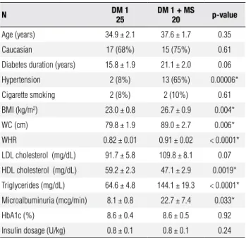
Table 1 shows the clinical characteristics of type 1 DM women with and without MS. Prevalence of MS using WHO criteria was 45%. There were no age or race di-fferences between the groups. Duration of type 1 DM was similar in subjects with and without MS, despite a non-statistic tendency (p = 0.06) of increased DM duration in subjects with MS. Mean BMI was 26.7 ± 0.9 kg/m2 in patients with MS and 23.0 ± 0.8 kg/m2
in patients without MS (p = 0.0004). None of the subjects in the study had obesity, deined by a BMI > 30 kg/m2. Patients with MS had increased waist
circum-ference (89.0 ± 2.7 cm vs. 79.8 ± 1.9 cm; p = 0.006) and waist-to-hip ratio (0.91 ± 0.02 cm vs. 0.82 ± 0.01 cm, p = 0.006). Metabolic syndrome was not associated with a worse glycemic control, since mean HbA1c were similar between the groups. Daily insulin dosage did not differ between the groups.
Body composition analysis by DEXA (Table 2) de-monstrated an increased central fat distribution in the type 1 DM patients with MS group, with a higher an-droid fat distribution and anan-droid-to-gynoid fat ratio (A/G) than patients without MS (41.9% ± 2.0% vs. 33.7% ± 1.8%, p = 0.004; and 0.9% ± 0.05% vs. 0.7% ± 0.03%, p = 0.0002). Total body fat and gynoid fat
dis-Table 1.Characteristics of type 1 DM women with and without metabolic syndrome
N DM 1
25
DM 1 + MS
20 p-value
Age (years) 34.9 ± 2.1 37.6 ± 1.7 0.35
Caucasian 17 (68%) 15 (75%) 0.61
Diabetes duration (years) 15.8 ± 1.9 21.1 ± 2.0 0.06
Hypertension 2 (8%) 13 (65%) 0.00006*
Cigarette smoking 2 (8%) 2 (10%) 0.61
BMI (kg/m2) 23.0 ± 0.8 26.7 ± 0.9 0.004*
WC (cm) 79.8 ± 1.9 89.0 ± 2.7 0.006*
WHR 0.82 ± 0.01 0.91 ± 0.02 < 0.0001*
LDL cholesterol (mg/dL) 91.7 ± 5.8 109.8 ± 8.1 0.07
HDL cholesterol (mg/dL) 59.2 ± 2.3 47.1 ± 2.9 0.0019*
Triglycerides (mg/dL) 64.6 ± 4.8 144.1 ± 19.3 < 0.0001*
Microalbuminuria (mcg/min) 8.1 ± 0.8 22.7 ± 7.4 0.033*
HbA1c (%) 8.6 ± 0.4 8.6 ± 0.5 0.92
Insulin dosage (U/kg) 0.8 ± 0.1 0.8 ± 0.1 0.24
BMI: body mass index; WC: waist circumference; WHR: waist-to-hip ratio; HbA1c: glycated hemoglobin. Values are expressed as mean ± SD, except for values related to hypertension and cigarette smoking which represent the absolute number of patients.
* Statistical significance (p < 0.05).
Table 2. Body composition by DEXA in type 1 DM women with and without metabolic syndrome
DM 1 DM 1 + MS p-value
Total body fat (%) 35.4 ± 1.4 38.4 ± 1.8 0.19
Android fat (%) 33.7 ± 1.8 41.9 ± 2.0 0.004*
Gynoid fat (%) 45.9 ± 1.2 45.1 ± 1.8 0.71
A/G 0.7 ± 0.03 0.9 ± 0.05 0.0002*
Values are mean ± SD. * Statistical significance (p < 0.05).
Total body fat (TBF), android and gynoid fat regions were expressed as a percentage of total body weight. Android fat and gynoid fat correspond to central and peripheric fat distribution, respectively. A/G: android-to-gynoid fat ratio.
tribution were not different between the groups with and without MS (38.4% ± 1.8% vs. 35.4% ± 1.4%, p = 0.19; and 45.1% ± 1.8% vs. 45.9% ± 1.2%, p = 0.71; respectively).
Type 1 diabetic patients with MS had a signi-icantly lower mean eGDR than patients without MS (4.99 ± 0.40 vs. 8.37 ± 0.39 mg.kg-1.min-1; p <
Cop
yright
© ABE&M t
odos os dir
eit
os r
eser
vados
.
Figure 2. Correlation between eGDR and android fat deposition.
Figure 1. Estimated eGDR (mg.kg-1.min-1) in type 1 DM women with and without metabolic syndrome.
Table 3. Correlation between android (central) fat deposition (%) and lipid profile (mg/dL) in women with type 1 DM
r p-value
LDL cholesterol 0.38 0.010*
HDL cholesterol -0.38 0.019*
Triglycerides 0.35 0.010*
* Statistical significance (p < 0.05).
Patients with type 1 DM and MS had signiicantly higher triglyceride levels (144.1 ± 19.3 mg/dL vs. 64.6 ± 4.8 mg/dL; p < 0.0001) and lower HDL levels (47.1 ± 2.9 mg/dL vs. 59.2 ± 2.3 mg/dL; p = 0.0019), with a tendency of higher LDL levels (109.8 ± 8.1 mg/dL vs. 91.7 ± 8.1 mg/dL; p = 0.07) (Table 1), when compared to those without MS. Android fat distribution was po-sitively correlated with LDL cholesterol (r = 0.38; p = 0.01) and triglycerides (r = 0.35; p = 0.019). A negative correlation was found with android fat distribution and HDL cholesterol (r = -0.38; p = 0.01). No correlation was found between gynoid fat distribution and LDL, HDL cholesterol or triglyceride levels (Table 3).
DISCUSSION
The present study demonstrated that non-obese type 1 diabetic women with predominant central fat deposition exhibit MS clinical parameters and increased insulin resis-tance, what might confer an increased cardiometabolic risk.
We observed a high prevalence of MS (45%) in young adult women with type 1 DM using the WHO criteria. These criteria appear to have the highest sen-sitivity to discriminate negative outcomes in patients with type 1 diabetes (10,13). A previous study from our group critically analyzed different criteria for MS in type 1 DM. It was observed that the WHO criteria were the preferred method to identify MS in this population, in comparison to IDF and NCEP criteria, and MS was found in 30% of women and 34% of men with type 1 DM (13). Prior studies also observed that the prevalen-ce of MS in type 1 DM is as high as 30%-45%, what can be linked with adverse outcomes (8,10-12,14).
Body composition evaluation was important to identify patients with clinical parameters of MS. We found that increased central fat deposition was a ma-jor determinant of MS in non-obese type 1 diabetic women. Type 1 DM with MS had increased WC and WHR (Table 1). Using DEXA, these patients also had a higher android (central) fat distribution and A/G ratio. Nonetheless, total body fat and gynoid (peripheric) fat distribution were not related to MS (Table 2). Some patients with MS were overweight, but none had obesi-ty. Thus, our data gives further evidence that predomi-nant central obesity is a risk factor for MS in type 1 DM. Insulin resistance has been recognized as an important feature in type 1 DM (9,11,25-27). It has been demons-trated that insulin resistance is a predictor of coronary ar-tery disease in type 1 diabetes (27). Clinically, it is often dificult to accurately identify insulin resistance in type 1 DM. Recently, eGDR has been developed and validated as an easy method of insulin resistance evaluation in type 1 diabetes (24). In the present study, type 1 DM patients with MS had decreased eGDR, which indicates dimini-shed insulin sensitivity (Figure 1). Central obesity was also associated with insulin resistance since there was an inverse correlation between eGDR and WC, WHR and android fat distribution (Figure 2). No correlation was found be-tween eGDR with peripheric fat distribution, represented by the gynoid fat region. Therefore, our data suggest that MS and body fat distribution, particularly central fat tissue, are signiicantly correlated with insulin resistance in type 1 DM. The Pittsburg Study also described a reduced eGDR in type 1 DM with MS (10). In that study, eGDR was the
10
eGDR
15
5
0
Yes
p < 0.0001
Not
Metabolic syndrome
50 60
40
30
20
10
0
0 2 4 6 8 10 12
eGDR (mg.kg-1.min-1)
Android (%)
Cop
yright
© ABE&M t
odos os dir
eit
os r
eser
vados
.
best predictor for renal failure and also predicted coronary artery disease and diabetes related mortality (10).
The presence of clinical parameters of MS in young adult women with type 1 diabetes was accompanied by increased microalbuminuria, hypertension and the worst lipid proile in this study (Table 1), which are well known cardiovascular risk factors. Central fat deposition, but not peripheric fat, was associated with higher LDL cholesterol and triglyceride con-tent and lower HDL cholesterol (Table 3). There-fore, type 1 DM with MS and central obesity ac-cumulates several atherogenic risk factors at young age, which will probably contribute for an adverse cardiovascular outcome (10-12,26,27).
In conclusion, central fat deposition in non-obese type 1 DM women was related to MS and insulin resistance. Fur-thermore, young adult women with MS and/or central obesity accumulate several cardiovascular risk factors, such as insulin resistance, microalbuminuria, hypertension and atherogenic lipid proile. Thus, body composition analysis with identiication of central fat deposition might be im-portant to identify non-obese type 1 DM patients with in-creased cardiometabolic risk. Further studies with a greater number of patients are necessary to corroborate these data.
Acknowledgements: Authors are grateful to Dr. Martha Cecilia Suárez-Mutis for the statistical analysis and to Andreia Maria do Amaral Bruni, Rosangela Adriano Barbosa and Patricia Cotilha de Andrade de Sena for their technical assistance.
Disclosure: no potential conlict of interest relevant to this article was reported.
REFERENCES
1. Reaven GM. Role of insulin resistance in human disease (syn-drome X): an expanded definition. Ann Rev Med. 1993;44:121-31. 2. Balkau B, Charles MA. Comment on the provisional report from
the WHO consultation. European group for the study of insulin resistance (EGIR). Diabet Med. 1999;16:442-3.
3. Laaksonen DE, Lakka HM, Niskanen L, Kaplan GA, Salonen JT, Lakka TA. Metabolic syndrome and development of diabetes mellitus: application and validation of recently suggested defi-nitions of the metabolic syndrome in a prospective cohort study. Am J Epidemiol. 2002;156:1070-7.
4. Picon PX, Zanatta CM, Gerchman F, Zelmanowitz T, Gross JL, Ca-nani LH. Analysis of the criteria used for the definition of metabo-lic syndrome in patients with type 2 diabetes. Arq Bras Endocri-nol Metabol. 2006;50:264-70.
5. Isomaa B, Almgren P, Tiinamaija T, Forsén B, Lahti K, Nissén M, et al. Cardiovascular morbidity and mortality associated with the metabolic syndrome. Diabetes Care. 2001;24:683-9.
6. Haffner SM. The metabolic syndrome: inflammation, diabetes mellitus, and cardiovascular disease. Am J Cardiol. 2006;97:3-11. 7. Teupe B, Bergis K. Epidemiological evidence for “double
diabe-tes”. Lancet. 1991;337:361-2.
8. Thorn LM, Forsblom C, Fegerudd J, Thomas MC, Petterson-Fer-nholm K, Saraheimo M, et al. Metabolic syndrome in type 1 dia-betes. Diabetes Care. 2005;28:2019-24.
9. Dib SA. Insulin resistance and metabolic syndrome in type 1 dia-betes. Arq Bras Endocrinol Metabol. 2006;50:250-63.
10. Pambianco G, Costacou T, Orchard TJ. The prediction of major outco-mes of type 1 diabetes: a 12-year prospective evaluation of three se-parate definitions of the metabolic syndrome and their components and estimated glucose disposal rate. Diabetes Care. 2007;30:1248-54. 11. Kilpatrick ES, Rigby AS, Atkin SL. Insulin resistance, the metabo-lic syndrome, and compmetabo-lication risk in type 1 diabetes. Diabetes Care. 2007;30:707-12.
12. Davis TME, Bruce DG, Davis WA. Prevalence and prognostic im-plications of the metabolic syndrome in community-based pa-tients with type 1 diabetes: the Fremantle Diabetes Study. Diabe-tes Res Clin Pract. 2007;78:412-7.
13. Santos CE, Schrank Y, Kupfer R. Critical analyses of WHO, IDF and NCEP criteria for metabolic syndrome among patients with type 1 diabetes mellitus. Arq Bras Endocrinol Metab. 2009;53:1096-102. 14. Kissebah AH, Vydelingum N, Murray R, Evans DJ, Hartz AJ,
Kalkhoff RK, et al. Relation of body fat distribution to metabolic complication of obesity. J Clin Endocrinol Metab. 1982;54:254-60. 15. Fujioka S, Matsuzawa Y, Tokunaga K, Tarui S. Contribution of intra--abdominal fat accumulation to the impairment of glucose and lipid metabolism in human obesity. Metabolism. 1987;36:54-9. 16. Ribeiro Filho FF, Mariosa LS, Ferreira SRG, Zanella MT. Gordura
visceral e síndrome metabólica: mais do que uma simples asso-ciação. Arq Bras Endocrinol Metabol. 2006;50:230-8.
17. Mazess RB, Barden HS, Bisek JP, Hanson J. Dual-X-ray-absorp-tiometry for total body and regional bone-mineral and soft-tissue composition. Am J Clin Nutr. 1990;51:1106-12.
18. Prior BM, Cureton KJ, Modlesky CM, Evans EM, Sloniger MA, Sa-unders M, et al. In vivo validation of whole body composition es-timates from dual-energy X-ray absorptiometry. J Appl Physiol. 1997;83:623-30.
19. Clasey JL, Bouchard C, Teates CD. The use of anthropometric and dual-energy X-ray absorptiometry (DXA) measures to estimate total abdominal and abdominal visceral fat in men and women. Obes Res. 1999;7:256-64.
20. Park YW, Hemsfield SB, Gallagher D. Are dual-energy X-ray ab-sorptiometry regional estimates associated with visceral adipose tissue mass? Int J Obes Relat Metab Disord. 2002;26:978-83. 21. Norcross J, Van Loan MD. Validation of fan beam dual energy
X-ray absorptiometry for body composition assessment in adults aged 18-45 years. Br J Sports Med. 2004;38:472- 6.
22. Lee CC, Glickman SG, Dengel DR, Brown MD, Supiano MA. Ab-dominal adiposity assessed by dual energy X-ray absorptiometry provides a sex-independent predictor of insulin sensitivity in ol-der adults. J Gerontol A Biol Sci Med Sci. 2005;60:872-8. 23. Friedwald WT, Levy RI, Fredrickson DS. Estimation of the
con-centration of low lipoprotein colesterol in plasma without use of preparative centrifugation. Clin Chem. 1972;18:499-502. 24. Williams K, Erbey J, Becker D, Arslanian S, Orchard T. Can clinical
factors estimate insulin resistance in type 1 diabetes? Diabetes. 2000;49:626-32.
25. DeFronzo RA, Hendler R, Simonson D. Insulin resistance is a proeminent feature of insulin-dependent diabetes. Diabetes. 1982;31:795-801.
26. Yip J, Mattock MB, Morocutti A, Sethi M, Trevisan R, Viberti G. Insulin resistance in insulin-dependent diabetic patients with mi-croalbuminuria. Lancet. 1993;342:883-7.

