C
a s eR
e p o Rt4 9 Arq Bras Oftalmol. 2017;80(1):49-51 http://dx.doi.org/10.5935/0004-2749.20170013
INTRODUCTION
Hypotrichosis with juvenile macular dystrophy (HJMD), OMIM(1),
is a rare autosomal recessive disorder characterized by hypotrichosis at birth, limited hair growth throughout life, and progressive dege-neration of the retina leading to blindness, potentially at an early
age(2), and is caused by a mutation in the cadherin 3 (CDH3) gene,
re sulting in abnormal expression of P-cadherin. Mutations in CDH3 are associated with ectodermal dysplasia, ectrodactyly, and macular dystrophy(3).
CASE REPORT
An 11-year-old boy of Iranian descent was born to parents who were first cousins (Figure 1). The patient was full term with no gesta-tional complications and was born via elective cesarean section. His birth weight was 5 pounds 8 ounces. There was a family history of two 20-year-old maternal-side female cousins with HJMD (Figure 1). The proband’s maternal grandfather had a color vision deficiency.
The proband was born with a missing left index fingernail and sparse scalp hair (Figure 2). The hair follicles were examined derma-topathologically, and no abnormalities were found. There were no other dermatologic conditions, except for mild eczema. The patient had multiple food allergies but no drug allergies. The patient’s mother reported some developmental delays initially, but at the time of examination, the subject had reached an appropriate developmental level. He had delayed primary teeth maturation.
ABSTRACT
Hypotrichosis with juvenile macular dystrophy is a rare autosomal recessive disorder characterized by sparse scalp hair caused by hair follicle abnormalities as well as progressive retinal degeneration leading to blindness in the second or third decade of life. It is associated with mutations of the cadherin 3 (CDH3) gene, which result in abnormal expression of P-cadherin. Mutations in CDH3 are related to ectodermal dysplasia, ectrodactyly, and macular dystrophy. In this report, we describe an 11-year-old Iranian boy born with a missing left index fingernail and sparse scalp hair who later displayed macular pigmentary changes. Genetic testing of the CDH3 gene revealed a homozygous gene variant at exon 6 (640A>T). This novel in-frame mutation converts a lysine to a premature stop codon, altering synthesis of P-cadherin on chromosome 16q22.
Keywords: Hypotrichosis; Macular degeneration; Cadherins/genetics; Humans; Case reports
RESUMO
Hipotricose com distrofia macular juvenil (HDMJ) é uma doença autossômica recessiva rara caracterizada por rarefação capilar por alteração nos folículos pilosos e dege-neracão progressiva da retina levando a cegueira na segunda e terceira década de vida. Associada a mutações no gene CDH3, resultando em expressão anormal de P-caderina. Mutações no gene CDH3 estão relacionados à displasia ectodérmica, ectrodactilia e distrofia macular. Neste relato descrevemos um menino Iraniano de 11 anos de idade, com ausência da unha na mão esquerda e rarefação capilar desde o nascimento, e que posteriormente apresentou alterações pigmentares maculares. Teste genético do gene
CDH3 revelou uma variação homozigótica no exon 6 (640A>T). Essa mutação in-frame
troca uma lisina por um codon de parada prematura, alterando a síntese da proteína P-caderina no cromossomo 16q22.
Descritores: Hipotricose; Degeneração macular; Caderinas/genética; Humanos;
Re-latos de casos
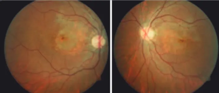
The proband began wearing glasses at 5 years of age when his teachers referred him for eye care because of trouble seeing the board. His visual acuity at the age of 8 years was 20/40 +2 for both eyes, which was stable for 2 years. The dilated fundus examination at that time showed stable, primarily macular pigmentary, changes (Figure 3).
Electroretinography (ERG; Espion; Diagnosys LLC)was performed
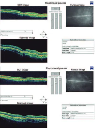
at the age of 9 years and showed decreased a and b wave amplitudes for the rod-, mixed rod-cone-, cone-, and flicker-mediated respon-ses for both eyes. Another ERG was performed 6 months later and showed decreased rod-mediated responses for OD and borderline responses for the OS. The mixed rod-cone-mediated responses were borderline for both eyes, and the cone and flicker responses for both eyes were diminished. An ERG performed 1 year later showed no downward progression of the responses compared with the previous year. Goldmann visual field testing demonstrated full peripheral isopters and a marginally enlarged blind spot in both eyes. Optical coherence tomography (OCT; Stratus; Carl Zeiss Meditec) testing de monstrated mild macular region attenuation (Figure 4). Both tests were conducted at the age of 9 years.
Sanger sequencing was performed to specifically screen for a
mutation in the CDH3 gene, which encodes for the P-cadherin
pro-tein. A homozygous in-frame nonsense mutation was found in exon 6 (640A>T), resulting in the substitution of a lysine for a premature stop codon, thus leading to the synthesis of a truncated protein unable to fulfill its physiologic functions.
Hypotrichosis with juvenile macular dystrophy: a case report with molecular study
Hipotricose com distroia macular juvenil: relato de caso com estudo molecular
Lucas Perez Vicente1, simone Finzi1, remo susanna Jr.1, terri L. Young2
Submitted for publication: October 20, 2015 Accepted for publication: March 16, 2016
1 Department of Ophthalmology, Universidade de São Paulo (USP), São Paulo, SP, Brazil. 2 Department of Ophthalmology and Visual Sciences, University of Wisconsin, Wisconsin, USA.
Funding: This study was supported by Educational Grant from Novartis for international rotation of residents from Department of Ophthalmology of Universidade de São Paulo.
Disclosure of potential conflicts of interest: None of the authors have any potential conflict of interest to disclose.
Hy p o t r i c H o s i sw i t Hj u v e n i l em a c u l a rd y s t r o p H y: ac a s er e p o rtw i t Hm o l e c u l a rs t u d y
5 0 Arq Bras Oftalmol. 2017;80(1):49-51
DISCUSSION
Several authors have described different families with this
con-dition found in remote areas of the Middle East(2,4-7). All the families
studied by these authors shared mutations in the CDH3 gene. Symptom
Figure 1. Pedigree chart.
Figure 2. Picture of proband with sparse scalp hair.
Figure 3. Primary macular pigmentary changes and atrophy of the retinal pigment epithelium.
severity varies for each individual, but all share common features of sparse scalp hair with limited growth throughout life and retinal de-generation.
Naqvi et al.(4) studied a family from a remote region of Pakistan.
The family was consanguineous, with six affected members delinea-ted in an autosomal recessive state. All affecdelinea-ted individuals had sparse scalp hair. Their finger and toenails, teeth, and skin were normal, with normal growth. The affected individuals had progressive vision loss starting at 12-20 years of age. Fundus examination revealed pigmentary abnormalities of the posterior pole as in the patient in our study. Both ERG and fundus inspections were consistent with macular dystrophy(4).
Jelani et al.(2) studied another large consanguineous pedigree
from Pakistan with nine affected individuals. They were born with sparse scalp hair but otherwise normal integumentary and teeth. Examina-tion showed degeneraExamina-tion of the macular pigmented epithelium. ERG testing of two of the affected individuals showed severe retinal
dysfunction(2). Despite a different genotype (IVS10-1 G→T), the
phe-notype was similar to that in the present report.
Sprecher et al.(5) studied four large consanguineous families from
Israel, with 11 affected individuals. At birth, they had seemingly nor-mal hair growth but developed alopecia at 3 months of age. Scalp skin biopsies were taken and showed “reduced ratio of terminal versus vellus hair follicles.” They observed “distinct structural aberrations of the hair shaft.” Progressive macular degeneration started between 3 and 21 years of age. Electrophysiological evaluation was also con-ducted and showed anomalies consistent with impaired macular
function(5). The authors identified a homozygous deletion in exon 8
(981delG) of the CDH3 gene in all families, identifying cadherin
mo-lecule pathogenesis resulting in a human hair and retinal disorder for the first time. In the present study, the age of onset was 8 years, and mild macular region attenuation on OCT was observed at 9 years.
Indelman et al. reported, in four members with HJMD of a consan-guineous family, a homozygous missense mutation (R503H) resulting in a single amino acid substitution at position 503 of the P-cadherin sequence. The affected individuals were born with sparse scalp hair. Retinal degeneration manifested in all of the affected individuals with decreased visual acuity during the first decade of life(6). These clinical
Vi c e n t e LP, e ta L.
5 1
Arq Bras Oftalmol. 2017;80(1):49-51
hypotrichosis with cone-rod dystrophy would be a more appropriate
name for this syndrome(7).
Genetic reports of rare diseases such as HJMD are important for prevalence estimation and to heighten awareness. Recent successes in stem cell and genetic therapies for ocular diseases suggest that the retinal aspects of this disorder may potentially be treatable(8,9).
REFERENCES
1. OMIM. Online Mendelian Inheritane in Man. Hypotrichosis congenital, with juvenile macular dystrophy; HJMD [Internet]. Johns Hopkins University [updated 21 March 2016; cited 2015 Jan 21]. Available from: http://www.omim.org/entry/601553 2. Jelani M, Salman Chishti M, Ahmad W. A novel splice-site mutation in the CDH3 gene
in hypotrichosis with juvenile macular dystrophy. Clin Exp Dermatol. 2009:34(1):68-73. 3. Balarin Silva V, Simões AM, Marques-de-Faria AP. EEM syndrome: report of a family and
results of a ten-year follow-up. Ophthalmic Genet. 1999:20(2):95-9.
4. Kamran-Ul-Hassan Naqvi S, Azzem Z, Ali G, Ahmad W. A novel splice-acceptor site muta-tion in CDH3 gene in a consanguineous family exhibiting hypotrichosis with juvenile macular dystrophy. Arch Dermatol Res. 2010:302(9):701-3.
5. Sprecher E, Bergman R, Richard G, Lurie R, Shalev S, Petronius D, et al. Hypotrichosis with juvenile macular dystrophy is caused by a mutation in CDH3, encoding P-cadherin. Nat Genet. 2001:29(2);134-6.
6. Indelman M, Bergman R, Lurie R, Richard G, Miller B, Petronius D, et al. A missense mutation in CDH3, encoding P-cadherin, causes hypotrichosis with juvenile macular dystrophy. J Invest Derm. 2002:119(5):1210-3.
7. Leibu R, Jermans A, Hatim G, Miller B, Sprecher E, Perlman I. Hypotrichosis with juvenile macular dystrophy: clinical and electrophysiological assessment of visual function. Ophthalmology. 2006:113(5):841-7.
8. Wiley LA, Burnight ER, Songstad AE, Drack AV, Mullins RF, Stone EM, Tucker BA. Patient-specific induced pluripotent stem cells (iPSCs) for the study and treatment of retinal degenerative diseases. Prog Retin Eye Res. 2015:44:15-35.
9. Nagel-Wolfrum K, Möller F, Penner I, Wolfrum U. Translational read-through as an alter-native approach for ocular gene therapy of retinal dystrophies caused by in-frame nonsense mutations. Vis Neurosci. 2014:31(4-5);309-16.
Figure 4. Optical coherence tomography demonstrating a mild decrease in macular thickness bilaterally.