AR
TIGO ORIGINAL / ORIGINAL AR
TICLE
INTRODUCTION
Obesity is considered a worldwide public health problem. Its incidence has been increasing in men, women and children from developed and in devel-opment countries(17,20,26). Nowadays, gastrointestinal
surgery is accepted as the most effective approach to reach weight loss in morbid obesity patients. Roux-en-Y gastric bypass (RYGB) is the most used surgical technique for the treatment of morbid obesity in the American continent(1).
H. PYLORI
INFECTION, ENDOSCOPIC,
HISTOLOGICAL ASPECTS AND CELL
PROLIFERATION IN THE GASTRIC MUCOSA
OF PATIENTS SUBMITTED TO ROUX-EN-Y
GASTRIC BYPASS WITH CONTENTION
RING: a cross sectional endoscopic and
immunohistochemical study
Thiago De Bortoli
NOGUEIRA
, Ricardo
ARTIGIANI NETO
,
Benedito
HERANI FILHO
and Jaques
WAISBERG
Received 16/6/2015 Accepted 25/11/2015
ABSTRACT - Background - Morbid obesity treatment through vertical gastroplasty Roux-en-Y gastric bypass initially used a contention ring. However, this technique may create conditions to the development of potentially malign alterations in the gastric mucosa. Although effective and previous-ly performed in large scale, this technique needs to be better evaluated in long-term studies regarding alterations caused in the gastric mucosa.
Objective - To analyze the preoperative and postoperative endoscopic, histological and cell proliferation indings in the gastric antrum and body mucosa of patients submitted to the Roux-en-Y gastric bypass with a contention ring. Methods - We retrospectively evaluated all patients submitted to Roux-en-Y gastric bypass with a contention ring with more than 60 months of postoperative follow-up. We compared the preoperative (gastric antrum and body) and postoperative (gastric pouch) gastric mucosa endoscopic indings, cell proliferation index and H. pylori prevalence. We evaluated cell proliferation through Ki-67 antibody immunohistochemical expression. Results - In the study period, 33 patients were operated with the Roux-en-Y gastric bypass using a contention ring. We found a chronic gastritis rate of 69.7% in the preoperative period (gastric antrum and body) and 84.8% in the postoperative (gastric pouch). H. pylori was present in 18.2% of patients in the preoperative period (gastric antrum and body) and in 57.5% in the postoperative (gastric pouch). Preoperative cell proliferation index was 18.1% in the gastric antrum and 16.2% in the gastric body, and 23.8% in the postoperative gastric pouch. The postoperative cell proliferation index in the gastric pouch was signiicantly higher (P=0.001) than in the preoperative gastric antrum and body. Higher cell proliferation index and chronic gastritis intensity were signiicantly associated to H. pylori presence (P=0.001 and P=0.02, respectively).
Conclusion - After Roux-en-Y gastric bypass with contention ring, there was a higher chronic gastritis incidence and higher cell proliferation index in the gastric pouch than in the preoperative gastric antrum and body. Mucosa inlammation intensity and cell proliferation index in the postoperative gastric pouch were associated to H. pylori presence and were higher than those found in the preoperative gastric antrum and body mucosa. HEADINGS - Morbid obesity. Gastroplasty. Roux-en-Y anastomosis. Ki-67 antigen. Helicobacter pylori.
Declared conflict of interest of all authors: none
Disclosure of funding: Coordination for the Improvement of Higher Education Personnel (CAPES) Departamento de Gastroenterologia Cirúrgica, Universidade Federal de São Paulo, São Paulo, SP, Brasil.
Correspondence: Thiago De Bortoli Nogueira. Av. Rei Alberto I, 341, ap. 122 - CEP 11030-380 - Santos, SP, Brasil. E-mail: [email protected]
Furthermore, the RYGB technique may create conditions that contribute to the development of potentially malignant alterations, such as: ulcers in gastrojejunal anastomosis(4,8);
gastric acidity, which may provoke lesions when the gastric pouch is larger or when gastrogastric istula is developed(3);
and H. pylori infection, which appears to be more frequent in the group submitted to RYGB(2,21,31), promoting inlammatory
alterations in the gastric mucosa with glandular loss, atrophy and gastritis and ulcer increase(30).
RYGB with a ring was performed in Brazil in large scale. However, there is scarcity of long-term studies evaluating macroscopic and histopathological alterations with a long period of patient follow-up. The objective of this study was to analyze the endoscopic and histological alterations in the gastric mucosa of patients submitted to RYGB with a contention ring. The hypothesis was that physiopathological alterations in the gastric mucosa after gastroplasty could pre-dispose chronic and proliferative inlammatory alterations.
METHODS
This is an observational, cross-sectional study, performed in a public university hospital and approved by the Institu-tional Research Ethics Committee. Patient informed consent forms were waived since this is a retrospective study based in archived material.
We searched records of all patients consecutively admitted to undergo open surgical treatment for morbid obesity with Roux-en-Y gastric bypass using a contention ring from 1997 to 2009. Therefore we selected the sample in the period before 2009 to allow a homogenous sample of patients operated with the same technique and long-term follow-up.
We included all consecutive adult patients submitted to ring Roux-en-Y gastric bypass who met the following criteria:
• submitted to preoperative upper digestive endoscopy; • submitted to preoperative gastric antrum and body
biopsy and postoperative gastric pouch biopsy; • submitted endoscopy again in the postoperative period
and after a minimum period of 60 months.
The exclusion criterion was the presence of neoplastic or pre-neoplastic gastric lesions, observed in the upper digestive endoscopy performed in the preoperative period.
We gathered patients’charts, pre and postoperative endos-copy reports and histopathological exams from preoperative gastric antrum and body biopsy and postoperative gastric pouch biopsy, in slides stained with haematoxylin and eosin (HE). We evaluated H. pylori presence in the gastric pouch archived slides and performed an immunohistochemical study to assess cell proliferation, as described next.
Histopathological study
The archived slides contained tissues from gastric biop-sies, ixed in 10% formalin and processed for histological analysis as the hospital usual routine, through paraffin embedding, with 4 mm histological sections and HE stain-ing. Modiied Giemsa staining was used to detect H. pylori
presence or absence in the gastric tissue.
For this study, a single pathologist reviewed the slides and evaluated atrophy, inlammation, intestinal metaplasia and dysplasia occurrence. The pathologist also reviewed
H. pylori diagnosis.
Immunohistochemical study
We performed an immunohistochemical study in the material obtained from the pre and postoperative gastric biopsies. We added the monoclonal antibody Ki-67 (Dako Cytomation, Carpinteria, CA, USA) at a 1:100 dilution in BSA (1% bovine serum albumin) to slides previously silanised with 4 mm histological sections (3-aminopropyltrietoxy-silane, Sigma Chemical Co., Saint Louis, MO, USA.) and maintained them in a stove at 60ºC for 24 hours.
We calculated Ki-67 expression index. Using a microscope with a 400x magniication, we evaluated the percentage of glandular epithelial cells with marked nuclei in four 100.000 μm2 areas. We considered as positive the cells marked by the
antibody, even if weakly stained. We classiied the Ki-67 mark-er expression as high cell prolifmark-eration level when the positive cells count was ≥25%, and low when <25%, based on the Ki-67 immunoreactivity in the stomach of normal individuals(18, 25).
Endoscopic findings
We analyzed the endoscopic reports and evaluated the preoperative indings in the esophagus, gastric chamber and duodenum, and the postoperative indings in the esophagus, gastric pouch, gastrojejunal anastomosis, and jejunal afferent and efferent loop.
We also evaluated the presence, absence or internal mi-gration of the contention ring in the gastric pouch. We used the Sydney classification(27) for inflammatory alterations
found in the preoperative gastric chamber mucosa and in the postoperative gastric pouch. We evaluated inlammatory indings from the esophagus according to the Los Angeles classiication(16). We also evaluated the jejunal afferent and
efferent loop endoscopically.
Statistical analysis
We measured the results though arithmetic mean and standard deviation (SD), and analyzed them through the paired t test (Student test), chi-square test and Fisher’s exact test. We considered P values <0.05 signiicant. We used the statistical program PASW Statistics, version 18.0 (IBM Corp. New York, NY, USA).
RESULTS
The hospital staff operated 33 patients with the ring tech-nique in the selected period. Thus, we included 33 patients in this study. The mean age was 42±9 years (22-55 years) and 22 of them (66.7%) were women. The mean postoperative follow-up was 91±21months (60-144 months).
We describe the pre and postoperative histological ind-ings indicative of gastritis in Table 3. We identiied intestinal metaplasia in the preoperative exams in two (6.0%) patients and mucosa atrophy in three (9.1%), two of them present-ing both indpresent-ings. In the postoperative stomach histological study, we identiied intestinal metaplasia in three (9.1%) pa-tients, and two (6.0%) of them also presented this alteration in the preoperative study. We found four (12.1%) patients with gastric mucosa atrophy.
We found no cases of gastric mucosa dysplasia in the pre and postoperative period. We found no statistically sig-niicant concordance (P=0.1) between the endoscopic and histological indings in the pre and postoperative period. The longer postoperative follow-up did not signiicantly inluence the indings of endoscopic (P=0.5) or histological (P=0.3) abnormalities.
Among the 33 patients, 12 (36.3%) were H. pylori nega-tive before and after the surgery; 4 (12.1%) were posinega-tive before and after. However, 15 (45.4%) were negative before the surgery and became positive after. Only two (6.0%) were positive before and became negative after the surgery. All patients with a positive preoperative H. pylori exam re-ceived speciic antibiotic therapy, and endoscopic biopsies conirmed the bacteria eradication. The histological gastritis intensity in the gastric pouch was associated to H. pylori
presence (P=0.02) (Table 4). The gastric pouch higher cell proliferation index were associated to Helicobacter pylori
presence (Table 5), whose infection was signiicantly higher in the gastric pouch of patients with greater postoperative follow-up period (Table 6).
The cell proliferation index in the gastric pouch (Ki67) was signiicantly higher (P=0.001) than in the preoperative gastric chamber (Table 7 and Figures 1 and 2).
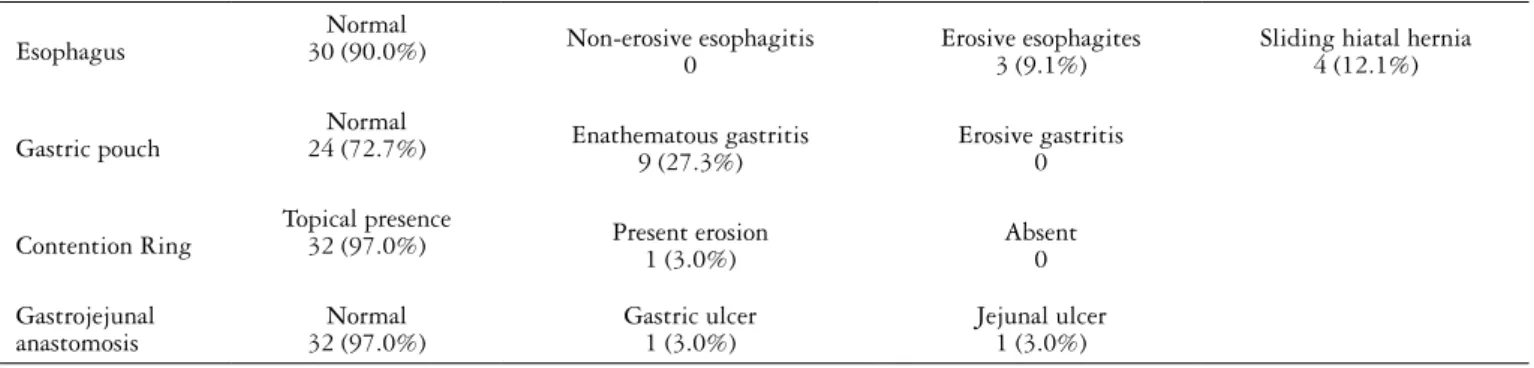
TABLE 2. Postoperative endoscopic indings in obese patients treated with Roux-en-Y gastric bypass with a restraining ring
Esophagus
Normal
30 (90.0%) Non-erosive esophagitis 0
Erosive esophagites 3 (9.1%)
Sliding hiatal hernia 4 (12.1%)
Gastric pouch
Normal
24 (72.7%) Enathematous gastritis
9 (27.3%)
Erosive gastritis 0
Contention Ring
Topical presence
32 (97.0%) Present erosion
1 (3.0%)
Absent 0
Gastrojejunal anastomosis
Normal 32 (97.0%)
Gastric ulcer 1 (3.0%)
Jejunal ulcer 1 (3.0%)
TABLE 1. Preoperative endoscopic indings in obese patients treated with Roux-en-Y gastric bypass with a restraining ring
Esophagus
Normal
29 (87.9%) Non-erosive esophagitis1 (3.0%) Erosive esophagites3 (9.1%) Sliding hiatal hernia4 (12.1%)
Gastric antrum
Normal
22 (66.6%) Enathematous gastritis 5 (15.1%)
Erosive gastritis 3 (9.1%)
Atrophic gastritis 3 (9.1%)
Gastric body Normal
23 (69.7%)
Enathematous gastritis 4 (12.1%)
Erosive gastritis 3 (9.1%)
Atrophic gastritis 3 (9.1%)
TABLE 5. Ki-67 expressions and Helicobacter pylori distribution in the gastric pouch of 33 morbid obesity patients submitted to Roux-en-Y gastric bypass with a restraining ring
Helicobacter pylori
P Positive (%) Negative (%) Total (%)
Gastric pouch (Ki-67)
< 25% 7 (21.2) 13 (39.4) 20 (60.6)
0.001*
≥ 25% 12 (36.4) 1 (3.0) 13 (39.4)
Total 19 (57.6) 14 (42.4) 33 (100)
* Signiicant; Fisher’s exact test
TABLE 4. Helicobacter pylori distribution in the gastric pouch of 33 morbid obesity patients submitted to Roux-en-Y gastric bypass with a restraining ring
Helicobacter pylori
P
Positive (%) Negative (%)
Gastritis
Mild 7 (21.2) 4 (12.1)
0.02*
Moderate 11 (33.3) 5 (15.1)
Intense 1 (3)
-* Signiicant; Fisher’s exact test.
TABLE 3.Gastric pre and postoperative histological indings of morbid obesity patients submitted to Roux-en-Y gastric bypass with a restraining ring
Histological aspect Preoperative Postoperative
Normal 10 (30.3%) 5 (15.1%)
Mild chronic gastritis 14 (42.4%) 11 (33.3%)
Moderate chronic gastritis 9 (27.3%) 16 (48.5%)
DISCUSSION
Nowadays the most used bariatric surgery technique for morbid obesity treatment is the Roux-en-Y gastric bypass (RYGB), in which a contention ring may be used(1). There
are no prevalence reports of gastric neoplasia after RYGB. However, symptoms such as abdominal pain, bleeding, uncontrollable vomiting and weight loss, which are common in gastric cancer, can also occur after RYGB. Therefore this procedure may theoretically contribute to late diagnosis of gastric cancer. Malignant gastric tumours after 5 to 22 years of the postoperative period have been described in 21 obese patients submitted to RYGB, and 2 of them were located in the gastric pouch(19,22). Due to reports of late neoplasia after
RYGB(19), there is a concern regarding gastric pouch evaluation.
A retrospective study(16) with 161 patients submitted to
preoperative gastric biopsy demonstrated alterations in 109 (68%) of them, mainly chronic gastritis. The alterations were signiicantly more frequent (94%) in the H. pylori positive cases when compared to the negative (51%). The present study identiied chronic gastritis in similar proportions and signiicantly more related to H. pylori infection cases.
Csendes et al.(6) evaluated 227 patients submitted to gastric
bypass, with a mean follow-up of 27 months after operation. The endoscopic exam showed no alterations in 225 (99%) pa-tients. One patient (0.4%) presented ulcer in the gastrojejunal anastomosis and in 96 (56%) cases the gastric pouch mucosa TABLE 7. Ki-67 expressions preoperatively and postoperatively in patients
submitted to Roux-en-Y gastric bypass with a restraining ring
Preoperative Postoperative
P
N% % N% %
Ki-67 SD SD
Gastric antrum 18.1 8.8
Gastric body 16.2 10.2 0.02*
Gastric pouch 23.8 16.2 0.001*
SD: standard deviation, *Signiicant; Student’s test.
TABLE 6. Helicobacter pylori distribution gastric pouch and postoperative follow-up period of patients submitted to Roux-en-Y gastric bypass with a restraining ring
Postoperative follow-up (months)
H. pylori
P
Positive Negative
N 14 19
Variation
(minimum-maximum) 60-108 84-144
< 0.001*
Median 72 96
Average
(standard deviation) 73.7 (13.2) 104.2 (14.4)
* Signiicant; Student’s test.
FIGURE 1. Photomicrographs of Ki-67 immunoexpression in the pre (A) and postoperative (B) gastric mucosa of operated patients. A – Normal nuclear immunostaining in the basal layer of the gastric body epithelium, 200 X. B – Ki-67 antibody immunoexpression in the gastric pouch, with increased cell proliferation index in the mucosa basal layer, until the surface epithelium, 400 X.
A
B
FIGURE 2. Photomicrographs of Ki-67 immunoexpression in the pre (A) and postoperative (B) gastric mucosa of operated patients. A – Normal nuclear immunostaining in the basal layer of the gastric body epithelium, 200 X. B – Ki-67 antibody immunoexpression in the gastric pouch, with increased cell proliferation index in the mucosa basal layer until the surface epithelium associated to intestinal metaplasia, 200 X.
A
was histologically normal. While 48 (28%) patients presented active chronic gastritis, it was associated to H. pylori presence in 43 (89.6%) cases. After 2 years of evaluation, 53 (31%) cases presented H. pylori positivity and 9 (5.5%) cases presented inactive chronic gastritis. These authors reported late marginal ulcer in 0.6% to 25% of operated patients(4,8). Our study found
one (3%) patient with gastrojejunal anastomotic ulcer only. Flickinger et al.(11) performed endoscopies 13 to 20 months
after the gastric bypass in 53 patients. These authors found the gastric pouch without detectable macroscopic alterations in 45 (85%) patients. On the other hand, 11% of patients had biliary stasis, probably due to the construction of an afferent loop shorter than usual in the Roux-en-Y. The gastric pouch histological analysis revealed the mucosa was normal in 45% of patients, with acute gastritis in 23%, chronic gastritis in 30% and with intestinal metaplasia in 13%. These results indicate no correlation between endoscopic appearance and histological indings, which may justify the necessity of histo-logical evaluation even when the gastric mucosa macroscopic appears to be normal. In the present study we observed a similar result: normal postoperative gastric pouch macro-scopic aspect in 24 (72.7%) cases and mucosa inlammatory process with histological evidence in 28 (84.8%) patients.
A factor that could explain the high histological gastritis index we found is H. pylori presence. H. pylori infection is more prevalent (61.3%) in the obese population in general, while in the obese patients submitted to bariatric surgery,
H. pylori presence varied from 24% to 70%(2,21,31).
Csendes et al.(5) found H. pylori in the preoperative period
in 47% of obese patients submitted to bariatric surgery. None of them received eradication treatment, since all had the distal gastric segment totally dissected. H. pylori was present in the antrum in 20% of patients and in the gastric fundus in 5%. Two years after operation, H. pylori infection rate in the gastric pouch was 31%. In only 50% of patients with
H. pylori in the gastric pouch the bacterium was present in the preoperative period. This inding suggests H. pylori
quickly colonizes this little gastric pouch with a minimum amount of parietal cells, probably since in this portion there is no acid secretion in great quantity, which contributes to the rapid H. pylori infection of the mucosa(24).
In the present study, H. pylori was present in the preopera-tive gastric antrum and body of six (18.2%) patients and in the postoperative gastric pouch of 19 (57.5%). All patients with pre-sent preoperative H. pylori received speciic antibiotic therapy and bacteria eradication conirmed by endoscopic biopsy. This result suggests that the H. pylori infection persistence in the postoperative period was probably due to reinfection episodes. As in the study from Csendes et al.(5), in the present study there
was a higher H. pylori prevalence in the postoperative gastric pouch. The bacteria greater permanence in the gastric pouch after gastric derivation could favor the appearance of long-term histological alterations in the gastric mucosa(10).
Kuga et al.(14), in a study with 40 patients submitted to
gastrojejunal derivation in Roux-en-Y and a 77.3-month follow-up, observed H. pylori gastric pouch presence in 34.3% of cases. The histological gastritis intensity of the gastric pouch was associated with H. pylori presence. In the present study, 84.8% of patients presented light or moderate
histological gastritis in the gastric pouch. And, as in the study from Kuga et al.(14), the gastritis level in the gastric pouch
was associated to H. pylori presence (P=0.02).
Usually an inflammatory response in the underlying mucosa accompanies H. pylori gastric mucosa colonization. This induces lymphocytes, plasma cells, neutrophils and monocytes inlammatory iniltrates and proinlammatory cytokines such as interleukins, interferon and tumor necrosis factor(15,28). H. pylori, besides being an important peptic ulcer
and gastritis etiological agent, is related to chronicity of these lesions and progression to premalignant conditions(7,10).
The Ki-67 used to evaluate cell proliferation allows a very approximate identiication of a cell population growth frac-tion. For that reason, we pioneerly used Ki-67 in this study to determine eventual histological alterations in the gastric mu-cosa of patients submitted to RYGB. Gerdes et al.(12); Verheijen
et al.(29) reported increased expression of this antigen with
progression of the cell cycle in normal and neoplastic tissues. Safatle-Ribeiro et al.(23) evaluated 35 patients submitted to
RYGB with a postoperative follow-up higher than 36 months and observed Ki-67 antigen expression in the gastric pouch and in the excluded stomach mucosa. They compared these results to the expression of this antigen in the gastric antrum and body of not operated obese patients. In operated patients, the cell proliferation index evaluated through Ki-67 in the gastric antrum was of 24.9%, in the body 24.7% and in the pouch 18.3%. In the control group, Ki-67 proliferation index in the gastric antrum was of 17.7%, and in the body 15%.
In our investigation, the proliferative index of epithelial cells through Ki-67 antigen expression increased in the post-operative period (23.8%) when compared to the prepost-operative (17.1%), mainly in cases with H. pylori presence. Safatle-Ribeiro et al.(23), observed a different result: gastric pouch cell
proliferation was of 18.3% (the authors mentioned a treatment for pathogen eradication, but presented no cure conirmation). This result discrepancy could be explained by the higher post-operative H. pylori infection rate we found in our study, mainly in patients with greater period of postoperative follow-up.
We conclude that, in patients submitted to surgical treatment for morbid obesity through vertical gastroplasty Roux-en-Y gastric bypass with a contention ring, histological findings indicated high chronic gastritis prevalence in the gastric pouch, unrelated to endoscopical indings. Furthermore, the gastric pouch inflammation intensity and higher cell proliferation index were associated to Helicobacter pylori
presence, whose infection was signiicantly higher in the gastric pouch of patients with greater postoperative follow-up period.
Authors’ contributions
REFERENCES
1. Arterburn DE, Courcoulas AP. Bariatric surgery for obesity and metabolic conditions in adults. BMJ. 2014;349:g3961.
2. Azagury D, Dumonceau JM, Morel P, Chassot G, Huber O. Preoperative work-up in asymptomatic patients undergoing Roux-en-Y gastric bypass: is endoscopy mandatory? Obes Surg. 2006;16:1304-11.
3. Capella JF, Capella RF. Gastro-gastric istulas and marginal ulcers in gastric bypass procedures for weight reduction. Obes Surg. 1999;9:22-7;discussion 28.
4. Coblijn UK, Goucham AB, Lagarde SM, Kuiken SD, van Wagensveld BA. Development of ulcer disease after Roux-en-Y gastric bypass, incidence, risk factors, and patient presentation: a systematic review. Obes Surg. 2014;24:299-309. 5. Csendes A, Smok G, Burgos AM. Endoscopic and histologic indings in the gastric
pouch and the Roux limb after gastric bypass. Obes Surg. 2006;16:279-83. 6. Csendes A, Smok G. Burgos AM, Canobra M. [Prospective sequential endoscopic
and histologic studies of the gastric pouch in 130 morbidly obese patients submitted to Roux-en-Y gastric by-pass]. ABCD Arq Bras Cir Dig. 2012;25:245-9. 7. de Vries AC, HaringsmaJ, Kuipers EJ. The detection, surveillance and treatment of
premalignant gastric lesions related to Helicobacter pylori infection. Helicobacter. 2007;12:1-15.
8. El-Hayek K, Timratana P, Shimizu H, Chand B. Marginal ulcer after Roux-en-Y gastric bypass: what have we really learned? Surg Endosc. 2012;26:2789-96. 9. Elias AA, Garrido-Junior AB, Berti LV, Oliveira MR, Bertin NTS, Malheiros
CA, et al. [Roux-en-Y gastric bypass with silicone ring for the obesity treatment: study of the complications related to the ring]. ABCD Arq Bras Cir Dig. 2011;24:290-5.
10. Erkan G, Gonul II, Kandilci U,Dursun A. Evaluation of apoptosis along with BCL-2 and Ki-67 expression in patients with intestinal metaplasia. Pathol Res Pract. 2012;208:89-93.
11. Flickinger EG, Sinar DR, Pories WJ, Sloss RR, Park HK, Gibson JH. The by passed stomach. Am J Surg. 1985;149:151-6.
12. Gerdes J, Lemke H, Baisch H, Wacker HH, Schwab U, Stein H. Cell cycle analysis of a cell proliferation-associated human nuclear antigen deined by the monoclonal antibody Ki-67. J Immunol. 1984;133:1710-5.
13. Ilias EJ. [What are the most frequent complications of Fobi-Capella bariatric surgery and how to treat them?] Rev Assoc Med Bras (1992). 2011;57:365-66.
14. Kuga R, Safatle-Ribeiro AV, Faintuch J, Ishida RK, Furuya CK, Garrido AB Jr, et al. Endoscopic indings in the excluded stomach after Roux-en-gastric bypass surgery. Arch Surg. 2007;142:942-6.
15. Misiewicz JJ. Current insights in the pathogenesis of Helicobacter pylori infection. Eur J Gastroenterol Hepatol. 1995;7:701-3.
16. Moraes Filho JPP, Hashimoto CL. I Consenso Brasileiro de Doença do Reluxo Gastroesofágico. Foz do Iguaçu; 2000. [accessed 2015 Jun 2]. Available from: http:// www.fbg.org.br/Arquivos/consenso32_1W2R03.pdf.
17. Ng M, Fleming T, Robinson M, Thomson B, Graetz N, Margono C, et al. Global, regional, and national prevalence of overweight and obesity in children and adults during 1980-2013: a systematic analysis for the Global Burden of Disease Study 2013. Lancet. 2014;384:766-81.
18. Olvera M, Wickramasinghe K, Brynes R, Bu X, Ma Y, Chandrasoma P. Ki67 expression in different epithelial types in columnar lined oesophagus indicates varying levels of expanded and aberrant proliferative patterns. Histopathology. 2005;47:132-40.
19. Orlando G, Pilone V, Vitiello A, Gervasi R, Lerose MA, Silecchia G, et al. Gastric cancer following bariatric surgery: a review. Surg Laparosc Endosc Percutan Tech. 2014;24:400-5.
20. Pimentel GD, Bernhard AB, Frezza MR, Rinaldi AE, Burini RC. Bioelectric impedance overestimates the body fat in overweight and underestimates in Brazilian obese women: a comparison with Segal equation 1. Nutr Hosp. 2010;25:741-5. 21. Ramaswamy A, Lin E, Ramshaw BJ, Smith CD. Early effects of Helicobacter pylori
infection in patients undergoing bariatric surgery. Arch Surg. 2004;139:1094-6. 22. Ribeiro MC, Lopes LR, Coelho Neto J de S, Tercioti Junior V Jr, Andreollo NA.
Gastric adenocarcinoma after gastric bypass for morbid obesity: a case report and review of the literature. Case Rep Med. 2013;2013:609727.
23. Safatle-Ribeiro AV, Petersen PA, Pereira Filho DS, Corbett CE, Faintuch J, Ishida R, et al. Epithelial cell turnover is increased in the excluded stomach mucosa after Roux-en-Y gastric bypass for morbid obesity. Obes Surg. 2013;23:1616-23. 24. Smith CD, Herkes SB, Behrns KE, Fairbanks VF, Kelly KA, Sarr MG. Gastric
acid secretion an vitamin B12 absorption after vertical Roux-en-Y gastric bypass for morbid obesity. Ann Surg. 1993;218:91-6.
25. Thomé JA, Fett-Conte AC, Cordeiro JA. [Morphologic and immunohistochemical evaluation of primary gastric lymphomas]. J Bras Patol Med Lab. 2005;41:117-24. 26. Toledo CC, Camilo GB, Guimarães RL, Moraes FR, Soares Júnior C. [Quality of life in the late postoperative period of patients undergoing bariatric surgery]. Rev APS. 2010;13:202-9.
27. Tytgat GN. The Sydney System: endoscopic division. Endoscopic appearances in gastritis/duodenitis. J Gastroenterol Hepatol. 1991;6:223-34.
28. Vaira D, Ricci C, Perna F, Gatta L, Tampieri A, Miglioli M. Diagnosis of Helicobacter pylori infection: which is the best test? The stool test. Dig Liver Dis. 2000;32:S193-5.
29. Verheijen R, Kuijpers HJ,vanDriel R, Beck JL, van Dierendonck JH, Brakenhoff GJ, Ramaekers FC. Ki-67 detects a nuclear matrix-associated proliferation-related antigen. II.Localization in mitotic cells and association with chromosomes. JCell Sci. 1989;92:531-40.
30. World Health Organization. International Agency for Research on Cancer. IARC Monographs on the evaluation of carcinogenic risks to humans. Schistosomes, liver lukes and Helicobacter pylori. Lyon: IARC; 1994. [accessed 2015 Jun 2]. Available from: http://monographs.iarc.fr/ENG/Monographs/vol61/mono61.pdf.
31. Yang CS, Lee WJ, Wang HH, Huang SP, Lin JT, Wu MS. The influence of Helicobacter pylori infection on the development of gastric ulcer in symptomatic patients after bariatric surgery. Obes Surg. 2006;16:735-9.
Nogueira TDB, Artigiani Neto R, Herani Filho B, Waisberg J. Infecção por H. pylori, aspectos endoscópicos, histológicos e da proliferação celular na mucosa gástrica de pacientes submetidos à gastroplastia em Y de Roux com anel de contenção: estudo transversal endoscópico e imuno-histoquímico. Arq Gastroenterol. 2016,53(1):55-60.
RESUMO - Contexto - O tratamento da obesidade mórbida através da gastroplastia vertical com derivação gastrojejunal em Y de Roux inicialmente utilizou o anel de contenção. No entanto, essa técnica pode criar condições para o desenvolvimento de alterações potencialmente malignas na mucosa gástrica. Apesar de eicaz e realizada anteriormente em grande escala, essa técnica precisa ser melhor avaliada em estudos de longo prazo em relação às alterações causadas na mucosa gástrica. Objetivo - Analisar os achados endoscópicos, histológicos e da proliferação celular na mucosa do antro e corpo gástricos no pré-operatório e no pós-operatório de pacientes submetidos à derivação gastrojejunal em Y de Roux com anel de contenção. Métodos - Avaliamos retrospectivamente todos os pacientes submetidos à derivação gastrojejunal em Y de Roux com anel de contenção e mais de 60 meses de seguimento pós-operatório. Comparamos os achados endoscópicos da mucosa gástrica, o índice de proliferação celular e a prevalência do H. pylori no pré-operatório (antro e corpo gástricos) e no pós-operatório (bolsa gástrica). Avaliamos a proliferação celular pela expressão imuno-histoquímica do anticorpo Ki67. Resultados - No período do estudo, 33 pacientes foram operados com a derivação gastrojejunal em Y de Roux usando anel de contenção. Encontramos a taxa de gastrite crônica de 69,7% no período pré-operatório (antro e corpo
gástrico) e 84,8% no pós-operatório (bolsa gástrica). O H. pylori estava presente em 18,2% dos pacientes no período pré-operatório (antro e corpo
gástrico) e em 57,5% no pós-operatório (bolsa gástrica). O índice de proliferação celular pré-operatório foi de 18,1% no antro gástrico e 16,2% no corpo gástrico, e de 23,8% na bolsa gástrica no pós-operatório. O índice de proliferação celular pós-operatório na bolsa gástrica foi signiicantemente
maior (P=0,001) do que no antro e corpo gástrico no pré-operatório. O maior índice de proliferação celular e a intensidade da gastrite crônica na
bolsa gástrica associaram-se signiicantemente à presença do H. pylori (P=0,001 e P=0,02, respectivamente). Conclusão - Após a derivação gastro-jejunal em Y de Roux com anel de contenção, houve maior incidência de gastrite crônica e maior índice de proliferação celular na bolsa gástrica do que no antro e corpo gástricos no pré-operatório. A intensidade da inlamação da mucosa e o índice de proliferação celular encontrados na bolsa gástrica no pós-operatório associaram-se à presença do H. pylori e foram maiores do que os encontrados na mucosa gástrica do antro e corpo gástricos no pré-operatório.