DOI: 10.1590/0004-282X20150029 ARTICLEVIEW AND REVIEW
Non-motor signs in Parkinson’s disease: a
review
Sinais não motores na doença de Parkinson: uma revisão
Renato P. Munhoz1, Adriana Moro2,3, Laura Silveira-Moriyama4, Helio A. Teive2
he traditional pathological description of PD as a pre-syn-aptic degenerative process afecting dopaminergic neurons in the substantia nigra pars compacta (SNc) of the midbrain with formation of Lewy bodies (LB) has changed dramatically in the last decade since functional and neuropathological stud-ies showed the occurrence of extensive extranigral involve-ment1
. Among these investigations, the seminal study of Braak et al.2
suggested that the degenerative process in the PD starts in the caudal regions of the brainstem and the olfactory bulb, progressing in the caudal-rostral direction. he involvement of the SNc occurs, according to this hypothesis, on the third of a six-stage process that begins in the dorsal motor nuclei of the glossopharyngeal and vagus nerves and the olfactory bulb. Although it demonstrates the occurrence of this peculiar path-ological distribution, the study fails to make the clinical corre-lation between any of these changes and motor or non-motor manifestations. On the other hand, even considering this
caveat, it was one of the studies that formed the basis and mo-tivated the dissemination of the concept of the existence of non-motor signs (NMS), especially dysautonomia, olfaction and sleep disorders, as part of the disease, sometimes even pre-ceding the classic motor symptoms of PD.
he motor aspects of PD typically have a slow progression and direct correlation with the presence of neuronal degen-eration in the SNc, but they do not represent the actual begin-ning of the degenerative process. his process is, in fact, in full progression, despite been apparently silent when one seeks only for motor signs3.
EXTRANIGRAL DEGENERATION
he notion of PD as a multisystemic disease, not restrict-ed to the nigrostriatal tract is not new, however the citrestrict-ed
1University of Toronto, Toronto Western Hospital, Movement Disorders Centre, Toronto ON, Canada;
2Universidade Federal do Paraná, Hospital de Clínicas, Unidade de Distúrbios do Movimento, Curitiba PR, Brazil;
3Associação Paranaense dos Portadores de Parkinsonismo, Curitiba PR, Brazil;
4Universidade Nove de Julho, Sao Paulo SP, Brazil.
Correspondence: Renato P Munhoz; 399 Bathurst Street; ML-7 Toronto, ON, Canada M5T 2S8; E-mail: renato.munhoz@uhn.ca
Conlict of interest: There is no conflict of interest to declare. Received 20 January 2015; Accepted 09 February 2015.
ABSTRACT
During the past decade the view of Parkinson’s disease (PD) as a motor disorder has changed significantly and currently it is recognized as a multisystem process with diverse non-motor signs (NMS). In addition to been extremely common, these NMS play a major role in undermin-ing functionality and quality of life. On the other hand, NMS are under recognized by physicians and neglected by patients. Here, we review the most common NMS in PD, including cognitive, psychiatric, sleep, metabolic, and sensory disturbances, discuss the current knowledge from biological, epidemiological, clinical, and prognostic standpoints, highlighting the need for early recognition and management.
Keywords: Parkinson’s disease, non-motor signs, dementia, hyposmia, psychosis.
RESUMO
No decorrer da última década, a visão da doença de Parkinson (DP) como um distúrbio de movimento puro mudou significativamente, sendo atualmente reconhecida como um processo multisistêmico com diversos sinais não motores (SNM). Além de serem extremamente comuns, estes SNM têm um impacto muito significativo na limitacão funcional e da qualidade de vida na DP. Por outro lado, os SNM são mal reconhecidos pelos clínicos e negligenciados pelos pacientes. Nessa revisão os SNM mais comuns na DP foram detalhados, incluindo distúrbios cognitivos, psiquiátricos, do sono, metabólicos, e sensitivos; e discutindo o conhecimento atual sob o ponto de vista biológico, epidemiólogico, clínico e prognóstico, destacando a necessidade de reconhecimento e abordagem terapêutica precoces.
study of Braak et al.2
not only showed that the degenerative process begins in non-dopaminergic structures of the brain stem, or even in the peripheral autonomic system, but also inluenced the way in which we interpret apparently nonspe-ciic signs that can be detected in the course of the disease. Another innovative aspect of this hypothesis is that the dis-ease process advances in a sequential and predictable man-ner. hus, when the process afects the SNc, components of other brainstem structures are already heavily afected. Also, from the moment when the motor signs appear, other brain-stem nuclei will also degenerate, until inally the degenera-tive process reaches the anteromedial temporal cortex and then the neo cortex, starting with associative and pre frontal, until inally sensory, motor and pre motor areas are afected.
his concept, however, does not escape criticism and be-tween 6% and 43% of the cases described in other pathologi-cal studies published since then do not it perfectly with this model, especially the sequential aspect4. For example, a study
that evaluated pathologically conirmed cases of PD with the intention of testing the Braak’s model, showed that the dis-tribution of the pathological markers is consistent with this pattern, but also showed that the pathologic density in the brainstem was diferent from the cortex, indicating that the accumulation of degeneration occurs in these two regions independently. Another important point was the lack of an adequate correlation between the clinical and pathological indings in terms of duration and severity of motor and NMS and between neuronal loss density and these same clinical variables5
. Finally, almost 10% of cases from other pathologi-cal series feature the degenerative process markers in the midbrain and cortex, but not in the most caudal areas of the brainstem, contrary to the model of an upward progression from the medulla.
Regardless of the lack of consonance of pathological stud-ies in PD, especially in terms of its dynamic distribution dur-ing the course of the disease, the literature is unanimous in regards to its progressive and difuse distribution, advancing in either phase of the disease to cortical and subcortical ar-eas, not restricted to the motor system.
PREVALENCE AND IMPACT OF NON-MOTOR SIGNS OF PARKINSON’S DISEASE
Most, if not all, patients with PD present NMS. A survey that included 1,072 patients with PD using a structured ques-tionnaire conducted in Italy showed that 98.6% of patients had at least one of the SNM studied, including more com-monly psychiatric, sensory and sleep-related symptoms. On average, in this study, each patient had about eight symp-toms, a igure that increased according to the duration of the disease and its severity6. hese NMS can occur at any
time in the course of the disease, however some are gener-ally described in the early stages, such as REM sleep behavior
disorder (RBD), olfactory disorders and constipation. Others tend to occur in the later stages, such as psychosis and cog-nitive deicits. A third group of these symptoms do not have a clear relationship with stages of the disease, including de-pression and forms of dysautonomia7.
he functional and the quality of life (QoL) related impact of NMS is very signiicant, equaling or even surpassing those imposed by common motor symptoms of PD. A comprehen-sive review of the literature assessing the main determinants of QoL in PD showed that this parameter tends to deteriorate in the later stages; however it does not correlate closely with disease duration and clinical assessment scores. On the other hand, the presence and severity of symptoms of depression, cognitive impairment and sleep problems have a strong cor-relation with QoL scores. his review also showed that in half of the studies, the main limitation of QoL was a NMS, similar to the motor signs classically considered refractory and dis-abling such as postural instability and gait disorders8. From a
diferent perspective, a study that evaluated the main limit-ing factors of activities of daily livlimit-ing (ADL) in patients under-going stereotactic surgery with deep brain stimulation for the treatment of PD, showed that after nine years since the inter-vention, non-axial signs, including tremor, rigidity and brady-kinesia, remained relatively stable. However, axial signs (pos-tural instability and gait disorder) and NMS began to exert a growing negative efect, relecting levels of functionality that were similar or worse than those observed before surgery9.
NMS represent some of the leading causes of disability and impairment in QoL in PD, therefore, the recognition of these aspects of the disease is essential to provide care that can potentially induce a decisive and comprehensive impact in these cases10. Despite its importance and frequency, NMS
are not adequately recognized in clinical practice. he impact and the ability to detect NMS in specialized consultations were the focus of the study by Gallagher et al.11
that evalu-ated cases divided into those with advanced disease and an-other group with a conirmed diagnosis of PD for less than least ive years, which was considered “recent diagnosis”. In both situations the NMS were common and associated with limitations in QoL that surpassed those caused by mo-tor dysfunction. Interestingly, the NMS with the greatest im-pact were the same in both groups: autonomic dysfunction and fatigue, added by depression for the group with more re-cent diagnosis. he ability to detect NMS is not uniform. In part this is due to the perception that certain NMS are not directly related to the DP, such as apathy and sphincter dys-functions, so patients do not spontaneously report and the complaints can be missed during routine clinical investiga-tions. Depression and hallucinations appear to be routinely investigated in some studies but not in others11
explanations for these diferences are the presence of false-positive responses to the questionnaires, lack of hope for the resolution of problems in spite of motor symptoms that are considered “legitimate” neurological, and insuicient time for consultation.
Another study analyzed the results of a series of scales and NMS-related questionnaires compared to the chance of identifying these same signs by the neurologist who treat-ed the patient with PD. his study showtreat-ed that depression, anxiety, fatigue, and sleep disturbances are not identiied in almost 50% of visits12
. A study with similar methodolo-gy assessed which were the NMS observed in PD patients among the ones listed in a speciic questionnaire compared to those cited by the same patients during a specialized routine medical consultation. Almost 43% of NMS noted in the questionnaire had not been mentioned in the consul-tation, including hallucinations, sleep disorders (insomnia and RBD), drooling and restless legs syndrome. he authors highlight that from the list of non-declared NMS, most were potentially treatable. his study also found more NMS among patients with the rigid akinetic form of PD13
. A study involving cases with pathological conirmation of PD retro-spectively seeking the irst signs of the disease, showed that nearly a quarter of the patients had NMS in the early stages and that the presence of such symptoms led to a greater chance of demand for orthopedic surgeons and rheumatol-ogists than neurolrheumatol-ogists14.
CLINICAL CHARACTERIZATION OF NON-MOTOR SIGNS IN PARKINSON’S DISEASE
We discuss below a few key points about each individual NMS. Autonomic dysfunction also comprehends an impor-tant and broad group of NMS that is not included in this re-view (except for constipation) due to length restrictions and also because we believe this particular topic deserves a re-view on its own.
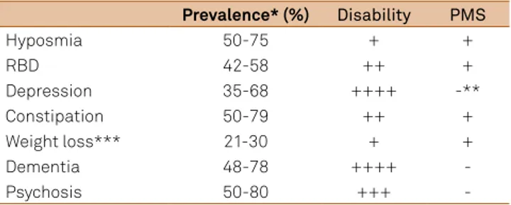
A summary of prevalence, its correlation with disease progression, level of functional disability and potential role as premotor signs of PD is presented in Table 1 and Figure.
Olfactory deficit
he irst description of olfactory deicits in PD was made almost 40 years ago15 and ive years later this signal was
de-scribed as an early feature and a “precursor of motor signs”16.
During the 80s, studies have shown that discrimination, de-tection threshold, and identiication of odors are abnormal in PD17,18. In addition, sequential studies have shown that
ol-factory deicits in these cases do not depend on odor sub-type, age of onset, duration and severity of the disease, motor laterality and phenotype, treatment, and presence of cogni-tive impairment19,20. Finally, a more recent study showed that
hyposmia does not luctuate during periods on and of21
. he
combination of these data indicates that this dysfunction is not directly related to the dopaminergic dysfunction or with the degenerative process responsible for motor signals of PD. In fact, a study that evaluated cholinergic and dopaminergic innervation concluded that the cholinergic deicit, especially located in the limbic cortex, represents a more robust deter-minant to olfactory deicits in PD than nigrostriatal dopami-nergic denervation22.
From a pathophysiological point of view, the presence of LB in the olfactory bulb seems to be a constant inding in patients with symptomatic PD, occurring in 100% of cases examined post-mortem. Congruent with the hypothesis de-scribed by Braak et al.2
cited previously, which proposes that the degenerative process in PD also begins in the olfactory bulb and anterior olfactory nuclei, loss of olfactory sensitiv-ity occurs in 70%-90% of patients with PD, including newly diagnosed cases who did not yet receive any form of treat-ment23
. Besides being an inherent part of the established disease, several evidences suggested and consolidated the hypothesis that hyposmia is an early sign of pre-motor DP, starting with the fact that the presence of LB in the SNc in-variably accompanied these pathological markers in the ol-factory structures. he opposite, however, is not always true, i.e., many cases have LB in the olfactory bulb with intact SNc. he length of time between the expression of hyposmia and the appearance of motor signs is still unknown and may be of several years24.
Since the early 1990s, it was demonstrated that olfactory deicits could distinguish sporadic PD from essential tremor, dystonic tremor and progressive supranuclear palsy (PSP)25
. Later, this same conclusion was determined with respect to multiple system atrophy (MSA), cortico basal degeneration and monogenic parkinsonism related to mutations in the PINK1 and PARK2 genes26,27 while cases with mutations in
the LRRK2 gene have intermediate results28,29.
he main longitudinal study showing that the olfactory def-icit may precede PD was held in Honolulu, Hawaii, by the group of Ross et al.30 who published a series of articles showing that
in the longitudinal follow-up of more than 2,000 neurologically
Table 1. Non-motor signs: prevalence, respective level of functional disability and potential role as premotor signs of Parkinson’s disease15,16,17,18,19,35,43,44,45,47,48,49,51,52,53,60.
Prevalence*(%) Disability PMS
Hyposmia 50-75 + +
RBD 42-58 ++ +
Depression 35-68 ++++ -**
Constipation 50-79 ++ +
Weight loss*** 21-30 + +
Dementia 48-78 ++++
-Psychosis 50-80 +++
healthy elderly individuals, those with proven olfactory deicit had a higher risk of developing symptomatic PD clinical and when they remained asymptomatic, these individuals present-ed LB in the SNc on post- mortem examination. Other studies that strengthen the idea that the olfactory disorder precedes the motor onset of PD include those by include Berendse et al.31
, and its follow-up after three years32 showing the
devel-opment of PD signals in cases of a group of “high risk”, which included family members of patients with PD who had hy-posmia, while family members without hyposmia remained asymptomatic after two years. Finally, a study that evaluated possible associations between olfactory deicit and the risk of developing dementia in PD with an observation period of three years using functional imaging techniques showed that chang-es in the areas associated with olfactory function have a close relationship with cognitive decline. In addition, the study also showed that more severe hyposmia represents a prominent factor for the development of dementia33.
REM sleep behavior disorder
Physiologically, patterned changes of neuronal activation and release of speciic neurotransmitters in the brain stem occur during the REM sleep phase, inducing, among other phenomena, motor inhibition and muscle atonia. RBD is a parasomnia characterized by the loss of this normal muscle atonia during this sleep stage34.
he gold standard for diagnosis requires polysomnog-raphy demonstrating excessive muscle activity, continued or intermittent tone of submentonian during REM sleep35
. Clinically, the diagnosis can also be established using his-torical data using instruments that range from formal cri-teria of the Manual of Disorders of Sleep of the American Academy of Sleep Medicine36 to the use of a single
ques-tion with a categorical “yes or no” type answer37. he
typi-cal clinitypi-cal picture includes a description of movements as-sociated with dream content and aggressive behavior with interpretation of such contents. Since the dream content in these cases tends to involve conlict situations, there is usu-ally sleep disruption of patients and/or their bed partner, who are occasionally victims to physical harm, including descriptions of trauma with signiicant severity35
. Patients with PD and RBD probably have a diferent distribution of the degenerative process compared to those without this parasomnia, once it’s the most probable substrate involves the pons tegmentum and the medial nuclei of the medul-la38. Clinically, RBD may be linked to particular
manifesta-tions such as worse scores of postural instability and gait, lower amplitude of motor response to levodopa, higher fre-quency of orthostatic hypotension and visual color percep-tion deicit. From a behavioral point of view, RBD is asso-ciated with vivid dreams and visual hallucinations, and is a warning sign for the occurrence of dementia and other
cognitive deicits involving speciic episodic verbal memo-ry, executive functions and visuospatial and visuoperceptu-al processing35.
Interestingly, a study that analyzed the quality of the movements during episodes of RBD showed that they do not have the standard asymmetric signs of parkinsonism ob-served during wakefulness, and are rapid, large, coordinated and symmetrical, however violent, ballistic and occasion-ally repetitive, suggesting that during REM sleep there may be a transient re-establishment of more physiological basal ganglia function. Alternatively, parkinsonism may disappear during REM sleep because at this stage the movements gen-erated follow directly to pyramidal tract neurons and sequen-tially to the peripheral motor neuron, bypassing the extrapy-ramidal system and the physiological barrier responsible for atonia during this sleep stage in the brainstem39.
RBD occurs in up to half of PD patients and, as in the case of hyposmia, is considered an established pre-motor symp-tom. Also, in almost half of PD patients and RBD, the sleep complication precedes the onset of parkinsonism by sever-al years. he mean intervsever-al from the onset of symptoms of RBD and motor signs is 11 to 12 years, but may range from 1 to up to 2035
. Moreover, cases of apparently idiopathic RBD are uncommon and have a chance of more than 50% to de-velop neurodegenerative diseases after 12 years of follow up, including more commonly PD, followed by LB dementia, Alzheimer’s disease and MSA40. Another important aspect is
that individuals with idiopathic RBD with the highest chance of developing PD have peculiar sleep characteristics, includ-ing higher proportion of REM sleep, more periodic lower limb movements per hour during of non-REM sleep and abnor-mal intensity of muscle atonia41. Finally, although RBD is
de-scribed in atypical forms of parkinsonism associated with the pathological background of synucleinopathies, particularly multiple system atrophy and LB dementia, their occurrence in PSP, although less common, is signiicant, indicating the concept that this parasomnia is not related to a speciic mo-lecular form, but rather to a speciic topographic progression of the degenerative process42.
Behavioral disorders
Mood disorders are common in PD, occurring in more than a quarter of newly diagnosed cases. Several studies have also shown that individuals with depression are 2.2 to 3.2 times more likely to develop PD compared to healthy con-trols43. Although the available data regarding this correlation
are less conclusive than for hyposmia and RBD, the onset of depressive symptoms may precede the motor signs, with a peak around 3-6 years before the diagnosis of PD. One of these studies evaluated 1,358 patients with depression and 67,570 healthy individuals matched for age and sex for an average period of 15 years, showing a chance of occurrence of PD in those with a history of depression 13.3 times higher than controls without depression. Another epidemiological
study found similar results with a chance of 2.95 times for the occurrence of PD in individuals with depression. In these studies and in the literature in general, depression is consid-ered a risk factor, as well as family history and life in a rural environment, but not necessarily a pre-motor symptom44.
In the course of the disease, depression is one of the ma-jor indicators of a poor quality of life and functional disability, occurring in about 40% of patients in cross-sectional stud-ies and almost 70% in longitudinal studstud-ies8. he diagnosis of
depression in PD may be complex because some of the signs of the disease itself may mimic it. Moreover, there are pecu-liar aspects regarding the presence and severity of depressive symptoms compared to the general population with depres-sion (Table 2) PD depresdepres-sion tends to manifest with more in-tense anxiety, pessimism, hopelessness, social withdrawal, self-depreciative thoughts, and introversion, but, on the oth-er hand, less felling of guilt, ideation of death and suicide8.
Depression in PD is probably multifactorial, involving a neurobiological substrate permeated by evidence of a reactive component related to living with a chronic neurodegenerative disease and its complications and limitations. For example, a study by Rickards et al.45 showed that the frequency of
depres-sion is high in the early stages of PD, when the patient is di-agnosed, decreasing when starting treatment, relecting the classic “honeymoon phase”, and increasing again with the pro-gression of symptoms in later stages. In addition, depression in PD is directly related to the severity of motor symptoms and may luctuate along with motor luctuations, relecting the pharmacological efect of levodopa. he biological basis of de-pression also has a pathological basis, related to the presence of PD’s own degeneration markers in serotonergic and norad-renergic structures as the raphe nuclei and limbic structures8.
Still under the behavioral aspect, a premorbid person-ality in PD has been observed, including a relatively uni-form description that often uses terms such as introversion,
Table 2. Clinical differentials in presentation of depression in
Parkinson’s disease (PD) versus the general population. More common in PD depression
Apathy Dysphoria Irritability Anxiety
Fluctuation of mood throughout the day Pessimism
Hopelesness Social withdrawal Introversion
Self-deprecating thoughts Uncommon in PD depression Sensation of guilt
behavioral inlexibility, diiculty with abstraction thought, more reliability, responsibility, loyalty, subordination, less tendency to leadership, conservative attitude with self-protection, intense self-control and diiculty expressing aggression. Unfortunately, these aspects are all very common in the general population and have a low speciicity for PD46.
Constipation
he clinical history of constipation is common at diagno-sis in cases of PD, and is also described before the emergence of motor signs. As a pre-motor symptom, it was described in a variable period that can reach up to 24 years before the on-set of parkinsonism in studies using diferent methodologies. In a longitudinal study of 6,790 males with constipation, the risk of occurrence of PD was 2.7 times higher than in those without this symptom. In this study, the mean time interval between the detection of constipation to diagnosis of PD was 12 years, ranging from two months to 24 years47.
he pathological support of this relationship comes from the observation that 9% to 17% of healthy subjects have alpha synuclein aggregates in the peripheral autonomic system, in-cluding abdominal-pelvic, cardiac and myenteric plexus of the esophagus, sympathetic ganglia and vagus nerve. hus, from the pathophysiological point of view, constipation may relect not only this peripheral involvement but also cen-tral mechanisms that cause pelvic loor dysfunction. Some of these individuals with peripheral autonomic disorder still present LB in the central nervous system and others have other pre-motor signs such as RBD, striatal abnormalities in functional tests or even pre-diagnostic PD as described above, in another words, the presence of a subtle cardinal sign that did not ill formal criteria for PD47,48.
Weight loss
Several studies have found that PD patients have low-er body mass index (BMI), compared to healthy controls matched for sex and age. he etiology seems to be multifac-torial, related to the presence of dyskinesias, change in eating habits, efect of medication (mainly levodopa), longer time for the ingestion of meals leading to lower energy intake and ear-lier satiety, in addition to being potentially related to intrinsic physiological changes of the neurodegenerative process. For example, the concentration of leptin, insulin-like growth fac-tor type 1 (IGF-1), and thyroid-stimulating hormone were in-vestigated in patients with PD and weight loss compared to controls with PD but no weight loss, inding lower levels of leptin and higher IGF-1 in the study group49,50.
he onset of weight loss in PD does not have a deined correlation with the stages of the disease, and may be an early expression or even precede the onset of motor signs. A pro-spective study that examined the changes in BMI in 468 pa-tients with PD using anthropometric data collected before the onset of motor symptoms, followed during the course of the disease showed that the average body mass remained
stable in most subjects up to a variable period before the on-set of motor signs. his period ranged from a few months up to four years. Interestingly, this study showed a tendency to the increase in caloric intake while the BMI decreased. In the course of the disease, the reduction of BMI followed a con-tinuous process with the same tendency of the pre-motor pe-riod, indicating that, regardless of the process that triggers this early change in BMI, it is relatively uniform and follows the disease progression51.
Cognitive deficit
Several degrees of cognitive impairment are common fea-tures of PD and, considering the model of disease progression described by Braak, may be a natural consequence of end-stage disease2
. he diagnosis of dementia is also especially common in PD, It has been described in about 20% of PD cases in cross-sectional studies52
, and almost 80% in longitudinal studies, with an incidence at least four times greater than in the gen-eral population53
. Furthermore, despite being more common and severe in the later stages, cognitive impairment that does not cause enough dysfunction to fulill diagnostic criteria for dementia may be present early in the course of the disease, with abnormalities in neuropsychological tests detected in up to 24% of patients at diagnosis of PD. Finally, those with the for-mal diagnosis of dementia will present it for an average period of four years during the course of the disease53.
Cognitive disorders tend to exacerbate the motor symp-toms disabilities in PD. In fact, the presence of cognitive im-pairment or dementia in PD patients is associated with loss of independence, poor quality of life and shortening of lifes-pan. Moreover, individuals in advanced stages of PD who de-velop dementia, often present a functional decline that fol-lows a stereotyped pattern, indicating that the beginning of this sign announces the need for a more intensive care and increased risk of mortality54. hus, the onset and progression
of cognitive impairment in PD have an important inluence on patient management and prognosis.
he phenomenology of speciic cognitive symptoms asso-ciated with PD varies and probably relects the heterogeneous underlying neuropathological substrate. he most common cognitive impairments are attention, executive behavior and visuospatial processing deicits, although patients may also exhibit diferent degrees of loss of memory, which could be, in some cases, a dysfunction secondary to a frontal (i.e., executive behavior) rescue information. In the same way, dementia in PD is not characterized by an intrinsic deicit of language, but may be associated with diiculty in processing and composition of phrases, which are also executive dysfunctions53
Among the risk factors for dementia in PD, age is the most important. On the other hand, the increased age-re-lated dementia risk does not seem to be associated with the possible inluence of age of onset of motor symptoms; in oth-er words, patients develop dementia in a similar mean age, regardless of when the motor signs appear54. Other studies
also showed that cognitive impairment in PD correlates with the severity of motor disabilities, male gender, low education, visual hallucinations and the presence of rigidity and axial bradykinesia55.
he pathological basis for dementia in PD is, in part, re-lated to the spread of LB to the cortex, which occurs in the later stages of disease2
. Moreover, other studies show that the pathology of the cognitive impairment in PD is proba-bly multifactorial, including LB and pathology characteristic of Alzheimer’s disease, along with factors related to age and other comorbidities55.
he efect of treatment must also be considered, since pharmacological and non-pharmacological interventions could worsen incipient cognitive impairment. he use of an-ticholinergics, for example, classically enhances the cognitive symptoms of PD, as well as treatment with deep brain stimu-lation. In these cases, although the motor function has a good response to surgery, quality of life tends to decline after the irst 6 months, probably, in part because of cognitive decline, which seems to be more signiicant when the target of this intervention is the subthalamic nucleus56.
Psychosis
Psychotic symptoms such as hallucinations and delu-sions are common in PD patients and occur from 10% to 80% of cases, depending on the deinition and the studied popula-tion. Risk factors includes cognitive impairment, dementia, age greater than 65 years, advanced disease, longer duration, presence of sleep disorders, depression, visual disturbances and use of most of the drugs needed to treat motor symp-toms of PD, the commonest culprits been dopamine agonists,
levodopa, amantadine and anticholinergics57. Psychosis is
associated with diferent pathological and molecular abnor-malities, however the most consistent indings suggest the presence of LB in the temporal/limbic cortex58.
Diagnostic criteria include the presence of one of the fol-lowing symptoms: illusions, false sense of presence, halluci-nations and delusions in the context of previous diagnosis of PD deined by Queen Square Brain Bank criteria. In addition, symptoms should be recurrent or continuous for at least one month59 visual hallucinations tend to be more common,
in-cluding a wide range of imagery, from simple indiscriminate shapes to well-formed entities with complex interaction with patients. Auditory hallucinations and other sensory func-tions are less common60.
As mentioned before, psychotic symptoms are more fre-quent in patients with cognitive impairment. In the case of a subclinical deicit, the presence of psychosis must be consid-ered as a predictor of imminent cognitive decompensation and dementia. hus, the so-called “benign” hallucinations are actually part of a gradual process with a potentially malig-nant prognosis60.
FINAL REMARKS
Despite the former cited observations about the func-tional and QoL related impact of NMS, until recently they re-ceived relatively little attention from clinicians. In part, that has happened due to unawareness and diiculties in con-ducting a more thorough and extensive assessment in a sys-tem that has limits of time and resources. Moreover, many patients and their caregivers tend to interpret NMS as being not “parkinsonian”, not deserving evaluation by a specialist in neurological diseases. On the other hand, our review shows that most of the NMS that have a signiicant impact on QoL and functionality are, at least in part, manageable and should be properly and promptly identiied.
References
1. Rajput AH, Rozdilsky B, Rajput A. Accuracy of clinical diagnosis in parkinsonism: a prospective study. Can J Neurol Sci. 1991;18(3):275-8. 2. Braak H, Del Tredici K, Rüb U, Vos RA, Steur ENJ, Braak E. Staging of
brain pathology related to sporadic Parkinson’s disease. Neurobiol Aging. 2003;24(2):197-211.
3. Jellinger KA. A critical evaluation of current staging of alpha-synuclein pathology in Lewy body disorders. Biochim Biophys Acta. 2009;1792:730-40. http://dx.doi.org/10.1016/S0197-4580(02)00065-9 4. Burke RE, Dauer WT, Vonsattel JP. A critical evaluation of the
Braak staging scheme for Parkinson’s disease. Ann Neurol. 2008;64(5):485-91. http://dx.doi.org/10.1002/ana.21541 5. Kingsbury AE, Bandopadhyay R, Silveira-Moriyama L, Ayling H,
Kallis C, Sterlacci W et al. Brain stem pathology in Parkinson’s disease: an evaluation of the Braak staging model. Mov Disord. 2010;25(15):2508-15. http://dx.doi.org/10.1002/mds.23305
6. Barone P, Antonini A, Colosimo C, Marconi R, Morgante L, Avarello TP et al. The PRIAMO study: a multicenter assessment of nonmotor symptoms and their impact on quality of life in Parkinson’s disease. Mov Disord. 2009;249(11):1641-9. http://dx.doi.org/10.1002/mds.22643
7. Chaudhuri KR, Odin P, Antonini A, Martinez-Martin P. Parkinson’s disease: the non-motor issues. Parkinsonism Relat Disord. 2011;17(10):717-23. http://dx.doi.org/10.1016/j.parkreldis.2011.02.018
8. Schrag A. Quality of life and depression in Parkinson’s disease. J Neurol Sci. 2006;248(1-2):151-7. http://dx.doi.org/10.1016/j.jns.2006.05.030 9. Zibetti M, Merola A, Rizzi L, Ricchi V, Angrisano S, Azzaro C et al.
10. Chaudhuri KR, Healy D, Schapira AHV. Non-motor symptoms of Parkinson’s disease: diagnosis and management. Lancet Neurol. 2006;5(3):235-45. http://dx.doi.org/10.1016/S1474-4422(06)70373-8 11. Gallagher DA, Lees AJ, Schrag A. What are the most important
nonmotor symptoms in patients with Parkinson’s disease and are we missing them? Mov Disord. 2010;25(15):2493-500. http://dx.doi.org/10.1002/mds.23394
12. Shulman LM, Taback RL, Rabinstein AA, Weiner WJ. Non-recognition of depression and other non-motor symptoms in Parkinson’s disease. Parkinsonism Relat Disord.2002;8(3):193-7.
http://dx.doi.org/10.1016/S1353-8020(01)00015-3
13. Chaudhuri KR, Prieto-Jurcynska C, Naidu Y, Mitra T, Frades-Payo B, Tluk S et al. The nondeclaration of nonmotor symptoms of Parkinson’s disease to health care professionals: an international study using the nonmotor symptoms questionnaire. Mov Disord. 2010;25(6):704-9. http://dx.doi.org/10.1002/mds.22868 14. O’Sullivan SS, Williams DR, Gallagher DA, Massey LA,
Silveira-Moriyama L, Lees AJ.Non motor symptoms as presenting complaints in Parkinson’s disease: a clinicopathological study. Mov Disord.2008;23(1):101-6. http://dx.doi.org/10.1002/mds.21813 15. Ansari KA, Johnson A. Olfactory function in patients with
Parkinson’s disease. J Chronic Dis. 1975;28(9):493-7. http://dx.doi.org/10.1016/0021-9681(75)90058-2 16. Korten JJ, Meulstee J. Olfactory disturbances in
Parkinsonism. Clin Neurol Neurosurg. 1980;82(2):113-8. http://dx.doi.org/10.1016/0303-8467(80)90005-0
17. Ward CD, Hess WA, Calne DB. Olfactory impairment in Parkinson’s disease. Neurology. 1983;33(7):943-6. http://dx.doi.org/10.1212/WNL.33.7.943 18. Quinn NP, Rossor MN, Marsden CD. Olfactory threshold in Parkinson’s
disease. J Neurol Neurosurg Psychiatry. 1987;50(1):88-9.
19. Doty RL, Deems DA, Stellar S. Olfactory dysfunction in parkinsonism: a general deficit unrelated to neurologic signs, disease stage, or disease duration. Neurology. 1988;38(8):1237-44.
20. Doty RL, Stern MB, Pfeiffer C, Gollomp SM, Hurtig HI. Bilateral olfactory dysfunction in early stage treated and untreated idiopathic Parkinson’s disease. J Neurol Neurosurg Psychiatry. 1992;55(2):138-42. http://dx.doi.org/10.1136/jnnp.55.2.138 21. Saifee T, Lees AJ, Silveira-Moriyama L. Olfactory
function in Parkinson’s disease in ON versus OFF states. J Neurol Neurosurg Psychiatry. 2010;81(11):1293-95. http://dx.doi.org/10.1136/jnnp.2009.182022
22. Bohnen NI, Muller ML, Kotagal V, et al. Olfactory dysfunction, central cholinergic integrity and cognitive impairment in Parkinson’s disease. Brain. 2010;133(6):1747-54. http://dx.doi.org/10.1093/brain/awq079 23. Hoyles K, Sharma JC. Olfactory loss as a supporting feature in the
diagnosis of Parkinson’s disease: a pragmatic approach. J Neurol. 2013;260(12):2951-8. http://dx.doi.org/10.1007/s00415-013-6848-8 24. Noyce AJ, Bestwock JP, Silveira-Moriyama L, Hawkes CH, Giovannoni
G, Lees AJ et al. Meta-analysis of early nonmotor features and risk factors for Parkinson disease. Ann Neurol. 2012;72(6):893-901. http://dx.doi.org/10.1002/ana.23687
25. Busenbark KL, Huber SJ, Greer G, Pahwa R, Koller WC. Olfactory function in essential tremor. Neurology. 1992;42(8):1631-2. 26. Kertelge L, Bruggemann N, Schmidt A, Tadic V. Wisse C, Dankert S et
al. Impaired sense of smell and color discrimination in monogenic and idiopathic Parkinson’s disease. Mov Disord. 2010;25(15):2665-9. http://dx.doi.org/10.1002/mds.23272
27. Alcalay RN, Siderowf A, Ottman R et al. Olfaction in Parkin heterozygotes and compound heterozygotes: the CORE-PD study. Neurology. 2011;76(4):319-26. http://dx.doi.org/10.1212/WNL.0b013e31820882aa 28. Silveira-Moriyama L, Munhoz RP, Carvalho MJ, Raskin
S, Rogaeva E, Aguiar PC et al. Olfactory heterogeneity in LRRK2 related Parkinsonism. Mov Disord. 2010;25:2879-83. http://dx.doi.org/10.1002/mds.23325
29. Marras C, Schule B, Munhoz RP, Rogaeva E, Langston JW, Kasten M et al. Phenotype in parkinsonian and nonparkinsonian LRRK2 G2019S mutation carriers. Neurology. 2011;77(4):325-33. http://dx.doi.org/10.1212/WNL.0b013e318227042d
30. Ross GW, Petrovitch H, Abbott RD, Tanner CM, Popper J, Masaki K et al. Association of olfactory dysfunction with risk for future Parkinson’s disease. Ann Neurol. 2008;63(2):167-73. http://dx.doi.org/10.1002/ana.21291
31. Berendse HW, Booij J, Francot CM, Bergmans PL, Hijman R, Stoof JC et al. Subclinical dopaminergic dysfunction in asymptomatic Parkinson’s disease patients’ relatives with a decreased sense of smell. Ann Neurol. 2001;50(1):34-41. http://dx.doi.org/10.1002/ana.1049 32. Ponsen MM, Stoffers D, Booij J, Eck-Smit BL, Wolters ECH, Berendse
HW. Idiopathic hyposmia as a preclinical sign of Parkinson’s disease. Ann Neurol. 2004;56(2):173-81. http://dx.doi.org/10.1002/ana.20160 33. Baba T, Kikuchi A, Hirayama K, Nishio Y, Hosokai Y, Kanno S et al.
Severe olfactory dysfunction is a prodromal symptom of dementia associated with Parkinson’s disease: a 3 year longitudinal study. Brain. 2012;135(1):161-9. http://dx.doi.org/10.1093/brain/awr321 34. Schenk CH, Mahowald MW. REM sleep behavior disorder: clinical,
developmental, and neuroscience perspectives 16 years after its formal identification in SLEEP. Sleep. 2002;25(2):120-38. 35. Postuma RB, Montplaisir J. Predicting Parkinson’s disease: why,
when, and how? Parkinsonism Relat Disord. 2009;15 Suppl 3:S105-9. http://dx.doi.org/10.1016/S1353-8020(09)70793-X
36. 36. American Academy of Sleep Medicine. ICSD - International classification of sleep disorders: diagnostic and coding manual. Westchester: American Sleep Disorders Association; 1990. 37. Postuma RB, Arnulf I, Hogl B, Iranzo A, Miyamoto T, Dauvilliers
Y et al. A single-question screen for rapid eye movement sleep behavior disorder: a multicenter validation study. Mov Disord 2012;27(7):913-6. http://dx.doi.org/10.1002/mds.25037 38. Postuma RB, Gagnon JF, Montplaisir J. Rapid eye movement
sleep behavior disorder as a biomarker for neurodegeneration: the past 10 years. Sleep Med 2013;14(8):763-7.
http://dx.doi.org/10.1016/j.sleep.2012.09.001
39. De Cock VC, Vidailhet M, Leu S, Teixeira A, Apartis E, Elbaz A et al. Restoration of normal motor control in Parkinson’s disease during REM sleep. Brain. 2007;130(2):450-6. http://dx.doi.org/10.1093/brain/awl363 40. Munhoz RP, Teive HA, Eleftherohorinou H, Coin LJ, Lees AJ,
Silveira-Moriyama L. Demographic and motor features associated with the occurrence of neuropsychiatric and sleep complications of Parkinson’s disease. J Neurol Neurosurg Psychiatry.
2013;84(8):883-7. http://dx.doi.org/10.1136/jnnp-2012-304440 41. Vendette M, Gagnon JF, Décary A, Massicotte-Marquez J, Postuma RB,
Doyon J et al. REM sleep behavior disorder predicts cognitive impairment in Parkinson disease without dementia. Neurology. 2007;69(19):1843-9. http://dx.doi.org/10.1212/01.wnl.0000278114.14096.74
42. Munhoz RP, Teive HA. REM sleep behaviour disorder: how useful is it for the differential diagnosis of parkinsonism? Clin Neurol Neurosurg. 2014;127:71-4. http://dx.doi.org/10.1016/j.clineuro.2014.09.014 43. Starkstein SE, Brockman S, Hayhow BD. Psychiatric syndromes
in Parkinson’s disease. Curr Opin Psychiatry. 2012;25(6):468-72. http://dx.doi.org/10.1097/YCO.0b013e3283577ed1
44. Leentjens AF, Van den Akker M, Metsemakers JF, Lousberg R, Verhey FR. Higher incidence of depression preceding the onset of Parkinson’s disease: a register study. Mov Disord. 2003;18(4):414-8. http://dx.doi.org/10.1002/mds.10387
45. Rickards H. Depression in neurological disorders: Parkinson’s disease, multiple sclerosis, and stroke. J Neurol Neurosurg Psychiatry. 2005;76 Suppl 1:i48-52. http://dx.doi.org/10.1136/jnnp.2004.060426
47. Abbott RD, Petrovitch H, White LR, Masaki KH, Tanner CM, Curb JD et al. Frequency of bowel movements and the future risk of Parkinson’s disease. Neurology. 2001;57(3):456-62. http://dx.doi.org/10.1212/WNL.57.3.456
48. Cersosimo MG, Benarroch EE. Autonomic involvement in Parkinson’s disease: pathology, pathophysiology, clinical features and
possible peripheral biomarkers. J Neurol Sci. 2012;313(1-2):57-63. http://dx.doi.org/10.1016/j.jns.2011.09.030
49. Cheshire Jr WP, Wszolek ZK. Body mass index is reduced early in Parkinson’s disease. Parkinsonism Relat Disord. 2005;11(1):35-8. http://dx.doi.org/10.1016/j.parkreldis.2004.07.001
50. Munhoz RP, Ribas CB. Body mass index in Parkinson’s disease. Parkinsonism Relat Disord. 2005;11(6):407. http://dx.doi.org/10.1016/j.parkreldis.2005.02.001
51. Chen H, Zhang SM, Hernán MA, Willett WC, Ascherio A. Weight loss in Parkinson’s disease. Ann Neurol. 2003;53(5):676-9. http://dx.doi.org/10.1002/ana.10577
52. Munhoz RP, Werneck LC, Teive HA. The differential diagnoses of parkinsonism: findings from a cohort of 1528 patients and a 10 years comparison in tertiary movement disorders clinics. Clin Neurol Neurosurg. 2010;112(5):431-5. http://dx.doi.org/10.1016/j.clineuro.2010.03.003
53. Aarsland D, Beyer MK, Kurz MW. Dementia in Parkinson’s disease. Curr Opin Neurol. 2008;21(6):676-82.
http://dx.doi.org/10.1097/WCO.0b013e3283168df0 54. Levy G, Schupf N, Tang MX, Cote LJ, Louis ED, Mejia H et al.
Combined effect of age and severity on the risk of dementia
in Parkinson’s disease. Ann Neurol. 2002;51(6):722-9. http://dx.doi.org/10.1002/ana.10219
55. Buter TC, Hout A, Matthews FE, Larsen JP, Brayne C, Aarsland D. Dementia and survival in Parkinson disease: a 12-year population study. Neurology. 2008;70(13):1017-22. http://dx.doi.org/10.1212/01.wnl.0000306632.43729.24 56. Witt K, Daniels C, Reiff J, Krack P, Volkmann J, Pinsker MO et al.
Neuropsychological and psychiatric changes after deep brain stimulation for Parkinson’s disease: a randomised, multicentre study. Lancet Neurol. 2008;7(7):605-14. http://dx.doi.org/10.1016/S1474-4422(08)70114-5 57. Munhoz RP, Espay AJ, Morgante F, Li JY, Teive HÁ, Dunn E et al.
Long-duration Parkinson’s disease: role of lateralization of motor features. Parkinsonism Relat Disord. 2013;19(1):77-80. http://dx.doi. org/10.1016/j.parkreldis.2012.07.008
58. Kalaitzakis ME, Christian LM, Moran LB, Graeber MB, Pearce RK, Gentleman SM. Dementia and visual hallucinations associated with limbic pathology in Parkinson’s disease. Parkinsonism Relat Disord. 2009;15(3):196-204. http://dx.doi.org/10.1016/j.parkreldis.2008.05.007
59. Ravina B, Marder K, Fernandez HH, Friedman JH, McDonald W, Murphy D et al. Diagnostic criteria for psychosis in Parkinson’s disease: report of an NINDS, NIMH work group. Mov Disord. 2007;22(8):1061-8. http://dx.doi.org/10.1002/mds.21382 60. Factor SA, Feustel PJ, Friedman JH, Comella CL, Goetz CF,