DOI: 10.1590/0004-282X20150161
ARTICLE
VIEW AND REVIEW
Clinical and genetic basis of familial
amyotrophic lateral sclerosis
Bases clínicas e genéticas da esclerose lateral amiotrófica familiar
Paulo Victor Sgobbi de Souza, Wladimir Bocca Vieira de Rezende Pinto, Marco Antônio Troccoli Chieia, Acary Souza Bulle Oliveira
Amyotrophic Lateral Sclerosis (ALS) or Lou Gehrig’s dis-ease is the main progressive adult-onset neurodegenerative motor neuron disease (MND), afecting primarily upper (UMN) and lower motor neurons (LMN) giving rise to its typical neurological manifestations. Overall prevalence of ALS is around two to seven cases per 100000 inhabitants1,2,
and incidence around one to two new cases per 100000 inhabitants per year2,3,4.
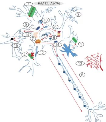
ALS arises as a consequence of multiple pathophysiologi-cal mechanisms and cellular dysfunctions (Figure 1), including protein misfolding and aggregation, altered RNA processing (mainly disturbed mRNA splicing and signaling), defects in axonal transport, abnormal metabolism and accumulation of reactive oxygen species, mitochondrial dysfunctions,
microg-lial neuroinlammatory mechanisms, direct glutamate excito
-toxicity by astrocytes, abnormal modulatory efects from other
glial cells, disturbances of autophagy, ubiquitine-proteosome system abnormalities and primary and secondary ion channel defects5,6,7,8. Environmental and toxic factors also represent
ma-jor factors, including traumatic sports mechanisms and toxic causes (including the ALS-parkinsonism/dementia of Guam)9.
Typical electrodiagnostic and clinical signs arise from a complex network of neuropathological changes includ-ing upper motor neuron degeneration in the frontal lobe (mainly Betz giant cells in the motor cortex), corticospinal and corticobulbar tracts (in the spinal cord, internal cap-sule and cerebral peduncles pathways), lower motor neu-rons in brainstem nuclei (motor nuclei of cranial nerves VII, X, XI and XII) and in anterior horn motoneurons of the spi-nal cord. Onufrowicz-Mannen’s nucleus and some cranial nerve motoneurons (motor nuclei of cranial nerves III, IV and VI) are generally spared, correlating with exceptionally
Universidade Federal de São Paulo, Departamento de Neurologia e Neurocirurgia, Divisão de Doenças Neuromusculares, Sao Paulo SP, Brazil.
Correspondence: Wladimir Bocca Vieira de Rezende Pinto; Departamento de Neurologia e Neurocirurgia - UNIFESP; Rua Estado de Israel, 899; 04022-002 São Paulo SP, Brasil; E-mail: wladimirbvrpinto@gmail.com
Conflict of interest: There is no conlict of interest to declare.
Received 08 March 2015; Received in inal form 27 June 2015; Accepted 17 July 2015.
ABSTRACT
Amyotrophic lateral sclerosis represents the most common neurodegenerative disease leading to upper and lower motor neuron compromise. Although the vast majority of cases are sporadic, substantial gain has been observed in the knowledge of the genetic forms of the disease, especially of familial forms. There is a direct correlation between the proile of the mutated genes in sporadic and familial forms, highlighting the main role of C9orf72 gene in the clinical forms associated with frontotemporal dementia spectrum. The different genes related to familial and sporadic forms represent an important advance on the pathophysiology of the disease and genetic therapeutic perspectives, such as antisense therapy. The objective of this review is to signal and summarize clinical and genetic data related to familial forms of amyotrophic lateral sclerosis.
Keywords: amyotrophic lateral sclerosis, motor neuron disease, neurogenetics, neurodegeneration, C9orf72 gene.
RESUMO
A esclerose lateral amiotróica representa a forma mais comum de doença neurodegenerativa com comprometimento do neurônio motor superior e inferior. Embora a maioria dos casos seja esporádica, ganho impressionante referente ao conhecimento das formas genética da doença foi observado, em especial das formas familiares. Há uma correlação direta entre o peril de genes mutados nas formas familiares e esporádicas, destacando-se o papel principal do gene C9orf72 nas formas clínicas associadas com espectro da demência frontotemporal. Os diferentes genes relacionados às formas familiares e esporádicas representam um importante avanço na isiopatologia da doença e perespectivas terapêuticas genéticas, como a terapia antisense. O objetivo desta revisão é apontar e resumir os principais dados clínicos e genéticos relacionados às formas familiares da esclerose lateral amiotróica.
rare compromises of facial and extrinsec ocular movements and sphincteral disturbances1,2. In microscopic evaluation,
cytoplasmic Bunina bodies and Lewy body-like are motor neuron neuropathological hallmarks of ALS, although each genetic subtype commonly presents with their signatures.
Neuropathological remarks include neuroilamentous swell
-ing of proximal axons with reduced calibre of distal axons and axonal wallerian degeneration, accumulation of neuro-ilament and peripherin in axons and perikarium, reactive glyosis, Lewy body-like cytoplasmic neuronal inclusions, perikarial inclusions with phosphorylated neuroilament and ubiquitin immunoreactivity and positive immunoreactivity for other biochemical markers depending on genetic basis of
the disease, mainly ubiquinated protein inclusions with posi-tivity for TAR (Transactive response) DNA-binding protein 43 (TDP-43)2. Furthermore, diferent authors believe there is a
regional spreading of intracellular misfolded pathogenic pro-teins SOD1 and TDP-43 involved in ALS in a prion-like prop-agation in an intercellular contiguous spreading fashion2,6,10.
Most cases of ALS present with asymmetric focal apen-dicular weakness progressing with bulbar dysfunction, quadriparesis and respiratory insuiciency leading to death in about two to three years after onset1. he combination of
upper motor neuron and lower motor neuron signs of com-promise is classically described with variable degrees of each component during disease progression1. Association with
cognitive and behavioural disturbances is common and sometimes present with the classical phenotype of behav-ioral variant (bv) frontotemporal dementia (FTD). Although
lability of afect commonly occurs, bvFTD, executive dysfunc
-tion and mood disorders occurs more frequently in some
speciic clinical and genetic conditions. A FTD clinical sus
-pection can be properly analyzed applying speciic criteria, including the Hodge’s and Neary criteria11. Other cognitive
compromise patterns also occurs including primary progres-sive nonluent aphasia, logopenic progresprogres-sive aphasia and semantic dementia. Around 5% of sporadic and familial ALS cases fullill properly diagnostic criteria for FTD11.
here are no speciic clinical, neuroimaging and serum and cerebrospinal luid biochemical markers to provide the deinite diagnosis of ALS and clinicians must be aware
about diferential diagnosis and red-lags1. Clinical and
elec-trodiagnostic indings are essential to guide the diagnosis process using both the revised El Escorial criteria and the Awaji-shima electrodiagnostic criteria. Although causative genes and susceptibility loci are well-established in sporadic and familial ALS (Table 1), presymptomatic testing does not represent a reliable and available diagnostic method in most neurological centers2,6.
Most cases of ALS are sporadic (90 to 95% of ALS cases) and generally occurs in patients between the ifth and seventh decades of life. Familial ALS cases are deined when there is a context of more than one afected family member (relative) from irst or second generation from the index or propositum case with the same disease presentation (despite the frequent occurence of intrafamilial clinical variability) and generally as-sociates with: (i) earlier age at disease onset; (ii) more com-monly symptom onset starts in the lower extremities; and (iii) longer or shorter disease duration, life expectancy and clini-cal progression, depending on particular genetic subtypes2,12,13.
Familial cases can occur in an autosomal recessive or dominant or dominant X-linked inheritance patterns. Most adult-onset cases of familial ALS have autosomal dominant inheritance pattern, while juvenile-onset cases have autosomal recessive pattern, in a similar way to the observed with autosomal re-cessive cerebellar ataxias and autosomal dominant spinocer-ebellar ataxias. Overall penetrance is age and gene-dependent 1. Direct glutamate astrocyte excitotoxicity (i.e. DAO, ALS2, HFE)
2. Altered RNA processing and metabolism (i.e. C9orf72, TARDBP, FUS, EWSR1, TAF15, ATXN2, HNRNPA1, SETX, ANG, SMN1, ELP3, MATR3)
3. Microglial neuroinlammatory activation (i.e. SOD1)
4. Secondary ion channel defects and transmembrane receptors (i.e. C9orf72, SIGMAR1, ERBB4, TRPM7, HFE)
5. Anterograde/retrograde axonal transport (i.e. SOD1, DCTN1, PRPH, SPG11, PRPH, CHMP2B, PFN1, KIFAP3)
6. Ubiquitin-proteosome system dysfunction (i.e. VCP, UBQLN2, SQSTM1, OPTN, FIG4)
7. Golgi-endosomal reticulum traficking (i.e. OPTN, FIG4, SIGMAR1, VAPB, VCP) 8. Direct DNA lesion repair (i.e. SPG11, SETX, FUS, APEX1)
9. Abnormal accumulation of reactive oxygen species (i.e. SOD1, ALS2, APEX1, HFE, PON)
10. Toxic protein aggregates and misfolding (i.e. SOD1, VCP, UBQLN2, DAO, OPTN, SQSTM1)
11. Mitochondrial dysfunctions (i.e. CHCHD10, CYP27A1, COX1, IARS2) 12. Cytoskeleton dynamics and architecture (i.e. TUBA4A, PFN1, DCTN1, NEFH, PRPH)
13. Angiogenesis (i.e. ANG, VEGF)
14. Endosomal and vesicular traficking (i.e. ALS2, VAPB, C9orf72, CHMP2B, OPTN, DCTN1, ATXN2, FIG4, VCP, UNC13A)
15. Disturbances of autophagy (i.e. FIG4, VCP, UBQLN2, SQSTM1)
Figure 1. Schematic representation of the main pathophysiological mechanisms involved in familial amyotrophic lateral sclerosis and the genetic changes involved in each case5,6,7,8.
1 2
7
3
5 10
1
9
4
6 8
EAAT2, AMPA
11 12
13 14
15
•OH
-•NO ONOO•O-2
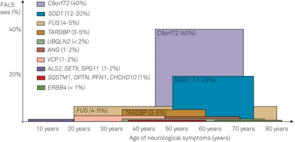
-in familial ALS, as nearly half of patients with SOD1 and FUS
genes mutations become symptomatic at their ifties and 90% at their seventies12. here is also a good correlation between
genetic subtypes and general age at onset (Figure 2). Genetic anticipation is exceptionally seen. here is also a tendency to progress with more prominent bulbar symptoms in familial ALS than in sporadic cases with the same age. It is important
to reiterate that no speciic clinical, cerebrospinal and neuro
-imaging parameters can rightly and reliably diferentiate fa -milial from sporadic ALS in cases without a signiicant fa-milial history of neurodegeneration2,12,13,14.
here are two other clinical situations which should be remembered in the context of familial and sporadic ALS: young-onset and juvenile ALS. Young-onset ALS starts before 45 years old, corresponds to 10% of all ALS cases and tends to present less commonly with bulbar-onset symptoms. Most cases arise in the context of sporadic ALS15. Juvenile ALS
rep-resents cases starting very early before 25 years of age with slowly progressive ALS, serving as a guide and key-element for clinical suspicion of autosomal recessive familial cases6,15.
Juvenile forms are frequently described in ALS2, ALS4, ALS5, ALS6 (rarely), ALS15 and ALS166.
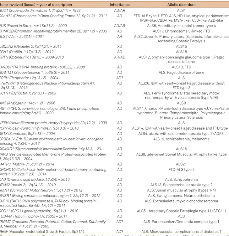
Table 1. Genetic causes of amyotrophic lateral sclerosis, year of description, pattern of inheritance and allelic conditions2,8,12,59.
Gene involved (locus) – year of description Inheritance Allelic disorders
SOD1 (Superoxide dismutase 1; 21q22.11) – 1993 AD/AR ALS1
C9orf72 (Chromosome 9 Open Reading Frame 72; 9p21.2) – 2011 AD FTD-ALS type 1, FTD, ALS, HD-like, atypical parkinsonism (PSP-like, CBD-like, MSA-like), CJD-like, AZD-like
FUS (Fused in Sarcoma; 16p11.2) – 2009 AD/AR ALS6, Hereditary essential tremor type 4
CHMP2B (Chromatin-modifying protein member 2B; 3p11.2) – 2006 AD ALS17, Chromosome 3-linked FTD
ALS2 (Alsin; 2q33.1) – 2001 AR ALS2, Juvenile Primary Lateral Sclerosis, Infantile-onset Ascending Spastic Paralysis
UBQLN2 (Ubiquilin 2; Xp11.21) – 2011 XD ALS15
PFN1 (Profilin 1; 17p13.2) – 2012 AD ALS18
OPTN (Optineurin; 10p13) – 2009/2010 AR/AD ALS12, primary open angle glaucoma type 1, Paget disease of bone
TARDBP (TAR DNA-binding protein; 1p36.22) – 2008 AD ALS10, FTD
SQSTM1 (Sequestosome 1; 5q35.3) – 2011 AD ALS, Paget disease of bone
PRPH (Peripherin; 12q13.12) – 2004 AD? ALS
HNRNPA1 (Heterogeneous Nuclear Ribonucleoprotein A1; 12q13.13) – 2013
AD ALS20, IBM with early-onset Paget disease without FTD type 3
DCTN1 (Dynactin 1; 2p13.1) – 2003 AD ALS, Perry syndrome, Distal hereditary motor neuronopathy with vocal paresis (type VIIB)
ANG (Angiogenin; 14q11.2) – 2006 AD ALS9
FIG4 (FIG4, S. cerevisiae, homolog of SAC1 lipid phosphatase domain containing; 6q21) – 2009
AD ALS11, Charcot-Marie-Tooth disease type 4J, Yunis-Varon syndrome, Bilateral Temporooccipital Polymicrogyria,
Primary Lateral Sclerosis
NEFH (Neurofilament protein, Heavy Polypeptide; 22q12.2) – 1999 AD ALS
VCP (Valosin-containing Protein; 9p13.3) – 2010 AD ALS14, IBM with early-onset Paget disease and FTD type 1
SETX (Senataxin; 9q34.13) – 2004 AD ALS4, ataxia with oculomotor apraxia type 2 (AOA2)
ERBB4 (V-Erb-B2 avian erythroblastic leucemia viral oncogene homolog 4; 2q34) – 2013
AD ALS19, schizophrenia, melanoma
SIGMAR1 (Sigma Nonopioid Intracellular Receptor 1; 9p13.3) – 2011 AR ALS16
VAPB (Vesicle-associated Membrane Protein-associated Protein B; 20q13.32) – 2004
AD ALS8, late-onset Spinal Muscular Atrophy Finkel type
MATR3 (Matrin-3; 5q31.2) – 2014 AD ALS21
CHCHD10 (Coiled-coil-helix-coiled-coil-helix-domain-containing protein 10; 22q11.23) – 2014
AD FTD-ALS type 2
DAO (D-amino acid oxidase; 12q24) – 2010 AD ALS, Schizophrenia
ATXN2 (ataxin 2; 12q24.12) – 2010 AD ALS13, Spinocerebellar ataxia type 2
SMN1 (Survival of Motor Neuron 1; 5q13.2) – 2012 AD ALS, Spinal muscular atrophy (types 1-4)
EWSR1 (Ewing sarcoma breakpoint region 1; 22q12.2) – 2012 AD ALS, Ewing sarcoma, Neuroepithelioma
TAF15 (TAF15 RNA polymerase II, TATA box-binding protein-associated factor, 68-kD; 17q12) – 2011
AD ALS, Extraskeletal myxoid chondrosarcoma
SPG11 (SPG11 gene/spatacsin; 15q21.1) – 2010 AR ALS5, Hereditary Spastic Paraplegia type 11 (SPG11)
TUBA4A (Tubulin, alpha-4A; 2q35) – 2014 AD ALS
TRPM7 (Transient Receptor Potential Cation Channel, Subfamily M, Member 7; 15q21.2) – 2005
CLINICAL AND GENETIC FORMS OF FAMILIAL ALS
From a historical perspective, Horton classiied fa
-milial forms of motor neuron disease in three main clinico-pathological phenotypes: (i) the irst with early-onset
motor symptoms, before ive years of age, with a rapidly pro
-gressive and lower motor neuron dominant ALS phenotype, with degeneration of corticospinal tracts and anterior horn motoneurons; (ii) the second form clinically similar, but also with posterior column and spinocerebellar tract degenera-tions; and (iii) a third form similar to the second, but with prolonged survival period of up to two decades16.
he genetic history of familial ALS can be summed up by the outstanding roles of SOD1 and C9orf72 genes. By 2011, the year of the initial description of the hexanucleotide re-peat expansion of C9orf72 gene, only 30% of familial cases had established their genetic etiology5,8,17,18,19. In the last
de-cade, more than 20 diferent loci were related to familial ALS. Despite the marked heterogeneity of familial ALS, it can be said that most cases relate to C9orf72, SOD1, FUS, TARDBP
and UBQLN2 genes6,8. hus, it is also possible to set speciic
genetic test batteries for recessive and autosomal dominant and X-linked forms, according to inheritance patterns, clini-cal comorbitidities, natural history and cliniclini-cal evolution, and population epidemiology. However, up to 32% of familial
cases and up to 11% of sporadic cases still do not have a dei
-nite genetic diagnosis of ALS2,7,8,19.
Establishing a definite diagnosis of familial ALS is complex. In an Italian study with 53 families, genetic screening for seven of the most important genes (SOD1, C9orf72, TARDBP, FUS, ANG, ATXN2, OPTN) disclosed only 25% of definite genetic diagnosis with 75% of them with two family members clinically affected and 17% with only one affected family member first and second degrees away14.
here is a lot of controversy regarding the role of most genes discovered nowadays in relation to ALS, as some of them do not have a unique causative mechanism (i.e. SOD1, C9ORF72, SETX, ANG, SPG11, FUS, TARDBP, VAPB, VCP, UBQLN2, OPTN, among others) but a disease-modifying func-tion or susceptibility loci (i.e. PGRN, HFE, NEFH, UNC13A, VEGF, among others)4,13. Further discussions regarding
ge-netic and clinical basis of familial ALS types will be provided forth (Tables 1 and 2).
ALS1
ALS1 (MIM #105400) represents the second most com-mon form of familial ALS, giving rise to up to 20% of cas-es with an autosomal dominant or reccas-essive inheritance.
SOD1 gene (superoxide dysmutase-1; 21q22.11) mutations give rise to abnormal function of copper/zinc superoxide dysmutase 1, responsible for converting superoxide free radical species from cytoplasm and inner intermembrane mitochondrial space into molecular oxygen and hydrogen peroxide. Secondary dysfunction of tyrosine phosphatases with phosphorilation inhibition by EGF (epidermal growth
factor), IGF-1 (insulin-like growth factor 1) and FGF-2 ( i
-broblast growth factor-2) by MAPK (mitogen-activated pro-tein kinase) pathway7,20.
By 2011, SOD1-associated ALS ( formerly the 21q-associated ALS) represented the most common form of familial and spo-radic ALS. Most cases present with adult-onset ALS without cognitive compromise and variable clinical outcomes de-pending on the genetic background involved in each case4,7,20.
Rapidly progressive forms of SOD1 familial ALS occur in USA with p.Ala4Val mutation. Slowly progressive cases corre-late to p.Asp90Ala mutation in Scandinavia and p.His46Arg in Japan. A LMN dominant ALS variant linked to SOD1 was described in cases of p.Ala4Val e p.Val148Gly mutations. A
Figure 2. Distribution of the most important genetic causes of familial ALS according to the age of onset of neurological signs and symptoms. The proportion of each gene in relation to all familial cases is also represented4,8.
FALS cases (%)
10 years 20 years 30 years 40 years 50 years 60 years 70 years 80 years
C9orf72 (40%) 40%
SOD1 (12-20%)
FUS (4-5%) TARDBP (3-5%) 20%
TARDBP (3-5%)
UBQLN2 (< 2%)
SOD1 (12-20%)
C9orf72 (40%)
VCP (1-2%)
SQSTM1, OPTN, PFN1, CHCHD10 (1%)
ALS2, SETX, SPG11 (1-2%)
FUS (4-5%)
ANG (1-2%)
ERBB4 (< 1%)
cerebellar ataxia variant with slow progression was described in Scandinavia in cases of p.Asp90Ala mutation7,20.
ALS2
ALS2 (MIM #205100) represents a rare autosomal reces-sive slowly progresreces-sive UMN-dominant juvenile ALS as a con-sequence of homozigosity mutations in ALS2 gene (2q33.1), coding alsin, involved in the membrane and endosomal intracellular traicking as guanine nucleotide exchange factor for Rac1 and Rab5 GTPases, in neurite outgrowth in hippocampus mediated by Rac1 activation and in prevention of glutamartegic excitoxicity mediated by Glur2 subunit of AMPA (α-amino-3-hydroxy-5-methyl-4-isoxazolepropionic acid) receptors15,21. It begins in preschool child until the
young adult ages (up to third decade) and has been described in japanese, turkish, tunisian, kuwaitian, saudi arabian, cy-priot and Amish population. ALS2 starts with lower limb and facial spasticity, moderate muscular atrophy, pseudobulbar signs, spastic dysathrophonia and bladder dysfunction evolv-ing statically after two decades. Important clinical overlap with allelic forms of Juvenile primary lateral sclerosis and in-fantile ascending hereditary spastic paralysis occurs13,15,21.
ALS3
ALS3 (MIM %606640) represents a rare autosomal domi-nant adult-onset familial ALS with lower limb onset associat-ed with 18q21 locus. Classicaly evolves to respiratory failure
and death ive years after onset22.
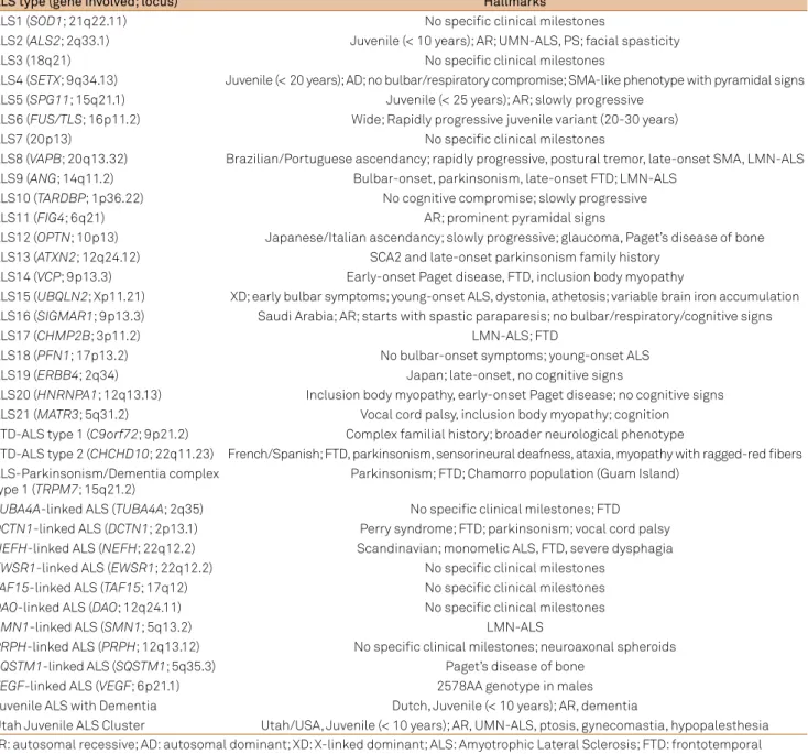
Table 2. Diagnostic cues and hallmarks of each type of familial ALS2,8,58.
ALS type (gene involved; locus) Hallmarks
ALS1 (SOD1; 21q22.11) No speciic clinical milestones
ALS2 (ALS2; 2q33.1) Juvenile (< 10 years); AR; UMN-ALS, PS; facial spasticity
ALS3 (18q21) No speciic clinical milestones
ALS4 (SETX; 9q34.13) Juvenile (< 20 years); AD; no bulbar/respiratory compromise; SMA-like phenotype with pyramidal signs ALS5 (SPG11; 15q21.1) Juvenile (< 25 years); AR; slowly progressive
ALS6 (FUS/TLS; 16p11.2) Wide; Rapidly progressive juvenile variant (20-30 years)
ALS7 (20p13) No speciic clinical milestones
ALS8 (VAPB; 20q13.32) Brazilian/Portuguese ascendancy; rapidly progressive, postural tremor, late-onset SMA, LMN-ALS ALS9 (ANG; 14q11.2) Bulbar-onset, parkinsonism, late-onset FTD; LMN-ALS
ALS10 (TARDBP; 1p36.22) No cognitive compromise; slowly progressive
ALS11 (FIG4; 6q21) AR; prominent pyramidal signs
ALS12 (OPTN; 10p13) Japanese/Italian ascendancy; slowly progressive; glaucoma, Paget’s disease of bone ALS13 (ATXN2; 12q24.12) SCA2 and late-onset parkinsonism family history
ALS14 (VCP; 9p13.3) Early-onset Paget disease, FTD, inclusion body myopathy
ALS15 (UBQLN2; Xp11.21) XD; early bulbar symptoms; young-onset ALS, dystonia, athetosis; variable brain iron accumulation ALS16 (SIGMAR1; 9p13.3) Saudi Arabia; AR; starts with spastic paraparesis; no bulbar/respiratory/cognitive signs
ALS17 (CHMP2B; 3p11.2) LMN-ALS; FTD
ALS18 (PFN1; 17p13.2) No bulbar-onset symptoms; young-onset ALS
ALS19 (ERBB4; 2q34) Japan; late-onset, no cognitive signs
ALS20 (HNRNPA1; 12q13.13) Inclusion body myopathy, early-onset Paget disease; no cognitive signs ALS21 (MATR3; 5q31.2) Vocal cord palsy, inclusion body myopathy; cognition FTD-ALS type 1 (C9orf72; 9p21.2) Complex familial history; broader neurological phenotype
FTD-ALS type 2 (CHCHD10; 22q11.23) French/Spanish; FTD, parkinsonism, sensorineural deafness, ataxia, myopathy with ragged-red ibers ALS-Parkinsonism/Dementia complex
type 1 (TRPM7; 15q21.2)
Parkinsonism; FTD; Chamorro population (Guam Island)
TUBA4A-linked ALS (TUBA4A; 2q35) No speciic clinical milestones; FTD
DCTN1-linked ALS (DCTN1; 2p13.1) Perry syndrome; FTD; parkinsonism; vocal cord palsy
NEFH-linked ALS (NEFH; 22q12.2) Scandinavian; monomelic ALS, FTD, severe dysphagia
EWSR1-linked ALS (EWSR1; 22q12.2) No speciic clinical milestones
TAF15-linked ALS (TAF15; 17q12) No speciic clinical milestones
DAO-linked ALS (DAO; 12q24.11) No speciic clinical milestones
SMN1-linked ALS (SMN1; 5q13.2) LMN-ALS
PRPH-linked ALS (PRPH; 12q13.12) No speciic clinical milestones; neuroaxonal spheroids
SQSTM1-linked ALS (SQSTM1; 5q35.3) Paget’s disease of bone
VEGF-linked ALS (VEGF; 6p21.1) 2578AA genotype in males
Juvenile ALS with Dementia Dutch, Juvenile (< 10 years); AR, dementia
ALS4
ALS4 (MIM #602433) represents a rare autosomal domi-nant slowly progressive juvenile ALS associated with muta-tions in SETX gene (9q34.13), coding the protein senataxin, a DNA/RNA helicase domain involved in RNA processing and metabolism, in DNA repair mechanisms and in RNA polymerase II-dependent transcription. A toxic gain of func-tion generally arises4,23. Very early-onset (generally before
6 years up to adolescence) with prominent distal muscular atrophy and eventually cerebellar ataxia are clues to diag-nosis, mimicking spinal muscular atrophy with pyramidal signs and some forms of hereditary distal motor neuropathy. Corticospinal tract and dorsal column changes may be seen in neuroimaging. Ataxia with oculomotor apraxia type 2 is an allelic condition4,23.
ALS5
ALS5 (MIM %602099) represents a rare autosomal reces-sive slowly progresreces-sive juvenile ALS, arising from missense and frameshift mutations or deletions in SPG11 gene (15q21.1), coding spatacsin, involved in axonal outgrowth and intracellular traicking. ALS5 represents solely the most important cause of autosomal recessive juvenile ALS4,24,25.
Prominent tongue fasciculations and amyotrophy in the irst two decades evolve in up to three decades with upper mo-tor neuron signs and spastic dysarthrophonia. Faster clinical courses were described in late-onset cases24,25. Allelic
condi-tion to Hereditary Spastic Paraplegia type 1125.
ALS6
ALS6 (MIM #608030) represents an autosomal reces-sive or dominant adult or late-onset LMN-dominant fa-milial ALS associated with heterozygous mutations in the
FUS/TLS gene (fused in sarcoma, translated in liposarcoma; 16p11.2), coding the FUS nucleoprotein, involved with DNA repair, transcription activation, and with RNA splicing and transport to the cytoplasm. FUS associates with TDP-43 pro-tein during the formation of SMN complex in spliceosome maintenance6,26,27. Up to 4% of familial and 1% of
sporad-ic ALS arise from mutations in FUS gene, most commonly in USA, Cape Verdean and Europe (Germany, Italy, France, French-Canadian, United Kingdom). A rare juvenile vari-ant with basophilic inclusions has also been described with a rapidly progressive motor phenotype starting during the second to third decade. Clinic and genetic correlations links p.Lys525Pro mutation with an aggressive rapidly progres-sive phenotype. Allelic disorders include hereditary essential tremor type 4 and FUS-related FTD6,26,27.
ALS7
ALS7 (MIM %608031) represents a rare autosomal dominant adult-onset familial ALS, occurring in USA in association with ALS7 locus (20p13) without FTD or other systemic signs28.
ALS8
ALS8 (MIM #608627) represents a rare autosomal dominant slowly progressive LMN-dominant famil-ial ALS, related to heterozygous mutation in VAPB gene (vesicle-associated membrane protein-associated protein B, synaptobrevin-associated membrane protein B; 20q13.32), coding the VAPB protein lnked to the response supression to unfolded protein accumulation in endoplasmic reticulum and microtubule-associated membrane transport,
presynap-tic neuronal terminal formation and vesicular traicking29.
A complex spectrum of neurological emerges, including: (i) a rapidly progreressive adult-onset severe LMN-dominant ALS; (ii) atypical slowly progressive ALS with postural trem-or; and (iii) late-onset spinal muscular atrophy type Finkel. Cases were described mainly in Brazilian families and portu-guese and United Kingdom ascending patients29.
ALS9
ALS9 (MIM #611895) represents a rare bulbar-onset au-tosomal dominant familial ALS linked to heterozygous mu-tation in ANG gene (angiogenin; 14q11.2), coding angiogenin protein (pancreatic ribonuclease A, superfamily 5), involved with angiogenic activity in motoneurons acting as a neovas-cularization inducer, ribosomal RNA formation and inducing cellular proliferation by VEGF (vascular endothelial growth factor). Cases were reported in Ireland, Scotland, Italy, USA, northeastern Europe and France. ANG-related ALS repre-sents near 2% of familial cases. Atypical parkinsonism with late-onset frontotemporal dementia, rapidly progressive vari-ants and LMN-dominant ALS with parkinsonism and FTD have also been described4,30.
ALS10
ALS10 (MIM #612069) represents a rare early-onset au-tosomal LMN-dominant familial ALS, linked to heterozy-gous missense mutations in TARDBP gene (TDP-43, trans-active response DNA binding protein 43 kDa; 1p36.22), coding the RNA-ligand TDP-43 (TAR DNA-binding protein 43-kD) ribonucleoprotein, involved in regulation of protein expression, transcription and translation, pre-mensager RNA alternative splicing and microRNA biogenesis6,20,31. ALS10
represent up to 5% of familial ALS cases and 2% of sporadic cases, being described in Italy (Sardinia), France, Germany, Japan, England, Australia and China. TARDBP-associated ALS also occur as long-standing symptoms in the upper limbs and with bulbar-onset in Asian patients. Clinical and genetical correlations are well-established: G298S mutations with rapidly progressive course, A315T and M337V muta-tions with longer survival periods6,20,31.
ALS11
5-phosphatase, Sac domain-containing inositol phosphatase 3; 6q21), coding the FIG4 protein, a 5-phosphatase acting on phosphatidyl-inositol-3,5-biphosphate involved in dynamic changes in endosomal membranes during ission and fusion processes in intracellular transport from lysossomes and late endosomes to the trans-Golgi system, giving rise to autho-phagia dysfunction and motorneuron vacuolization in ante-rior horn. FIG4-related disorders include a broader spectrum including Yunis-Varon syndrome, Charcot-Marie-Tooth type 4J and bilateral temporooccipital polimicrogyria6,32.
ALS12
ALS12 (MIM #613435) represents a rare autosomal re-cessive or dominant slowly progressive familial ALS sith dominant UMN signs, resulting from homozygous or hetero-zygous mutations in OPTN gene (optineurin;10p13), coding optineurin protein related to nuclear factor-kappa B (NF-κB) inhibition or activation by TNF-α pathways, Golgi complex manteinance, autophagy induction and interacting with huntingtin, Rab8 and transcription factor IIIA6,33. Cases were
described in japanese and italian families starting in the third
or ifth decades with bulbar compromise at early and moder
-ate disease stages. Allelic disorders include Paget disease of bone and primary open-angle glaucoma type 14,6,33.
ALS13
ALS13 (MIM #183090) represents an autosomal domi-nant form of familial ALS in families with spinocerebellar ataxia phenotypes, related to intermediate repeat lengths of CAG trinucleotide (27-33 repeats, generally more than 31) in the 5-prime end of the coding region in the exon 1 of the
ATXN2 gene (ataxin 2; 12q24.12), coding ataxin-2, involved
with EGF receptor traicking as a negative regulator of en
-docytosis by interactions with endophilins A1 and A3, forms a RNA-dependent complex with TDP-43, and interacts with PABP protein (poly-A-binding-protein 1) in motoneurons6,34.
Cases were described in Belgium, Netherlands, France and Canada. Intermediate repeat lengths also correlated with late-onset Parkinson’s disease and Progressive Supranuclear Palsy. More than 33 repeats are described in spinocerebellar ataxia type 2 (SCA2)6,34.
ALS14
ALS14 (MIM #613954) forms a rare autosomal domi-nant form of familial ALS resulting from heterozygous mu-tations in VCP gene (valosin-containing protein; 9p13.3), coding the AAA+-ATPase valoscontaining protein, in-volved in substrate extraction in ubiquitin-proteosome systems, in Golgi complex biogenesis, in chlatrin-mediated membrane trafficking in endocytosis and Golgi complex, regulates protein degradation at the outer mitochondrial membrane, peroxysomal assembly, autophagosome mat-uration, and regulates cell cycle4,35. Up to 2% of familial
ALS are VCP-related, occur in italian and north-american
families and starts in the fourth to sixth decades with spine-onset ALS. Allelic disturbances include inclusion body myopathy, early-onset Paget disease, and frontotem-poral dementia type 1, and distal myopathy4,35.
ALS15
ALS15 (MIM #300857) represents the rare dominant X-linked form of familial ALS with incomplete penetrance related to mutations in UBQLN2 gene (ubiquilin 2; Xp11.21), coding ubiquilin-2, linked to ubiquilin regulator family of ubiquitin-proteosome system and autophagy6,36. Up to 2%
of familial ALS cases have a X-linked pattern of inheritance, most commonly in the irst third to ifth decades, and starts with bulbar-onset disease evolving with FTD, dystonia, athe-tosis and spastic tetraparesis. Neuroimaging unveils cortical and basal ganglia atrophy, rarely with brain iron accumula-tion in basal ganglia6,36.
ALS16
ALS16 (MIM #614373) represents an autosomal reces-sive UMN-dominant juvenile ALS (starting in preschool and early infancy), linked to homozygous E102Q mutations in the SIGMAR1 gene (sigma nonopioid intracellular receptor 1; 9p13.3) in Saudi Arabia, coding the S1R receptor, an endoplas-mic reticulum chaperone of the cholinergic post-synaptic membrane, which binds neurosteroids, psychostimulants and involved in potassium chanell regulation and calcium signal-ing by IP3 receptors in cortical and spinal cord motoneurons. Ubiquitin-proteosome system abnormalities and abnormal motoneuron apoptosis have also been described6,37.
ALS17
ALS17 (MIM #614696) represents an autosomal dominant form of lower motor neuron dominant familial ALS (some-times associated with FTD) related to missense heterozy-gous mutations in the CHMP2B gene (Chromatin-modifying protein 2B or charged multivesicular body protein 2B; 3p11.2), coding the VPS2B protein (vacuolar protein sorting 2), in-volved with the endosomal sorting complex ESCRT-III linked to degradation of surface receptors to the trans-Golgi and lysosomal networks, formation of multivesicular endocytic bodies, axonal transport, protein translation and MAPK in-tracellular pathways38.
ALS18
ALS18 (MIM #614808) represents a rare autosomal domi-nant form of spinal-onset familial ALS linked to heterozygous
missense mutations in the PFN1 gene (proilin 1; 17p13.2),
coding the proilin-1 protein, which inhibits actin polymer
-ization and regulates the outgrowth of the ilamentous por
ALS19
ALS19 (MIM #615515) represents a rare autosomal dominant form of late-onset slowly progressive famil-ial ALS described in japanese and canadian families, re-sulting from heterozygous missense mutations in ERBB4
gene (V-ERB-B2 avian erythroblastic leucemia viral onco-gene homolog 4; 2q34), coding the HER4 protein ligand for NDF/heregulin and neurregulin-ERBB4 pathways, involved in synaptic plasticity, cell proliferation and diferentiation, glutamatergic hypofunction, and inhibition of NMDA cur-rents and raise of AMPA curcur-rents40.
ALS20
ALS20 (MIM #615426) represents an extremely rare auto-somal dominant form of ALS with late-onset motor symptoms linked to heterozygous missense mutations in the HNRNPA1
gene (heterogeneous nuclear ribonucleoprotein A1; 12q13.13), coding the heterogeneous nuclear ribonucleoprotein A1, in-volved with splicing and processing of pre-messenger RNA and further metabolism and associates with core proteins of the protein moiety of the nuclear 40S ribonucleoprotein particle with RNA polymerase II transcripts. Allelic disorders are rep-resented by inclusion body myopathy with early-onset Paget’s disease without frontotemporal dementia type 341.
ALS21
ALS21 (MIM #606070) represents a rare slowly-progressive autosomal dominant familial ALS linked to heterozygous missense mutations in the MATR3 gene (matrin3; 5q31.2), coding the matrin-3 protein, an internal matrix nuclear pro-tein which interacts with TDP-43 participating in the aber-rant processing of RNA and stabilizing messenger RNA42,43.
ALS21 ( formerly the vocal cord and pharyngeal dysfunc-tion with distal myopathy type 2) presents with adult-onset dystal myopathy with inclusion body myopathy-like fea-tures and vocal cord and pharyngeal weakness, occasional-ly with lower limbs brisk relexes and tongue fasciculations and death after 15 years of symptom-onset. Other European cases were described in assocation with dementia and MND. Other Indian patient presented with adult-onset ALS and hyper-CKemia42,43.
ALS22
ALS22 (MIM #616208), a recently described rare form of adult-onset familial ALS, correlates with TUBA4A gene (Tubulin, Alpha-4A; 2q35), coding the tubulin-alpha-4A protein, involved with microtubule network stabilization
and reducing repolimerization dynamics. his form pres
-ents with late-onset familial spinal-dominant ALS with or without FTD44.
FTD-ALS type 1
FTD-ALS type 1 (MIM #105550) represents the most common form of familial ALS. It occurs in an autosomal
dominant inherited pattern in association with FTD and is associated with hexanucleotide repeat expansion (GGGGCC) in the non-conding intronic region of 5’ regu-latory region of C9orf72 gene (chromosome 9 open reading frame 72; 9p21.2), coding the C9orf72 protein, involved in multiple intracellular mechanisms45,46.
Loss of function by happloinsuiciency and tox
-ic gain-of-function mechanisms are both present.
C9orf72-related disorders result from abnormal membrane and endosomal traicking (related to ubiquilin-2, HNRNPA1 and HNRNPA2B1 proteins), abnormal RNA processing and metabolism (with toxic RNA foci and gain-of-function), ab-normal functions of ubiquitin-proteosome system, abnor-mal regulation of Rab guanine-nucleotide exchange factors involved with autophagia, and secondary axonal transport disturbances. Abnormal expansion repeats form RNA G-quadruplexes with distinct structures and promote for-mation of DNA/RNA hybrid R-loops with linkage of abort-ed transcripts to the expandabort-ed regions in ribonucleoproteins leading to nucleolar stress and neuronal apoptosis. Another mechanism involves the translation of expansion transcripts without the ATG start codon, the so-called RAN process (repeat-associated non-ATG translation), generating poli-peptides with poli-glycine-arginine and poli-proline-arginine (also called dipeptide repeat proteins), which leads to neu-toxicity by direct efects to DNA and binding to HNRNPA2 and changing RNA biogenesis8,46,47,48,49.
Some neuropathological and molecular signatures from
C9orf72-related disorders diferentiate them from other
ALS and FTD genetic forms. Spinal motor neurons and gli-al cells may present with TDP-43 positive cytoplasmic inclu-sions in association with cortical and spinal cord intraneuro-nal cytoplasmic ubiquitin-positive, Tau-negative inclusions, FUS-negative and Ubiquilin 2 positive inclusions12. he same
way, a nearly pathognomonic neuromolecular marker of
C9orf72-related disorder is the presence of TDP-43 nega-tive and p62-posinega-tive intraneuronal intracytoplasmic inclu-sions with dipeptide repeat proteins in extraspinal regions (dentate gyrus granule cells in the CA4 pyramidal cells of the hippocampus, frontal neocortex, and granule cells of the cer-ebellum). hese dipeptide repeat proteins result from sense and antisense repeat associated non ATG-initiated transla-tion of the expanded repeat noncoding region, previously de-scribed. Furthermore, the loss of dopaminergic neurons in substantia nigra has also been described in C9orf72-related ALS with parkinsonism, p62-positive inclusions and
α-synuclein-negative Lewy bodies12.
C9orf72-associated ALS represents the main discovery in neuromuscular genetics since 2011 ( former chromosome 9p-linked FTD with ALS)48,49 and the major cause of ALS
ranging from one third up to 46% of familial cases and from 6% up to 21% of sporadic cases20. here is a clear tendency
detected in Finland (with a founder efect), Sweden, United Kingdom, Netherlands, Greece and USA, and high familial rates in Belgium, Sweden, Greece, Finland, Ireland, France and United Kingdom46,47.
Neurological symptomatic cases arise with 250 up to 1600 expansion repeats, as healthy people most commonly present with 2 to 19 repeats46,47. he clinical picture of C9orf72-related
disorders is expanding its phenotypical spectrum. Family his-tory of FTD-ALS patients may unveil dementia (with FTD, dementia with difuse Lewy bodies or Alzheimer’s disease), atypical parkinsonism (with rapidly progressive progressive supranuclear palsy and corticobasal degeneration), complex movement disorders (Huntington’s disease-like), and psychi-atric disorders (mainly late-onset psychosis). Intense intra-familial clinical variability makes diicult the recognition of this neurodegenerative complex46,47.
hus, C9orf72-related ALS must be investigated in cases
of adult-onset familial ALS, mainly in non-Asian patient cas-es with FTD phenotype, or in cascas-es with a complex familial network of neurodegenerative disorders, including adult and late-onset chorea, atypical parkinsonism or isolated psychi-atric syndromes.
FTD-ALS type 2
FTD-ALS type 2 (MIM #615911) represents a rare au-tosomal dominant form of familial ALS with frontotem-poral dementia, resulting from heterozygous mutations in the CHCHD10 gene (Coiled-coil-helix-coiled-coil-helix domain-containing protein 10; 22q11.23), coding the CHCHD10 protein, involved in oxidative phosphorylation and maintenance of cristae morphology in the inner inter-membranous mitochondrial space. In France and Spain pa-tients around the ifth decade of life present with complex neurological phenotypes involving frontotemporal demen-tia, cerebellar ataxia, myopathy with ragged-red ibers and COX-negative ibers, motor neuron disease with progressive bulbar dysfunction, and rarely with akinetic-rigid parkinson-ism and sensorineural deafness50.
UNCLASSIFIED FORMS OF FAMILIAL ALS
Several genetic forms of familial ALS, well-recognized by specialists, are not characterized as a single familial ALS form in the Monogenic Muscle Gene Table classiication (World Muscle Society)51. he most distinguished forms are
discussed here.
Familial forms associated to NEFH gene (neuroilament,
heavy polypepeptide; 22q12.2) are involved in monomelic ALS with dementia and severe dysphagia in Scandinavia. NEFH gene codes the heavy polypeptide neuroilament, the most important
neuron-speciic intermediate ilament of cytoskeleton in myelin
-ated axons, involved in maintenance of cytoskeleton and axonal architecture in proximal axonal region of spinal motoneurons51,52.
PRPH-associated familial ALS represents an adult-onset ALS with Lewy body-like inclusions and neuroaxonal spher-oids in proximal axonal region of spinal motoneurons. It is as-sociated with mutations in PRPH gene (peripherin; 12q13.12), coding peripherin, an intermediate ilament type 3 neuronal
cytoskeleton similar to neuroilament51,53.
DCTN1-related disorders are involved in a complex spectrum of neurodegenerative allelic disorders, includ-ing hereditary distal motor neuropathy type VIIB, FTD-like phenotypes and atypical parkinsonism, like PSP and Perry syndrome. A lower motor neuron dominant slowly pro-gressive ALS with vocal cord and facial palsy arises as a consequence of G59S heterozygous mutation in DCTN1
(dynactin 1; 2p13.1), coding dynactin-1, involved with dy-nactin complex connection with microtubules and cyto-plasm dynein for axonal transport54.
DAO (D-aminoacid oxidase; 12q24.11) gene mutations are also involved with very rare adult-onset familial ALS. Dysfunction of D-aminoacid oxidase gives rise to abnor-mal downregulation of D-serine, a norabnor-mal co-agonist of ex-citatory NMDA glutamate receptors, as a consequence of abnormal cerebellar and spinal oxidative deamination of D-aminoacids. here is association with schizophrenia in canadian patients6,55.
SQSTM1-associated ALS correlates with adult-onset au-tosomal dominant familial ALS associated with Paget dis-ease of bone, representing up to 1% of familial cases. SQSTM1
gene (Sequestosome 1; 5q35.3) mutations are involved with abnormal coding of p62 protein, involved in ubiquitination and authophagy by activation of NF-κB pathway4,6.
TAF15 (TATA Box-binding protein-associated factor RNA polymerase II; 17q12) and EWSR1 (Ewing sarcoma breakpoint region 1; 22q12.2) genes are involved with ex-tremely rare adult-onset familial ALS, both participating directly in transcription processes with other activators or repressors: the former coding RBP56 protein (RNA-binding protein 56), allelic to extraskeletal mixoid condrosarcoma; and the last coding EWS protein, allelic to neuroepithelio-ma and Ewing sarconeuroepithelio-ma4,6.
A rare autosomal recessive juvenile ALS form (MIM %20 5200) has been described in Dutch and Amish patients with early juvenile-onset dementia during the irst decade with distal muscular atrophy and death after up to two decades of symptom-onset56.
Another recently described familial ALS form has been described in Utah, USA, involving autosomal recessive in-heritance and clinically manifest with upper motor neuron dominant juvenile ALS, with slowly progressive motor symp-toms starting in the lower limbs and bulbar region in the irst decade of life and evolving with partial eyelid ptosis, gyneco-mastia and mild distal hypopalesthesia2,3.
Amyotrophic lateral sclerosis-parkinsonism/de-mentia complex type 1 results from mutations in the
subfamily M, member 7; 15q21.2), coding the transient re-ceptor potential cation channel subfamily M member 7, in-volved in regulation of oxidative stress metabolism and in the phosphorylation of diferent intracellular substrates. Susceptibility from direct exposure to neurotoxic efects β-methylamino-L-alanine found in local Guam species of
lying fox (Pteropus tokudae) was described in patients
from the Chamorro population from Guam Island in USA which posteriorly become called the Lytico-bodig disease57.
Most authors do not include Lytico-bodig disease as famil-ial ALS. However, this apparently narrowed distribution of this ALS form has also been linked to another form of ALS described in the Kii Peninsula of Japan, also known as Muro disease with a complex neurological phenotype with de-mentia and movement disorders (including parkinsonism, dystonia and myoclonus), in which TRPM7, C9orf72 and
SOD1-related mechanisms have been described58.
FAMILIAL AND SPORADIC FORMS ASSOCIATED WITH OTHER SUSCEPTIBILITY LOCI
In many of the so-called sporadic cases, there is an important role of genetic factors usually associated with incomplete penetrance and the complex stochastic pre-disposing events and the individual and environmental
risk factors59. Much data on susceptibility loci results
from linkage analysis studies, candidate-gene associa-tion studies, genome-wide associaassocia-tion studies and meth-ylation analysis, copy number variants and chromo-somal rearrangements studies, and whole-exome or genome-sequencing studies2,3,59. Most clinical
descrip-tions are related to single nucleotide polymorphisms (SNPs), polyglutamine repeats, missense mutations, ab-normal copy number of genes, gene promoter SNPs, and insertions and deletions7. More than 20 different gene loci
have been described so far.
CYP27A1 gene (Cytochrome P450, Subfamily XXVIIA, Polypeptide 1; 2q35) mutations have been described in ce-rebrotendinous xanthomatosis (CTX) (MIM #213700) and as a susceptibility loci for sporadic ALS60. In our experience,
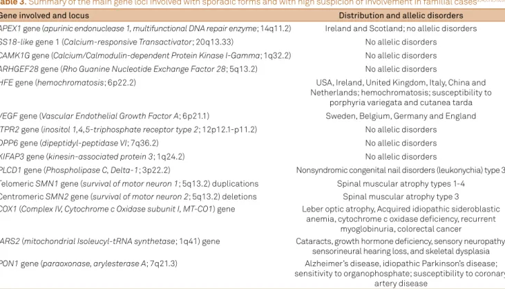
CTX may present with an early-onset lower motor domi-nant juvenile ALS phenotype in two Brazilian sisters with severe bulbar compromise, orofacial dyskinesia and gen-eralized epilepsy. One of them presented also with typical systemic signs of CTX, including chronic diarrhea, juvenile bilateral cataracts and early-onset premenopausal osteo-penia (unpublished data). Other gene loci are also involved with sporadic forms described as susceptibility loci (most as SNP associations or demonstrated by genome-wide asso-ciation studies) for which there is some degree of suspicion related to familial cases (Table 3)2,4,7,12,13,59.
Table 3. Summary of the main gene loci involved with sporadic forms and with high suspicion of involvement in familial cases2,4,7,12,13,59.
Gene involved and locus Distribution and allelic disorders
APEX1 gene (apurinic endonuclease 1, multifunctional DNA repair enzyme; 14q11.2) Ireland and Scotland; no allelic disorders
SS18-like gene 1 (Calcium-responsive Transactivator; 20q13.33) No allelic disorders
CAMK1G gene (Calcium/Calmodulin-dependent Protein Kinase I-Gamma; 1q32.2) No allelic disorders
ARHGEF28 gene (Rho Guanine Nucleotide Exchange Factor 28; 5q13.2) No allelic disorders
HFE gene (hemochromatosis; 6p22.2) USA, Ireland, United Kingdom, Italy, China and Netherlands; hemochromatosis; susceptibility to
porphyria variegata and cutanea tarda
VEGF gene (Vascular Endothelial Growth Factor A; 6p21.1) Sweden, Belgium, Germany and England
ITPR2 gene (inositol 1,4,5-triphosphate receptor type 2; 12p12.1-p11.2) No allelic disorders
DPP6 gene (dipeptidyl-peptidase VI; 7q36.2) No allelic disorders
KIFAP3 gene (kinesin-associated protein 3; 1q24.2) No allelic disorders
PLCD1 gene (Phospholipase C, Delta-1; 3p22.2) Nonsyndromic congenital nail disorders (leukonychia) type 3 Telomeric SMN1 gene (survival of motor neuron 1; 5q13.2) duplications Spinal muscular atrophy types 1-4
Centromeric SMN2 gene (survival of motor neuron 2; 5q13.2) deletions Spinal muscular atrophy type 3
COX1 (Complex IV, Cytochrome c Oxidase subunit I, MT-CO1) gene Leber optic atrophy, Acquired idiopathic sideroblastic anemia, cytochrome c oxidase deiciency, recurrent
myoglobinuria, colorectal cancer
IARS2 (mitochondrial Isoleucyl-tRNA synthetase; 1q41) gene Cataracts, growth hormone deiciency, sensory neuropathy, sensorineural hearing loss, and skeletal dysplasia
PON1 gene (paraoxonase, arylesterase A; 7q21.3) Alzheimer’s disease, idiopathic Parkinson’s disease; sensitivity to organophosphate; susceptibility to coronary
artery disease
CONCLUSION
Although most cases of ALS are sporadic, familial ALS cases represent important clinical, genetic and
neuropatho-logical keys to understand the natural history of diferent ge
-netic forms, clinical and ge-netic correlations, and allow new
genetic targeted therapies for ALS. Despite the current trend of ALS diagnoses are limited to syndromic clinical descrip-tions, in the near future it will be essential to establish the genetic types associated with the diferent clinical subtypes and presentations, especially in familial cases and in complex neurological phenotypes.
References
1. Oliveira ASB, Pereira RDB. Amyotrophic lateral sclerosis (ALS): three letters that change the people’s life. For ever. Arq Neuropsiquiatr. 2009;67(3A):750-82. doi:10.1590/S0004-282X2009000400040
2. Leblond CS, Kaneb HM, Dion PA, Rouleau GA. Dissection of genetic factors associated with amyotrophic lateral sclerosis. Exp Neurol. 2014;262(Part B):91-101. doi:10.1016/j.expneurol.2014.04.013
3. He J, Mangelsdorf M, Fan D, Bartlett P, Brown MA. Amyotrophic Lateral Sclerosis genetic studies: from genome-wide association mapping to genome sequencing. Neuroscientist. 2014;pii:1073858414555404. Forthcoming. doi:10.1177/1073858414555404
4. Su XW, Broach JR, Connor JR, Gerhard GS, Simmons Z. Genetic heterogeneity of amyotrophic lateral sclerosis: implications for clinical practice and research. Muscle Nerve. 2014;49(6):786-803. doi:10.1002/mus.24198
5. Ferraiuolo L, Kirby J, Grierson AJ, Sendtner M, Shaw PJ. Molecular pathways of motor neuron injury in amyotrophic lateral sclerosis. Nat Rev Neurol. 2011;7(11):616-30. doi:10.1038/nrneurol.2011.152
6. Iguchi Y, Katsuno M, Ikenaka K, Ishigaki S, Sobue G. Amyotrophic lateral sclerosis: an update on recent genetic insights. J Neurol. 2013;260(11):2917-27. doi:10.1007/s00415-013-7112-y
7. Chen S, Sayana P, Zhang X, Le W. Genetics of amyotrophic lateral sclerosis: an update. Mol Neurodegener. 2013;8(1):28. doi:10.1186/1750-1326-8-28
8. Renton AE, Chiò A, Traynor BJ. State of play in amyotrophic lateral sclerosis genetics. Nat Neurosci. 2014;17(1):17-23. doi:10.1038/nn.3584
9. Turner MR, Swash M. The expanding syndrome of amyotrophic lateral sclerosis: a clinical and molecular odyssey. J Neurol Neurosurg Psychiatry. 2015;86(6):667-73. doi:10.1136/jnnp-2014-308946
10. Kanouchi T, Ohkubo T, Yokota T. Can regional spreading of amyotrophic lateral sclerosis motor symptoms be explained by prion-like propagation? J Neurol Neurosurg Psychiatry. 2012;83(7):739-45. doi:10.1136/jnnp-2011-301826
11. Strong MJ, Grace GM, Freedman M, Lomen-Hoerth C, Woolley S, Goldstein LH et al. Consensus criteria for the diagnosis of frontotemporal cognitive and behavioural syndromes in amyotrophic lateral sclerosis. Amyotroph Lateral Scler 2009;10(3):131-46. doi:10.1080/17482960802654364
12. Al-Chalabi A, Jones A, Troakes C, King A, Al-Sarraj S, Berg LH. The genetics and neuropathology of amyotrophic lateral sclerosis. Acta Neuropathol. 2012;124(3):339-52. doi:10.1007/s00401-012-1022-4
13. Ticozzi N, Tiloca C, Morelli C, Colombrita C, Poletti B, Doretti A et al. Genetics of familial Amyotrophic lateral sclerosis. Arch Ital Biol. 2011;149(1):65-82. doi:10.4449/aib.v149i1.1262
14. Conte A, Lattante S, Luigetti M, Del Grande A, Romano A, Marcaccio A et al. Classiication of familial amyotrophic lateral sclerosis by family history: effects on frequency of genes mutation. J Neurol Neurosurg Psychiatry. 2012;83(12):1201-3. doi:10.1136/jnnp-2012-302897
15. Turner MR, Barnwell J, Al-Chalabi A, Eisen A. Young-onset amyotrophic lateral sclerosis: historical and other observations. Brain. 2012;135(9):2883-91. doi:10.1093/brain/aws144
16. Horton WA, Eldridge R, Brody JA. Familial motor neuron disease: evidence for at least three different types. Neurology. 1976;26(5):460-5. doi:10.1212/WNL.26.5.460
17. Al-Chalabi A, Hardiman O. The epidemiology of ALS: a conspiracy of genes, environment and time. Nat Rev Neurol. 2013;9(11):617-8. doi:10.1038/nrneurol.2013.203
18. Van Langenhove T, Zee J, Van Broeckhoven C. The molecular basis of the frontotemporal lobar degeneration-amyotrophic lateral sclerosis spectrum. Ann Med. 2012;44(8):817-28. doi:10.3109/07853890.2012.665471
19. Blitterswijk M, v Es MA, Hennekam EA, Dooijes D, Rheenen W, Medic J et al. Evidence for an oligogenic basis of amyotrophic lateral sclerosis. Hum Mol Genet. 2012;21(17):3776-84. doi:10.1093/hmg/dds199
20. Ince PG, Highley JR, Kirby J, Wharton SB, Takahashi H, Strong MJ et al. Molecular pathology and genetic advances in amyotrophic lateral sclerosis: an emerging molecular pathway and the signiicance of glial pathology. Acta Neuropathol. 2011;122(6):657-71. doi:10.1007/s00401-011-0913-0
21. Shirakawa K, Suzuki H, Ito M, Kono S, Uchiyama T, Ohashi T et al. Novel compound heterozygous ALS2 mutations cause juvenile amyotrophic lateral sclerosis in Japan. Neurology. 2009;73(24):2124-6. doi:10.1212/WNL.0b013e3181c67be0
22. Hand CK, Khoris J, Salachas F, Gros-Louis F, Lopes AA,
Mayeux-Portas V et al. A novel locus for familial amyotrophic lateral sclerosis, on chromosome 18q. Am J Hum Genet. 2002;70(1):251-6. doi:10.1086/337945
23. Chen YZ, Bennett CL, Huynh HM, Blair IP, Puls I, Irobi J et al. DNA/RNA helicase gene mutations in a form of juvenile amyotrophic lateral sclerosis (ALS4). Am J Hum Genet. 2004;74(6):1128-35. doi:10.1086/421054
24. Hentati A, Ouahchi K, Pericak-Vance MA, Nijhawan D, Ahmad A, Yang Y et al. Linkage of a commoner form of recessive amyotrophic lateral sclerosis to chromosome 15q15-q22 markers. Neurogenetics 1998;2(1):55-60. doi:10.1007/s100480050052
25. Daoud H, Zhou S, Noreau A, Sabbagh M, Belzil V, Dionne-Laporte A et al. Exome sequencing reveals SPG11 mutations
causing juvenile ALS. Neurobiol Aging. 2012;33(4):839:e5-9. doi:10.1016/j.neurobiolaging.2011.11.012
26. Hewitt C, Kirby J, Highley JR, Hartley JA, Hibberd R, Hollinger HC et al. Novel FUS/TLS mutations and pathology in familial and sporadic amyotrophic lateral sclerosis. Arch Neurol. 2010;67(4):455-61. doi:10.1001/archneurol.2010.52
27. Bäumer D, Hilton D, Paine SM, Turner MR, Lowe J, Talbot K et al. Juvenile ALS with basophilic inclusions is a FUS proteinopathy with FUS mutations. Neurology. 2010;75(7):611-8. doi:10.1212/WNL.0b013e3181ed9cde
28. Sapp PC, Hosler BA, McKenna-Yasek D, Chin W, Gann A, Genise H et al. Identiication of two novel loci for dominantly inherited familial amyotrophic lateral sclerosis. Am J Hum Genet. 2003;73(2):397-403. doi:10.1086/377158
30. Millecamps S, Salachas F, Cazeneuve C, Gordon P, Bricka B, Camuzat A et al. SOD1, ANG, VAPB, TARDBP, and FUS mutations in familial amyotrophic lateral sclerosis: genotype-phenotype correlations. J Med Genet. 2010;47(8):554-60. doi:10.1136/jmg.2010.077180
31. Corcia P, Valdmanis P, Millecamps S, Lionnet C, Blasco H, Mouzat K et al. Phenotype and genotype analysis in amyotrophic lateral sclerosis with TARDBP gene mutations. Neurology. 2012;78(19):1519-26. doi:10.1212/WNL.0b013e3182553c88
32. Chow CY, Landers JE, Bergren SK, Sapp PC, Grant AE, Jones JM et al. Deleterious variants of FIG4, a phosphoinositide phosphatase, in patients with ALS. Am J Hum Genet. 2009;84(1):85-8. doi:10.1016/j.ajhg.2008.12.010
33. Maruyama H, Morino H, Ito H, Izumi Y, Kato H, Watanabe Y et al. Mutations of optineurin in amyotrophic lateral sclerosis. Nature. 2010;465(7295):223-6. doi:10.1038/nature08971
34. Van Damme P, Veldink JH, Blitterswijk M, Corveleyn A, Vught PW, Thijs V et al. Expanded ATXN2 CAG repeat size in ALS identiies genetic overlap between ALS and SCA2. Neurology. 2011;76(24):2066-72. doi:10.1212/WNL.0b013e31821f445b
35. Johnson JO, Mandrioli J, Benatar M, Abramzon Y, Van Deerlin VM, Trojanowski JQ et al. Exome sequencing reveals VCP mutations as a cause of familial ALS. Neuron. 2010;68(5):857-64. doi:10.1016/j.neuron.2010.11.036
36. Deng HX, Chen W, Hong ST, Boycott KM, Gorrie GH, Siddique N et al. Mutations in UBQLN2 cause dominant X-linked juvenile and adult-onset ALS and ALS/dementia. Nature. 2011;477(7363):211-5. doi:10.1038/nature10353
37. Al-Saif A, Al-Mohanna F, Bohlega S. A mutation in sigma-1 receptor causes juvenile amyotrophic lateral sclerosis. Ann Neurol. 2011;70(6):913-9. doi:10.1002/ana.22534
38. Cox LE, Ferraiuolo L, Goodall EF, Heath PR, Higginbottom A, Mortiboys H et al. Mutations in CHMP2B in lower motor neuron predominant amyotrophic lateral sclerosis (ALS). PLoS One. 2010;5(3):e9872. doi:10.1371/journal.pone.0009872
39. Wu CH, Fallini C, Ticozzi N, Keagle PJ, Sapp PC, Piotrowska K et al. Mutations in the proilin 1 gene cause familial amyotrophic lateral sclerosis. Nature 2012;488(7412):499-503. doi:10.1038/nature11280
40. Takahashi Y, Fukuda Y, Yoshimura J, Toyoda A, Kurppa K, Moritoyo H et al. ERBB4 mutations that disrupt the neuregulin-ErbB4 pathway cause amyotrophic lateral sclerosis type 19. Am J Hum Genet. 2013;93(5):900-5. doi:10.1016/j.ajhg.2013.09.008
41. Kim HJ, Kim NC, Wang YD, Scarborough EA, Moore J, Diaz Z et al. Mutations in prion-like domains in hnRNPA2B1 and hnRNPA1 cause multisystem proteinopathy and ALS. Nature. 2013;495(7442):467-73. doi:10.1038/nature11922
42. Feit H, Silbergleit A, Schneider LB, Gutierrez JA, Fitoussi RP, Réyès C et al. Vocal cord and pharyngeal weakness with autosomal dominant distal myopathy: clinical description and gene localization to 5q31. Am J Hum Genet. 1998;63(6):1732-42. doi:10.1086/302166
43. Johnson JO, Pioro EP, Boehringer A, Chia R, Feit H, Renton AE et al. Mutations in the Matrin 3 gene cause familial amyotrophic lateral sclerosis. Nat Neurosci. 2014;17(5):664-6. doi:10.1038/nn.3688
44. Smith BN, Ticozzi N, Fallini C, Gkazi AS, Topp S, Kenna KP et al. Exome-wide rare variant analysis identiies TUBA4A mutations associated with familial ALS. Neuron. 2014;84(2):324-31. doi:10.1016/j.neuron.2014.09.027
45. Souza PV, Pinto WB, Oliveira AS. C9orf72-related disorders: expanding the clinical and genetic spectrum of neurodegenerative
diseases. Arq Neuropsiquiatr. 2015;73(3):246-56. doi: 10.1590/0004-282X20140229
46. Woollacott IO, Mead S. The C9ORF72 expansion mutation: gene structure, phenotypic and diagnostic issues. Acta Neuropathol. 2014;127(3):319-32. doi:10.1007/s00401-014-1253-7
47. Cooper-Knock J, Shaw PJ, Kirby J. The widening spectrum of C9ORF72-related disease; genotype/phenotype correlations and potential modiiers of clinical phenotype. Acta Neuropathol. 2014;127(3):333-45. doi:10.1007/s00401-014-1251-9
48. Renton AE, Majounie E, Waite A, Simón-Sánchez J, Rollinson S, Gibbs JR et al. A hexanucleotide repeat expansion in C9ORF72 is the cause of chromosome 9p21-linked ALS-FTD. Neuron. 2011;72(2):257-68. doi:10.1016/j.neuron.2011.09
49. DeJesus-Hernandez M, Mackenzie IR, Boeve BF, Boxer AL, Baker M, Rutherford NJ et al. Expanded GGGGCC hexanucleotide repeat in noncoding region of C9ORF72 causes chromosome 9p-linked FTD and ALS. Neuron. 2011;72(2):245-56. doi:10.1016/j.neuron.2011.09.011
50. Bannwarth S, Ait-El-Mkadem S, Chaussenot A, et al. A mitochondrial origin for frontotemporal dementia and amyotrophic lateral sclerosis through CHCHD10 involvement. Brain. 2014;137(8):2329-45. doi:10.1093/brain/awu138
51. Kaplan JC, Hamroun D. The 2015 version of the gene table of monogenic neuromuscular disorders (nuclear genome). Neuromuscul Disord. 2014;24(12):1123-53. doi:10.1016/j.nmd.2014.11.001
52. Al-Chalabi A, Andersen PM, Nilsson P, Chioza B, Andersson JL, Russ C et al. Deletions of the heavy neuroilament subunit tail in amyotrophic lateral sclerosis. Hum Molec Genet. 1999;8(2):157-64. doi:10.1093/hmg/8.2.157
53. Leung CL, He CZ, Kaufmann P, Chin SS, Naini A, Liem RK et al. A pathogenic peripherin gene mutation in a patient with amyotrophic lateral sclerosis. Brain Path. 2004;14(3):290-6. doi:10.1111/j.1750-3639.2004.tb00066.x
54. LaMonte BH, Wallace KE, Holloway BA, Shelly SS, Ascaño J, Tokito M et al. Disruption of dynein/dynactin inhibits axonal transport in motor neurons causing late-onset progressive degeneration. Neuron. 2002;34(5):715-27. doi:10.1016/S0896-6273(02)00696-7
55. Millecamps S, Da Barroca S, Cazeneuve C, Salachas F, Pradat PF, Danel-Brunaud V et al. Questioning of the role of D amino acid oxidase in familial amyotrophic lateral sclerosis. Proc Natl Acad Sci U S A. 2010;107(26):E107. doi:10.1073/pnas.1006190107
56. Staal A, Went LN. Juvenile amyotrophic lateral sclerosis-dementia complex in a Dutch family. Neurology. 1968;18(8):800-6.
doi:10.1212/WNL.18.8.800
57. Morris HR, Al-Sarraj S, Schwab C, Gwinn-Hardy K, Perez-Tur J, Wood NW et al. A clinical and pathological study of motor neurone disease on Guam. Brain. 2001;124(11):2215-22. doi:10.1093/brain/124.11.2215
58. Shindo A, Ueda Y, Kuzuhara S, Kokubo Y. Neuropsychological study of amyotrophic lateral sclerosis and parkinsonism-dementia complex in Kii peninsula, Japan. BMC Neurol. 2014;14(1):151. doi:10.1186/1471-2377-14-151
59. Marangi G, Traynor BJ. Genetic causes of amyotrophic lateral sclerosis: new genetic analysis methodologies entailing new opportunities and challenges. Brain Res. 2014;1607:75-93. doi:10.1016/j.brainres.2014.10.009