677
https://doi.org/10.1590/0004-282X20170117IMAGES IN NEUROLOGY
Dural sinus malformation presenting with
seizure and treated by combined arterial and
venous embolization
Malformação dos seios durais apresentando-se com crises convulsivas e tratada com
embolização arterial e venosa combinada
Ulysses C. Batista
1,2, Thiago Giansante Abud
1,2,3, Carlos Eduardo Baccin
1,2,3, Renato Tavares Tosello
1,2,
Aron Athayde Diniz
1,2, Benedito Jamilson Araújo Pereira
1, Ronie Leo Piske
1,2,31Hospital Beneficência Portuguesa de São Paulo, Centro de Neuroangiografia (CNA), São Paulo SP, Brasil;
2Hospital Alvorada, Neuroradiologia Intervencionista, São Paulo SP, Brasil;
3Hospital Albert Einstein, Neuroradiologia Intervencionista, São Paulo SP, Brasil.
Correspondence: Ulysses C. Batista; Hospital Beneficência Portuguesa de São Paulo, Neuroradiologia Intervencionista; Rua Maestro Cardim, 769; 01323-900 São Paulo SP, Brasil; E-mail: ulyssescbatista@gmail.com
Conflict of interest: There is no conflict of interest to declare.
Received 19 October 2015; Receive in final form 29 August 2016; Accepted 31 March 2017.
A newborn with a prenatal diagnosis of dural sinus
mal-formation, which was conirmed by an MRI (Figure 1). He was
followed and at the irst seizure (third month), a CT angiogra
-phy was performed, which showed a partial thrombosis and
an increase of the mass efects (Figure 2).
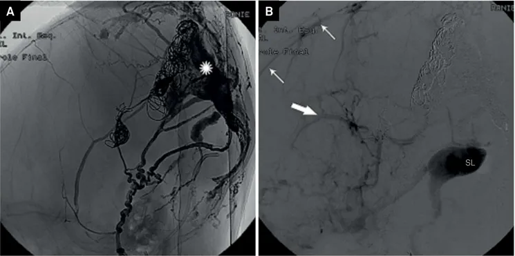
We performed three endovascular procedures (one
per week) combining arterial and venous embolization
,
without complications (Figures 3 and 4). The patient was
discharged without deficits.
A dural sinus malformation is a serious condition that can
evolve into seizures and hemorrhage. It is characterized by
a giant dural sinus lake associated with slow low arteriove
-nous istulae
1,2,3.
A
B
678
Arq Neuropsiquiatr 2017;75(9):677-679B
A
SS
Figure 2. Axial (A) and sagittal (B) contrast enhanced CT angiography showing the extension of thrombosis (white asterisks) and the mass effect in the posterior sagittal sinus malformation (black asterisks). Note the communication between the straight sinus and the posterior superior sagittal sinus and the vein of Galen (black arrow). The anterior superior sagittal (white arrow) and inferior sagital sinus (large white arrow) are preserved.
SS
A
B
Figure 3. Lateral view of early (A) and late phase (B) occipital selective artery angiography showing a high flow dural arteriovenous fistula fed by meningeal branches from the occipital artery and feeding the superior sagittal sinus malformation (white
679
Batista UC et al. Dural sinus malformationSL
A
B
Figure 4. Lateral view final cast (A) of combining arterial and venous with liquid embolic and coil embolization filling the superior sagittal sinus malformation (white asterisk) and late phase of internal carotid digital angiography (B) after the endovascular embolization in the superior sagittal showing the lateral sinus (SL), the superior sagittal sinus (white arrows) and the internal cerebral vein (large white arrow) demonstrating that there was an improvement of the deep venous congestion.
References
1. Lasjaunias P, Magufis G, Goulao A, Piske R, Suthipongchai S, Rodesch R et al. Anatomical aspects of dural arteriovenous shunts in children. Intervent Neuroradiol. 1996;2(3):179-91.
2. Benoit Jenny, et al. Giant dural venous sinus ectasia in neonates Report of 4 cases. J Neurosurg Pediatrics. 2010;5(5):523-8.
3. Rossi A, De Biasio P, Scarso E, Gandolfo C, Pavanello M, Morana G et al. Prenatal MR imaging of dural sinus malformation: a case report. Prenat Diagn. 2006;26(1):11-6.


