Midline facial defects with hypertelorism (MFDH) is the name suggested for a rare and heterogeneous group of craniofacial disorders mainly character-ized by ocular hypertelorism and bifid nose. Sever-al denominations have been used for this condition, such as median cleft face syndrome1
, frontonasal syndrome2, frontonasal dysostosis3; and malforma-tive frontonasal sequence4
. Frontonasal dysplasia5 is the name most commonly accepted; however, after a critical review of this clinical condition based upon dysmorphology concepts the same authors proposed
the denomination frontonasal malformation6. The existence of different denominations can be easily attributed to the clinical complexity of this condi-tion, which has been described from different points of view, according to the professional experience of each author. Considering all these particularities, the descriptive name herein proposed (MFDH) could be a real possibility of an integrative denomination for different health professionals. In the future, it could facilitate the descriptions concerning this heteroge-neous group. Besides differences among
denomina-A CLINICdenomina-AL STUDY OF 31 INDIVIDUdenomina-ALS WITH
MIDLINE FACIAL DEFECTS WITH HYPERTELORISM
AND A GUIDELINE FOR FOLLOW-UP
Vera Lúcia Gil-da-Silva-Lopes, Andréa Trevas Maciel-Guerra
ABSTRACT - In order to contribute to clinical delineation of midline facial defects with hypertelorism (MFDH) and to etiologic diagnosis of the isolated form, 31 patients with MFDH unaffected by known syndromic as-sociations were evaluated. Group A included patients personally examined by the authors, while Group B included those previously evaluated by other geneticists. Among the 14 patients from Group A, there were 7 with distinct pictures of multiple congenital anomalies. In Group B, 5 of the 17 patients also exhibited a distinct pattern of defects. Among isolated MFDH, there was association with anomalies of the skull and facial bones (13/14), otorhinologic (11/16), central nervous system (9/16), and ocular (6/7), and audiologic (3/16); 1/3 of the cases had a relevant gestational intercurrences. Isolated FNM may have involvement of environmental components in some cases; the possibility of a syndromic picture should be extensive inves-tigated. Follow-up of such patients must include the examinations herein performed.
KEY WORDS: craniofacial anomalies, facial clefts, ocular hypertelorism, frontonasal dysplasia, frontona-sal process, follow-up.
Estudo clínico de 31 indivíduos com defeitos de linha média facial com hipertelorismo e dire-trizes para seguimento clínico
RESUMO - Objetivando contribuir com o delineamento clínico de defeitos de linha média facial com hiper-telorismo (DLMFH) e com o diagnóstico etiológico das formas isoladas, foram avaliados 31 indivíduos com DLMFH sem condições clínicas definidas. O Grupo A constituiu-se de pacientes examinados pessoalmente e o Grupo B, inicialmente, por outro geneticista. Entre os 14 pacientes do Grupo A, detectou-se 7 novos qua-dros de anomalias múltiplas (AM). No Grupo B, 5 dos 17 pacientes exibiram um quadro clínico único e pecu-liar. Nos casos de DLMFH isolados, detectou-se associação com anomalias de ossos de crânio e face (13/14), otorrinolaringológicas (11/16), de sistema nervoso central (9/16), oculares (6/7), e audiológicas (3/16); hou-ve antecedentes gestacionais relevantes em 1/3. Existem evidências de envolvimento de fatores ambientais em parte dos casos de formas isoladas de DLMFH, devendo-se atentar para a possibilidade de um quadro distinto de AM. Todas as investigações realizadas são úteis para avaliação e seguimento clínico.
PALAVRAS-CHAVE: anomalias craniofaciais, fendas faciais, hipertelorismo, displasia frontonasal, processo frontonasal, seguimento clínico.
Departamento de Genética Médica, Faculdade de Ciências Médicas, Universidade Estadual de Campinas (UNICAMP), Campinas SP, Brasil.
Received 17 August 2006, received in final form 24 November 2006. Accepted 5 February 2007.
tions, pathogenesis is still incompletely understood. Failure of formation of the nasal capsule during em-bryogenesis, abnormalities on mesenchymal migra-tion from neural crest cells and unbalanced blood flow to the frontonasal process region could be im-plicated in causation of this condition7. Clinical classi-fication of facial clefts varies from those based upon clinical and radiological data1, 8
, or involving embryo-logical aspects5, 9
. There is also a specific facial classifi-cation for frontonasal dysplasia5, but is seldom men-tioned.
In view of the clinical variability of MFDH, cur-rent classification and diagnostic criteria are still not appropriated. Different diagnostic criteria are men-tioned, with some overlapping between them. Af-fected individuals should have two or more of the following features: true ocular hypertelorism, broad-ening of nasal root, median face cleft affecting the nose or both nose and upper lip and, at times, the palate, unilateral or bilateral clefting of the alae nasi, lack of formation of the nasal tip, and anterior crani-um bifidcrani-um6. Another classification considered ocu-lar hypertelorism, broad nasal root, and variable de-gree of median nasal groove as the main diagnos-tic signs10. After extensive review11, it was suggest-ed that a diagnosis of MFDH should be made for individuals presenting ocular hypertelorism (which leads to broadening of the nasal root) and medial and (or) lateral nasal cleft. These authors also sug-gest that the use of these criteria could lead to bet-ter knowledge of this anomaly and the need for a new classification.
Clinical presentation of MFDH includes isolated cases as well as those in which it is part of syndromes with different etiologies, such as craniofrontonasal dysplasia12,13, acromelic frontonasal dysplasia14 , and oculofrontonasal spectrum15,16.
The rarity of isolated cases, the different termi-nologies, classifications and emphasis in the reports, as well as the absence of detailed clinical and famil-ial history do not allow enough insight into the real etiologic and clinical profile of MFDH.
Despite that, it is known that there is no devia-tion of sex ratio, and most cases are sporadic. In view of its rarity, it is not possible to verify the existence of racial variability on prevalence or incidence. Heri-tability also could not be established, as the few re-ported cases of twinning belong to different popula-tions and times. Chromosomal aberrapopula-tions are rare-ly reported17-20
. A submicroscopic deletion of 22q11 was observed in a particular group presenting MFDH and tetralogy of Fallot21. In 2 patients presenting a
nasal dimple and 22q11.2 microdeletion, it was sug-gested that this picture should not be confused with the nasal abnormalities seen in frontonasal dyspla-sia22
. After careful review, familial recurrence of the isolated form could be characterized in just two fam-ilies23,24
, but it is was not possible to distinguish be-tween an autosomal dominant or X-linked pattern of inheritance.
The aims of this study were to establish the main clinical features of MFDH and to identify the main etiological factors related to isolated MFDH.
METHOD
The group was obtained from May, 1992 to November, 1996, and most of the individuals have been followed since them. It was composed by 31 individuals with MFDH (17F, 14 M) whose ages varied from 2 months to 29 years, select-ed through pictures and mselect-edical records from the Depart-ment of Medical Genetics / FCM / UNICAMP and specialized craniofacial hospitals.
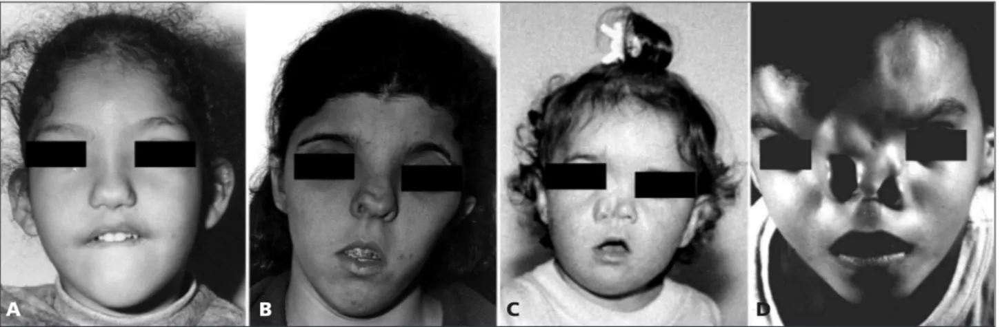
Inclusion criteria were ocular hypertelorism with me-dian and (or) lateral nasal cleft; patients with well-known syndromes were excluded (Figs 1,2,3,4). Group A was com-posed of 14 individuals personally examined by the first author, while sample B included those previously evaluat-ed by another clinical geneticist with expertise on cranio-facial anomalies. After that, they were evaluated by one of the authors (VLGSL) through pictures and clinical exam-ination (17 individuals).
Procedures – Patients in group A were evaluated by a specific investigation protocol including clinical history and dysmorphologic examination and, whenever possible, skull and facial X-rays, computerized tomography of brain, oph-thalmologic and otorhinologic evaluation and GTG band-ing karyotype. Medical records were reviewed for comple-mentary informations about Group B. A dysmorphological approach was performed for elucidation of diagnosis in all cases.
Based upon clinical evaluation, patients from each group were considered to be isolated (or associated with non-specific clinical signs) (samples A1 e B1) or associated with multiple congenital anomalies involving different de-velopment fields (samples A2 e B2).
Data from groups A1 and B1 were compared by t test, chi-square test and Fisher’s test. Syndromic pictures which could be characterized in samples A2 and B2 during this study were described.
This study was approved by the Research Ethics Com-mittee (protocol number 488/2002).
RESULTS
Bone abnormalities in skull / facial X-rays were detected in 11/12. When bone abnormalities which could only be detectable by computerized tomogra-phy (CT) of skull and face are added, 13/14 individu-als had some abnormality in these evaluations.
Considering central nervous system (CNS) abnor-malities, CT was abnormal in 9/16, including corpus callosum anomalies (6/16) (lipoma 2/16, agenesis 1/16, dysgenesis 2/16); encephalocele (2/16) and complex CNS abnormality (1/16); in another case, an ethmoid-al encephethmoid-alocele was suspected.
Otorhinologic abnormalities were found in 11/16; in 5 cases, anomalies could only be detected by a spe-cialist; audiometric abnormalities were observed in 3/14.
Ophthalmic findings were described in 6/7, with predominance of strabismus (3/7) and partial lens opacity (2/7); in one case there were severe abnor-malities of palpebral fissures, extrinsic eye muscles and lachrymal ducts.
deviate significantly from 1:1, mean maternal age was 28.4 years (SD=5.9), mean paternal age was 31.6 years (SD=7.3) and mean inbreeding coefficient was 0.00082; 1 case of discordant twinning was recog-nized.
Problems during gestation were mentioned in 12/18, including epilepsy (1/18), alcohol ingestion (1/18), hyperemesis (1/18), symptomatic myomatosis (1/18), bleeding (2/18), twinning (1/18), oral anticon-ceptional use (2/18), unspecified “poison” ingestion (1/18) , hypothyroidism (1/18) and “abnormal” preg-nancy (1/8).
There was no evidence of intrauterine growth re-tardation (mean birth weight: 3.1 kg; SD= 0.6). There were no abnormalities of anthropometric data at dif-ferent ages in comparison with normal patterns, as well as in neurological development, according to in-formation given by the families. Learning disabilities were mentioned in 3/9 individuals who were more than 7 years old.
Table. Clinical description of patients studied.
Sex Maine clinical findings Pattern of
transmition
Denomination proposed
Reference
Group A2
Male Mental retardation, coronal craniosynostosis, alar and lip/palate clefts, nystagmus, corneal leukoma, cataract, severe visual loss
? Isolated case
Crânio-oculo-fronto- nasal malformation
Gil-da-Silva-Lopes,et al, 1997
2 Female Mental retardation, MFDH, Fissure / dysgenesis of
corpus callosum, extensive mongolian spots, rugose
labia majora, Hypoplastic labia minora
Anteriorized anus, Precocious pubarche*, Pyelo-cal-cyeal duplication **
? Unrelated cases
MFDH corpus callosum
anomalies, extensive mongolian spots and mild ano-genital abnor-malities
Still in follow-up, unpublished data
2 Female MFDH, Hypoplastic labia majora, abnormal implan-tation of clitoris, asymmetric lower limbs, disloca-tion of hips ***, bone cyst in femur ** , Neuromo-tor delay ****
AD? Mother and daughter
FNM, mild ano-genital anomalies and skeletal alterations
Still in follow-up, unpublished data
Male MFDH, Pre / post natal macrosomia, normal bone age, prominent ears, blepharoptosis, epicanthus and strabismus, bifid uvula, dental exfoliation, inguinal hernia, hypospadia, hypotonia, neuropsychomotor delay, cafe-au-lait spots
? Isolated case
Midline defects, macro-somia, mental retarda-tion and dental abnor-malities
Still in follow-up, unpublished data
Male MFDH, narrow and anteverted nostrils, bifid uvu-la, atrial sept defect, aortic stenosis, diaphragmatic hernia, abnormal vertebra, peno-scrotal inversion, normal intelligence
? Isolated case
Midline defects, ocular abnormalities and nor-mal intelligence
Still in follow-up, unpublished data
Group B2 4 Male and 1 Female
MFDH, median cleft lip, blepharoptosis, agenesis / dysgenesis of the corpus callosum, basal encephalo-cele, “mild neuropsychomotor delay” (?) #
? Unrelated cases
Frontonasal dysplasia with optic disc abnor-malities and other mid-line craniofacial defects
Lees et al., 1998; Richieri-Costa and Guion- Almeida, 2004
Chromosomal analyses on GTG banding were nor-mal in 14 / 14 individuals.
Table presents the main clinical features of patients with undescribed syndrome pictures (Figure) which were identified in this sample (groups A2 and B2).
DISCUSSION
Although there are some important papers about MFDH1,5,6,25, as well as an interesting review about this condition from a rhinologic perspective26, this seems to be the first report of a large sample of MFDH in which the patients were selected using homoge-neous diagnostic criteria. A careful dysmorphologic evaluation allowed delineation of 5 pictures of mul-tiple congenital anomalies in group A. In six of them, after extensive search in the literature, the authors decided to maintain the follow-up before any con-clusion; during this study, one case were published by the authors, as a new condition27
. In group B, 5 individuals presented MFDH associated with median cleft lip, blepharoptosis, agenesis / dysgenesis of the corpus callosum, basal encephalocele and mild neu-ropsychomotor delay. This clinical picture has some similarity with others previously described28,29, but they are still on genetics investigation.
There were neither familial recurrence nor chro-mosome aberrations which could be diagnosed with usual cytogenetic techniques. The mean inbreeding coefficient of this sample was lower than that esti-mated for the Brazilian population (0.001), and ma-ternal and pama-ternal age average were also not differ-ent from that observed in the Brazilian population by the same authors (maternal age: 25.49 years, SD= 6.43; paternal age: 30.20 years, SD=8.67)30. These data did not indicate a genetic etiology (chromosomal, monogenic or polygenic) for MDFH in this sample.
Unfortunately, the finding of only one pair of dis-cordant twins of unknown zygosity did not allow an estimate of the heritability of MFDH. However, it is interesting to point out that craniofacial anomalies in general (caused by either malformation or defor-mation) seem to be more frequent in monozygotic twins31. It was also suggested that twining, per se, could be considered a congenital malformation32
. Cohen et al.33 affirmed that the frequency of twinning in families with a case of MFDH would be higher than that of the general population, which could not be verified in this study. The authors com-mented that some clinicians imagined that anomalies of the frontonasal process could be the result from an incomplete twinning of the head. However, con-sidering that this defect is caused by anterior duplica-tion of the notochord, it would be possible supposed that a mildest form would result on the duplication of the hypophysis, and, in the most severe case, di-prosopia. As there was no evidence of this spectrum of anomalies in MFDH, this hypothesis would not be supported. In fact, there was a unique description of a hypophyseal duplication in a MFDH34
.
MFDH was described in discordant dizygotic twins by some authors1,35-38. Discordant monozygotic twins were also described1,37
. Brazilian concordant mono-zygotic twins were described39. We observed that 2/3 of the nonsyndromic MFDH individuals had a history of gestational problems, which can be considered relevant in 1/3. In these cases, the possibility of an environmental influence may be considered, espe-cially in view of the recognized external influence on developmental genes activities. Unfortunately, information was not detailed enough to allow defi-nite conclusions. One of the patients was exposed to high doses of alcohol during gestation. Interestingly, craniofacial and CNS manifestations in fetal alcohol
Figure. Individuals presenting MFDH. Note the clinical variability.
syndrome, in which some of the facial findings re-semble MFDH had been reported40.
Most secondary anomalies associated with non-syndromic MFDH involved the midline, reinforcing the hypothesis of a developmental field defect4,6
. A history of developmental delay was not detect-ed in this study. However, interesting results, mainly involving cerebellar features, were obtained using a specific neurological protocol41. Learning disabili-ties, which were detected in 1/3 of MFDH individuals, could be due to an intrinsic mental impairment, com-plicated by low vision and hearing loss. Self – image disturbances are also a problem in the social life of an individual with craniofacial anomaly42-44. A specific study still in course about neuropsychological and neurological aspects in MFDH has been conducted by our group and preliminary results showed a hetero-geneous but important correlation between them, reinforcing the idea of an intrinsic CNS abnormality in this condition (unpublished data).
Complementary evaluations indicated the associa-tion of MFDH with skull and facial bone abnormali-ties (13/14), as well as CNS defects (9/16). Facial bone defects could be explained based upon disturbance of the embryonic development of the nasal capsule and the frontonasal process45
.
Abnormalities of corpus callosum, particularly li-pomas and calcification, are the most common CNS defect associated with MFDH1,37,42; the angular analy-sis of corpus callosum of MFDH individuals suggested that positional anomalies of this structure are intrin-sically related to this condition46
. The advent of mag-netic resonance image (MRI) brings new possibilities for a structural investigation. Using this technique, other structural abnormalities and errors of neuronal migration were detected in a large sample of MFDH individuals47
. In syndrome patients, MFDH was de-scribed in association with bilateral periventricular nodular heterotopia and mental retardation48 and with multiple pericallosal lipomas in 2 siblings49
. Ophthalmic (6/7) and otorhinologic (11/16) abnor-malities were also important findings, and audiomet-ric problems were less common. An isolated case of MFDH with optic nerve colobomata and nystagmus was reported50
. In 1994, 9 individuals affected by MFDH were evaluated before surgical procedures. Two had mild facial defects, and refraction errors, strabismus and amblyopia. In 7 patients with severe facial involvement, 71% had significant refraction er-rors, 51% had strabismus and 27% severe structural ocular anomalies. The authors conclude that the high
incidence of strabismus could be associated with dif-ficulties of ocular accommodation related to ocular hypertelorism51. These findings are very similar to the sample herein described, except for the frequency of severe refraction errors, and they indicate that a complete ophthalmic evaluation should be part of routine investigation of MDFH.
Audiometric findings have not been securely doc-umented until now. About 30% of MFDH patients evaluated, including syndromic and isolated cases, had hearing loss 51. In our study, this feature was de-tected in 1/5 of cases, which indicates that audiomet-ric examination and otorhinologic evaluation should always be done.
In conclusion, in order to establish guidelines for follow-up of MFDH, considering that MFDH is often part of a syndromic picture, this fact should be tak-en into account during clinical evaluation. Isolated MFDH is usually associated with skeletal abnormali-ties of the cranium and face, as well as anomalies of CNS, and ophthalmic and otorhinologic abnor-malities. Considering these findings, evaluation and clinical follow-up of a patient with MFDH should be multidisciplinary and include: skull and facial X-rays, computerized tomography / MRI of the cranium, and ophthalmic, otorhinologic and audiometric evalua-tion.
Finally, in view of etiological heterogeneity of iso-lated MFDH, an appropriated and detailed clinical description, high resolution chromosomal analysis and other techniques, including studies of mutations on developmental genes in affected individuals, may add more information in some cases.
Acknowledgements – We would like to thank the Hospital de Reabilitação de Anomalias Craniofaciais (HRAC, Bauru – USP and Sociedade Brasileira de Pesquisa e Reabili-tação para Anomalias CranioFaciais (SOBRAPAR, Campinas, SP) and the patients and their families for their coopera-tion. We also thank Professor Maria Leine Guion-Almeida for clinical evaluation patients from Group B. This paper is dedicated to Professor Robert J Gorlin for his contribution during the first steps of this work and for his carefully revi-sion of this manuscript.
REFERENCES
1. DeMyer W. The median cleft face syndrome: differential diagnosis of cranium bifidum occultum, hypertelorism, and median cleft nose, lip, and palate. Neurology 1967;17:961-971.
2. Rosasco AS, Masa, JL. Frontonasal syndrome. Brit J Plast Surg 1968; 21:244-249.
3. Gollop TR. Frontofacionasal dysostosis: a new autosomal recessive syn-drome. Am J Med Genet 1981;10:409-412.
�. Sedano H�, �ohen Jr MM, Jirasek J, Gorlin RJ. Frontonasal dysplasia.�. Sedano H�, �ohen Jr MM, Jirasek J, Gorlin RJ. Frontonasal dysplasia.Frontonasal dysplasia. J Pediatr 1970;76:906-913.
6. Sedano H�, Gorlin RJ. Frontonasal malformation as a field defect and in syndromic associations. �ral Surg �ral Med �ral Pathol 1988;6�:704-1988;6�:704- 6�:704-710.
7. Gil-da-Silva-Lopes VL. A malformação frontonasal: aspectos patogêni-cos, etiológipatogêni-cos, clínicos e diagnóstico diferencial. Dissertação. �ampi-nas, 199�.
8. Tessier P. Anatomical classification of facial, cranio-facial and latero-fa-cial clefts. J �ral Maxillofac Surg 1976;4:69-92.
9. Van Der Meulen, J�, Mazzola R, Vermej-Keers �, Stricker M, Paphael B. A morphogenetic classification of craniofacial malformation. Plast Reconstr Surg 1983;71:�60-�72.
10. Wilroy Jr RS, Buyse ML. Median cleft face syndrome. In: Birth Defects Encyclopedia. New York: Blackwell Scientific Publications, Inc. 1990 11. Gil-da-Silva-Lopes VL. Pathogenical, etiological and clinical aspects
of frontonasal malformation and its differential diagnosis. Braz J Gen 199�b;18:708.
12. �nline Mendelian Inheritance in Man, �MIM (TM). Johns Hopkins Uni-versity, Baltimore, MD. MIM Number: {MIM122929}: {MIM 3/19/1997}. World Wide Web URL: http://www.ncbi.nlm.nih.gov/omim/ 13. �nline Mendelian Inheritance in Man, �MIM (TM). Johns
Hop-kins University, Baltimore, MD, USA. MIM Number: {MIM*304110}: {MIM10/16/2003}. World Wide Web URL: http://www.ncbi.nlm.nih. gov/omim/
14. �nline Mendelian Inheritance in Man, �MIM (TM). Johns Hopkins University, Baltimore, MD. MIM Number: {MIM 603671}: {10/27/1999}. World Wide Web URL: http://www.ncbi.nlm.nih.gov/omim/ 1�. Toriello H, Higgins JV, Mann R. �culoauriculofrontonasal
syndrome;report of another case and review of differential diagnosis. �lin Dysmorphol 199�;4:338-346.
16. Guion-Almeida ML, Gil-da-Silva-Lopes VL. �culoauriculofrontonasal spectrum: report on a Brazilian male and review of the literature. �lin Dysmorphol 1997;6:2�1-2��.
17. Fryns JP. Frontonasal malformation and reciprocal translocation t(1�;22)(q22;q13). �lin Genet 1993;44:46-47.
18. �hen H. An approach to work-up of dysmorphic patients: clinical, cy-togenetic, and molecular aspects. Keio J Méd 1994;43:98-107. 19. Stevens �A, Qumsiyeh MB. Syndromal frontonasal dysostosis in a child
with a complex translocation involving chromosomes 3, 7, and 11. Am J Med Genet 199�;��:494-497.
20. Gorlin RJ, �ohen Jr MM, Levin LS. Syndromes of the head and neck, 4.Ed. New York: �xford University Press, 2001
21. Stratton R, Payne RM. Frontonasal malformation with tetralogy of Fal-lot associated with a submicroscopic deletion of 22q11. Am J Med Gen-et 1997;69:287-289.
22. Gripp KW, McDonald-McGinn DM, Driscoll DA, Reed La, Emanuel BS, Zackai EH. Nasal dimple as part of the 22q11.2 deletion syndrome. Am J Med Genet 1997;69:290-292.
23. Fryburg JS, Persing JA, Lin KY. Frontonasal dysplasia in two succes-sive generations. Am J Med Genet 1993;46:712-714.
24. Nevin N�, Leonard AG, Jones B. Frontonasal dysostosis in two suces-sive generations. Am J Med Genet 1999;26:2�1-2�3
2�. Guion-Almeida ML, Richieri-�osta A, Saavedra D, �ohen MM Jr. Fron-tonasal dysplasia: analysis of 21 cases and literature review. Int J � Maxillofac Surg 1996;2�:91-97.
26. Genç E, Derbent M, Ergin NT. A mild case of frontonasal dysplasia: the rhinologic perspective. Int J Ped �torhinolarongol 2002;6�:7�-83. 27. Gil-da-Silva-Lopes VL, �ampos NLV, Maciel-Guerra AT.
�ranio-oculo-fronto-nasal malformation: a new M�A condition? �lin Dysmorphol 1997;6:2�-29.
28. Lees MM, Hodgkins P, Reardon W, et al. Frontonasal dysplasia with optic disc anomalies and other midline craniofacial defects: a report of six cases. �lin Dysmorphol 1998;7:1�7-162.
29. Richieri-�osta A, Guion-Almeida ML. The syndrome of frontonasal dysplasia, callosal agenesis, basal encephalocele, and eye anomalies - phenotypic and aetiological considerations. Int J Med Sci 2004;1:34-42.
30. Pilotto RF, Magna LA, Beiguelman BB. Factors influencing human weight in normal pregnancy: a prospective study in a Brazilian uni-versity hospital. Rev Bras Genet 1993;16:4�7-469.
31. Keusch �F, Mulliken JB, Kaplan L�. �raniofacial anomalies in twins. Plast Reconst Surg 1991;87:16-23.
32. Schinzel AAGL, Smith DW, Miller JR. Monozigotic twinning and struc-tural defects. J Ped 1979;76:916-913.
33. �ohen MM Jr, Sedano H�, Gorlin RJ, Jirasek, JE. Frontonasal dyspla-Frontonasal dyspla-sia (median cleft face syndrome): coments on etiology and pathogene-sis. Birth defects: White Plains: �AS 1971;7:117-119.
34. Hori A. A brain with two hypophyses in median cleft face syndrome. Acta Neuropathol 1983;�9:1�0-1�4.
3�. Webster JP, Deming EG. The surgical treatment of the bifid nose. Plast Reconstr Surg 19�0;6:1-37.
36. Sauvegrain J, Nahun H. Hypertelorisme Essentiel. J Radiol Electrol Med Nucl 1962;43:�28-�31.
37. Naidich TP, �sborn RE, Bauer B, Naidich, MJ. Median cleft face syn-drome: MR and �T data from 11 children. J �omp Assist Tomogr 1988;12:�7-64.
38. Mohammed S N, Swan M�, Wall S A, Wilkie A�M. Monozygotic twins discordant for frontonasal malformation. Am J Med Genet 2004;130:384-388.
39. Aguiar MJB, Pena SDJ. Gêmeas monozigóticas concordantes para dis-plasia frontonasal. In: Anais da Reunião Anual da Sociedade Brasilei-ra de Genética �línica, 1994. AbstBrasilei-ract (3�), Vitória, BBrasilei-razil.
40. Johnson VP, Swayze VW II, Sato Y, Andreasen N�. Fetal alcohol syn-drome: craniofacial and central nervous system manifestations. Am J Med Genet 1996;61:329-339.
41. Giffoni SDA, Gonçalves VMG, Zanardi VA, Gil-da-Silva-Lopes VL. �er- �er-ebellar involvement in midline facial defects with ocular hypertelorism. �left Palate �raniofac J 2006;43:466-470.
42. Pascual-�astroviejo I, Pascual-Pascual SI, Péréz-Higueras A. Fron-to-nasal dysplasia and lipoma of the corpus callosum. Eur J Pediatr 198�;144:66-71.
43. Lefebvre A, Barclay S. Psychosocial impact of craniofacial deformities before and after recostructive surgery. �an J Psychiatry 1982;27:�79-�83.
44. Pertschuk MJ, Whitaker LA. Psychosocial considerations in craniofa-cial deformity. �lin Plast Surg 1987;14:163-168.
4�. Moore KL, Persaud TVN. The developing human: clinically oriented embryology, 7. Ed. Philadelphia: WB Saunders �ompany, 2002 46. Giffoni SDA, Gonçalves VMG, Zanardi VA, Gil-da-Silva-Lopes VL.
An-gular analysis of corpus callosum in 18 patients with frontonasal dys-plasia. Arq Neuropsiquiatr 2004;62:19�-198.
47. Gil-da-Silva-Lopes VL, Giffoni DAS. MRI and �T detect �NS abnormal-ities on midline facial defects with hypertelorism. Arq Neuropsiquiatr (in press).
48. Guerrini R, Dobyns WB. Bilateral periventricular nodular heteroto-pia with mental retardation and frontonasal malformation. Neurolo-gy 1998;�1:499-�03.
49. Alzoum MA, Alorainy IA, Husain NA, Ruhaimi KA. Multiple peri-calosal lipomas in two soblings with Frontonasal Dysplasia. Am J Neu-roradiol 2002;23:730-731.
�0. Bardelli, AM, Lasorella G, Barbieri L, Vanni M. �cular manifestations in Kniest syndome, Smith-Lemli-�pitz syndrome, Hallermann-Streiff-François syndrome, Rubinstein-Taybi syndrome and median cleft face syndrome. �phthalmic Paediatr Genet 198�;6:343-347.