SYSTEMATICS, MORPHOLOGY AND PHYSIOLOGY
Alimentary Canal and Reproductive Tract of Hypothenemus hampei
(Ferrari) (Coleoptera: Curculionidae, Scolytinae)
JOSÉ D. RUBIO G.
1, ALEX E. BUSTILLO P.
2, LUIS F. VALLEJO E.
3, JOSÉ R. ACUÑA Z.
1 ANDPABLO BENAVIDES M.
21Disciplina de Mejoramiento Genético. Centro Nacional de Investigaciones de Café, Cenicafé, AA 2427 Manizales, Colombia
2Disciplina de Entomología. Centro Nacional de Investigaciones de Café, Cenicafé, AA 2427, Manizales, Colombia 3Facultad de Agronomía, Universidad de Caldas, Calle 65 n° 26-10, Manizales, Colombia
Neotropical Entomology 37(2):143-151 (2008)
Morfología General del Tracto Digestivo y Sistema Reproductivo de Hypothenemus hampei (Ferrari) (Coleoptera: Curculionidae, Scolytinae)
RESUMEN - El estudio morfológico del tracto digestivo y el sistema reproductor de machos y hembras deHypothenemus hampei Ferrari se describe a continuación. El tracto digestivo presenta un arreglo típico descrito para otros insectos. Ahora las diferencias morfológicas se encontraron en el buche del estomodeo, en donde se observaron estructuras espinosas y en el mesenteron se observo un número reducido de ciegos gástricos. Se evidenció por primera vez el comportamiento de la alimentación de las hembras de H. hampei, que requiere aparentemente de la ingesta del endosperma del café para poder producir huevos viables durante la oviposición. La presencia de burbujas de aire al interior del ventrículo anterior se cree que puede ser debido al hambre, más que a la respuesta al medio ambiente durante el vuelo como lo indicaban anteriores observaciones. El sistema reproductor de la hembra presenta dos ovarios y un arreglo estructural común a especímenes de la familia Curculionidae. El sistema reproductor masculino mostró una diferencia signifi cativa con respecto a otros miembros de la familia Curculionidae e incluso a otra especie de la tribu Scolytinae presentando un aedeagus esclerotizado compuesto de varios orifi cios en la porción terminal, por donde el esperma se expulsa durante el apareamiento.
PALABRAS-CLAVE: Broca del café, morfología interna, comportamiento de alimentación, reproducción
ABSTRACT - The alimentary canal and the reproductive tract of males and females of Hypothenemus hampei Ferrari are described. The alimentary canal of H. hampei showed the crop with several spine-like structures and the midgut with few gastric caeca. We evidenced for the fi rst time that adult females need to feed on coffee in order to produce viable eggs before and during oviposition period. The presence of air bubbles inside the anterior midgut may be due to starvation rather than the response of the environment during fl ying as previously reported. Two ovaries and the same structures and arrangements common to individuals of the Curculionidae beetles composed the female reproductive system. The male reproductive tract showed a signifi cant difference with respect to other Curculionidae and even other Scolytinae species as it showed a sclerotized aedeagus with several pore-like structures in the terminal portion where sperm is released during mating.
KEY WORDS: Coffee Berry Borer, internal morphology, feeding behavior, reproduction
Coffee berry borer, Hypothenemus hampei Ferrari is one of the most severe insect pests of coffee in the world and feeds exclusively on coffee plants (Le Pelley 1968, Johanneson & Mansingh 1984). Adults of H. hampei damage coffee berries when reproducing inside the endosperm (Corbett 1933, Bustillo et al. 1998). Adult females of H. hampei bore into coffee berries, make an ovipositing chamber and start laying eggs in groups of 2-3 per day for 20 days (Bergamin 1956).
The alimentary canal in Curculionidae beetles can be divided in three regions: the anterior stomodaeum or foregut that is of ectodermal origin, the mesenteron or midgut that is of endodermal origin, and the posterior proctodaeum or hindgut that is also of ectodermal origin. All these regions are involved in ingestion, storage, digestion, absorption and of food and water balance (Borror et al. 1976, Calder 1989, Romoser & Stoffolano 1998). There gross morphology and histology of the Scolytinae beetles Scolytus multistriatus Marsham (Baker & Estrin 1974), Dendroctonus group (Díaz et al. 1998, 2003; Silva-Olivares et al. 2003), Trypodendron lineatum Oliv., Gnathotrichus retusus LeConte and G. sulcatus LeConte (Schneider & Rudinsky 1969), and Ips pini Say (Coleoptera: Curculionidae) (Hall et al. 2002) reported some features of alimentary canal which are useful for taxonomy of these beetles (Lopez-Buenfi l et al. 2001).
The reproductive tract of the female produces and stores eggs, and receives and stores spermatozoa. This system is usually formed by the ovaries, which are comprised of a number of ovarioles, the lateral and the common oviducts, the vagina, the accessory glands and the spermathecae. The male reproductive tract produces, stores, and releases spermatozoa, and is formed by the testes, the vas deferens, the seminal vesicle, the ejaculatory duct, the penis or aedeagus, and the gonopore, as well as the accessory gland (Snodgrass 1935, Wigglesworth 1950, Blum 1985).
The internal morphology of beetles is now commonly used for taxonomic characterization of insect species in the same genus, or different insect genus in the same family (Russo 1926, Williams 1945, Robertson 1961).
Here, we describe for the fi rst time the alimentary canal and the reproductive tract of males and females H. hampei in order contribute for the better understanding of this insect´s feeding habits and reproductive behavior.
Materials and Methods
This research was carried out at the National Center for Coffee Research – Cenicafé in Chinchiná, Colombia and the Universidad Nacional de Colombia in Manizales, Colombia. Single H. hampei infested coffee berries were collected in the fi eld and then individualized in glass vials. These samples were stored at 23qC during the evaluation of the adult insects. Twelve insects were cold immobilized and dissected in presence of Ringer´s solution pH 7.2 and the alimentary and reproductive tract isolated.
Different techniques for the description of the internal structures of H. hampei were used. The morphology was analyzed on H. hampei females under a Zeiss Axiophot light microscope equipped with a measuring device in order to record size and length. The internal structures were previously stained with toluidine blue and eosin.
In order to reveal the histology of different organs of H. hampei, we fi xed adults of H. hampei in a graded series of ethanol and then polymerized them with Spurr resin for further analysis at the light microscope following the procedure by Mercer & Birbek (1979). Tissues sections 1-3 Pm thick were obtained through cuts performed with the ultramicrotome LKB BROMMA Ultrotome-V 2088
with glass knife and then stained with Toluidine blue 2%. Another set of samples were fi xed in 2% glutaraldehyde in PSB (phosphate saline buffer) and further dehydration in a graded ethanol series for analyses in a Philips XL 30 TMP Environmental Scanning Electron Microscope (ESEM).
Results
Morphology of the alimentary canal. The main structures at the alimentary canal of H. hampei are easily distinguishable (Fig.1). The length of the whole alimentary canal is three folds that of the female adult (Table 1). The foregut has occupies 16%, the midgut 44% and the hindgut 40% of the total length of the alimentary canal.
The foregut begins at the mouth (Fig. 2A) followed by the pharynx and the esophagus (Figs. 2B, C, D). This region is responsible for the transport of food to the crop (Fig. 2E) whose function is the temporal storage of food. The external surface of the crop shows several spine-like structures (Fig. 2E).. Followed by the crop, we fi nd the proventriculus (Fig. 2F), which is the hardest and sclerotized section of the alimentary canal. The proventriculus is a spherical organ formed by eight sclerotized plates that are disposed in edge, and functions as gizzard and fi lter (Fig. 2G). Muscles that are responsible for the constriction of the proventriculus during the mortaring and fi ltering of food can be visualized (Fig. 2H). The cardiac valve is located at the end of the foregut (Fig. 2I). Circular muscles (Fig. 2J) that do not allow food to return from the midgut to the foregut surround this organ. The midgut comprises two distinguishable structures, the anterior and the posterior midguts (Fig. 3E). Both the anterior and posterior midguts are of the same length; however, the diameter of the posterior midgut is reduced by a third (Table 1). Closely to the midgut walls, we observed a great number of tracheae (Fig. 3A). The epithelium of the midgut is a single layer of cells surrounding the whole midgut (Fig. 3B, D). The food inside the midgut lumen is surrounded by the peritrophic envelope. This envelope is of Type II that is secreted around the cardiac valve and is projected along the midgut (Fig. 3B). The posterior midgut is characterized by the presence of two gastric caeca (Fig. 3C).
colon is easily visualized since it is enlarged and shows the insect’s excrements (Fig 4A). The colon is surrounded by a group of muscles that aid in the peristaltic movement. The hindgut ends with the rectum, which is a more enlarged and sclerotized section, right before the opening of the alimentary canal or anus (Fig. 4C, D). Attached to the rectum we observed the female reproductive system, which is projected to the anterior part of the insect and is reposed on the ileum (Fig. 4B).
Morphology of the reproductive tract of the female
H. hampei. The average length of the female H. hampei reproductive tract is about 76% that of the adult insects body (Table 2) and it is composed of two ovaries with two conical ovarioles each (Fig. 5A). Each ovary has a lateral oviduct that ends at the common oviduct (Fig. 5A). Even
Fig. 1. Morphology of the alimentary canal of Hypothenemus hampei. (ph) pharynx, (es) esophagus, (cr) crop, (pv) proventriculus, (ca) cardiac valve, (av) anterior midgut, (pv) posterior midgut, (gc) gastric caeca, (pa) pyloric ampulla, (mt) malpighian tubule, (il) ileum, (co) colon; (rt) rectum, (an) anal sac.
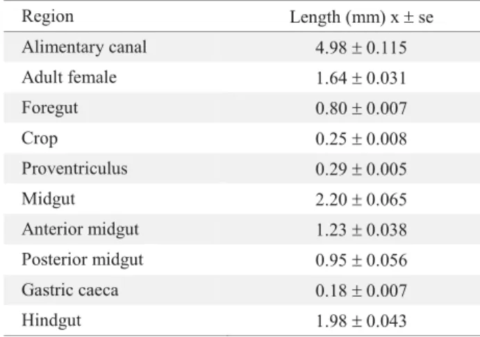
Region Length (mm) x r se Alimentary canal 4.98 r 0.115 Adult female 1.64 r 0.031 Foregut 0.80 r 0.007
Crop 0.25 r 0.008
Proventriculus 0.29 r 0.005 Midgut 2.20 r 0.065 Anterior midgut 1.23 r 0.038 Posterior midgut 0.95 r 0.056 Gastric caeca 0.18 r 0.007 Hindgut 1.98 r 0.043
Table 1. Average length of parts of the alimentary canal ofH. hampei. (n = 12)
ph
ca
av gc
pa mt
rt
an es cr
pv
il pv
Foregut Midgut Hindgut
co
though there must be a separation between the common oviduct and the vagina, the later is not differentiated in H. hampei. At the dorsal side of the base of the vagina, we observed the accessory organs, which are the spermatheca and one accessory gland (Fig. 5A). The spermatheca, unlike the accessory gland, is strongly sclerotized (Fig. 5B). The vagina ends at the pygidium, which is the last abdominal segment that is exposed after the elytra (Fig. 5C). The ovarioles contain oogonia at the gemarium that eventually will become primary oocytes (Fig. 5A). The oocytes mature at the vitellarium, which is the basal part of the ovariole (Fig.5A, D). The four ovarioles present in H. hampei are asynchronous, since there are not mature oocytes at the same time. We found oocytes at different developmental stages, so when one ovariole was forming an oocyte, other was maturing another oocyte, other contained a matured one that was expelled to the lateral oviduct, and the other ovariole is empty (Fig. 5D). The eggs pass through the lateral oviduct to the common oviduct, then trough the vagina before being fertilized and expelled (Fig. 5E). The common oviduct and the vagina showed a muscle layer that contributes to the movement of the eggs to the exterior.
movement of the aedeagus during mating. We found the ejaculatory duct after the aedeagus, which is a long thin structure that is connected to two seminal vesicles (Fig. 6D). There are two mesadenia (accessory glands) located between the seminal vesicles and the vas deferens. The vasa deferentia are between the seminal vesicles and the testes. The two testes are the biggest structures in the male reproductive system (Fig. 6A).
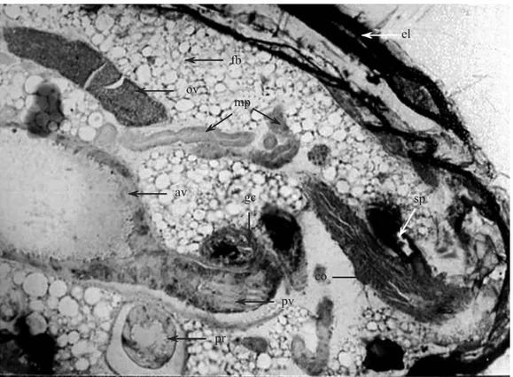
A brief hystological examination of the abdomen revealed the spatial organization of the different organs that composed the alimentary canal and the reproductive system in H. hampei (Fig. 7).
Discussion
This paper attempts to describe the morphology of the alimentary canal and the reproductive system of H. hampei,
the most destructive insect pest on coffee crops. To our knowledge, this research gave the fi rst description of these systems, and was aimed at providing biologists and scientists with basic knowledge of H. hampei internal structure. In general, the alimentary canal of H. hampei was similar to others Scolytinae beetles such as the Dendroctonus spp. (Díazet al. 2000, 2003; Silva-Olivares 2003), Trypodendron lineatum,Gnathotrichus retusus and Gnathotichus Sulcatus (Schneider & Rudinsky 1969), Ips pini (Hall et al. 2002) andScolytus multistriatus (Baker & Estrin 1974). The length proportion was also similar among Scolytinae. In H. hampei the foregut occupied around 16% of the alimentary canal, the midgut 44% and the hindgut 40%. These proportions were alike in species of Dendroctonus, in which the foregut occupied 15 to 25%, the midgut 42 to 52% and the hindgut 30 to 40% (Díaz et al. 1998).
The foregut begins at the mouth followed by the pharynx and the esophagus. This region is responsible for the transport
A B C D
E F G
H I J
mo ma
of food to the crop whose function is the temporal storage of food. The spine-like structures in the crop surface may be involved in water absorption or may help in the contraction and expansion of the crop as a response during food storage. This can also be used as a characteristic in taxonomy to further resolve any given identifi cation dispute within the genusHypothenemus.
The midgut has an anterior and posterior region, which is common in all other described Curculionidae beetles (Wigglesworth 1950, Crowson 1981, Macgown & Sikorowski 1981). The gastric caeca can be used as a taxonomic tool since their position and number change according to the genus. Therefore,H. hampei had two gastric caeca in the middle portion of the posterior midgut while Dendroctonus has several gastric caeca at the terminal portion of the posterior
midgut (Díaz et al. 2000). Rhynchophorus palmarum L. (Coleoptera: Curculionidae) have several gastric caeca in the anterior midgut (Sánchez et al. 2000), and Metamasius hemipterus L. and M. hebetatus Gyllenhal (Coleoptera: Curculionidae) that showed several gastric caeca in both anterior and posterior midgut (Rubio & Acuña 2006).
The fact that adults of H. hampei do feed from coffee beans do not offer any explanation about fecundity of this insect. However, Perez-Mendoza et al. (2004) indicated that there is oosorption in starved Sitophilus oryzae L. (Coleoptera: Curculionidae). This situation may indicate thatH. hampei would require feeding from coffee beans to produce viable eggs.
The presence of bubbles inside the midgut after the H. hampei adults were maintained three days without
A B C
D E
F G
Ļ
Ļ Ļ
av
gc pv
any food, are believed to be a consequence of starvation, and not a response from the environment during fl ight as suggested by Díaz et al. (2000) in Dendroctonus frontalis Zimmermann.
The number of ovaries in the reproductive system of H. hampei females is similar to other Curculionidae, but there are differences among families within Coleoptera. In Buprestidae, Buprestis aurulenta L., there is only one ovary, but in Staphylinidae can be found six and almost two
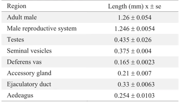
Region Length (mm) xrse Adult male
Male reproductive system
Testes
Seminal vesicles
Deferens vas
Accessory gland
Ejaculatory duct
Aedeagus
1.26r0.054 1.246r0.0054 0.435r0.026 0.375r0.004 0.165r0.0023
0.21r0.007 0.33r0.0063 0.254r0.0103 Table 3. Average length of parts of the male reproductive system of H. hampei. (n = 12)
Fig. 4. Morphology of the hindgut of Hypothenemus hampei. A) view of the hindgut indicating the (pi) pyloric valve; (mt) three out of six Malpighian tubules; (il) ileum; (co) colon; (r) rectum and (a) anus B) lateral view of the hindgut and the H. hampei female reproductive tract; C) ESEM colon, rectum, (as) anal sac and anus; D) ESEM lateral view of the anal orifi ce.
A B
C D
Ļ
Ļ
Ļ
Ļ
Ļ
Ļ Ļ
il
pi
mt
co r
a
Ļ
Ļ
Ļ
co
r as
a
sr
Region Length (mm) x r se Adult female 1.64 r 0.031 female reproductive system 1.24 r 0.020 Ovariole 0.43 r 0.015 Common oviduct 0.46 r 0.007 Lateral Oviduct 0.04 r 0.001 Spermatheca 0.13 r 0.005 Accessory Gland 0.10 r 0.003
Table 2. Average length of parts of the female reproductive system of H. hampei. (n = 12)
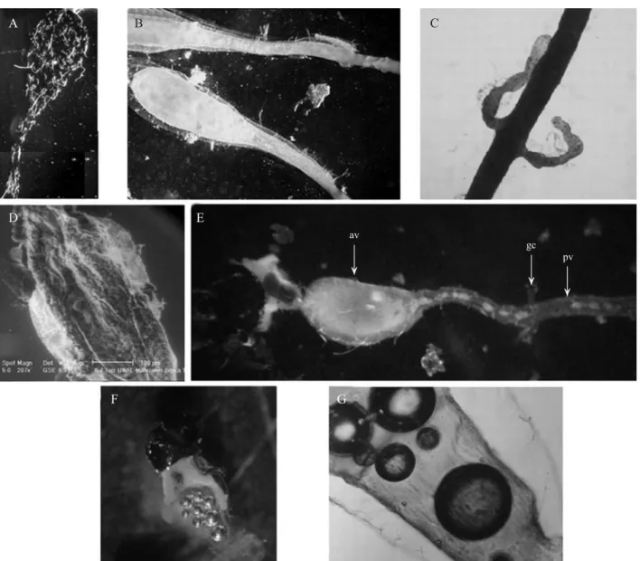
ov
lo co
ag sp
vi ge
D E
A
co
ag sp
B C
tf
Fig. 5. Morphology of female reproductive tract of Hypothenemus hampei. A) General view of the reproductive tract, (ov) ovaries; (ge) gemarium; (vi) vitelarium; (lo) lateral oviduct; (co) common oviduct; (sp) spermatheca and (ac) accessory gland, (tf) terminal fi lament. B) Lateral view of the distal region indicating the spermatheca; accessory gland and common oviduct. C) ESEM General view of the genital opening at the last dorsal segment named pygidium. D) Lateral view of the maturation oocytes in the ovariole in different stages. E) Female H. hampei ovipositing.
Fig. 6. Morphology of male reproductive tract of Hypothenemus hampei. A) General view of the reproductive tract of H. hampei, (ae) aedeagus, (ed) ejaculatory duct, (sv) seminal vesicles, (ag) accessory glands, (dv) deferens vas and (tes) testes. B) Lateral view of the aedeagus, where there are two apodemes (indicated with arrows).C) Amplifi cation of the small pores in the distal region of the aedeagus; D) Lateral view of the middle portion of the male reproductive tract showing the ejaculatory duct, the seminal vesicles and the accessory glands.
D
B
C
A
tes
dv
ag
sv
ed
ae
ag
sv
The form of the aedeagus in the male reproductive system is very unique and has not been observed or documented in other coleopterans. This aedeagus does not have a single opening at the terminal portion, but several small pores in a sclerotized structure instead. These pores must serve as canals for the sperm ejaculation.
In summary, we described for the first time the morphology of the alimentary canal and the reproductive tract of H. hampei. The results showed typical structures and arrangements of these systems found in other beetles of the same family. However, we documented the feeding behavior of the adults of this insect-pest, the difference in number and position of the gastric caeca, the production of bubbles inside the alimentary canal as a consequence of starvation, and the unique structure of the aedeagus of H. hampei that shows several pore-like structures.
Acknowledgments
The authors express their appreciation to the Colombian National Coffee Research Center - Cenicafé, also to the Biologist Gloria Camayo and to the Universidad Nacional de Colombia, Manizales for the ESEM analysis. This research was carried out with funds of the Colombian Coffee Growers Federation, FNC, Project No. ENT0299 approved in May 2002.
References
Baker, W.V. & C.L. Estrin. 1974. The alimentary canal of Scolytus multistriatus (Coleoptera – Scolytidae). A histological study. Can. Entomol. 106: 673-686.
Bergamin, J. 1956. Pragas do café. Boletim da superintendencia dos servicos do café (Brasil) 31: 7-9.
Blum, M.S. 1985. Fundamentals of insect physiology. 1. Ed., John Wiley & Sons, New York, 616p.
Borror, D.J., D.M.Delong & C.A. Triplehorn. 1976. An introduction to the study of insects. 4a. ed., Holst, Rinehart and Winstorn, New York, 852p.
Brun, L.O., J.J. Stuart, V. Gaudichon, K. Aronstein & R.H. Ffrench. 1995. Functional haplodiploidy: A mechanism for the spread of insecticide resistance in an important international insect pest. Proc. Nat. Acad. Sci. 92: 9861-9865.
Bustillo, A.E., M.R. Cardenas, D.A. Villalba, P. Benavides, J. Orozco & F. Posada. 1998. Manejo integrado de la broca del caféHypothenemus hampei (Ferrari) en Colombia. Chinchiná, Cenicafé, 133p.
Calder, A.A. 1989. The alimentary canal and nervous system of Curculionoidea (Coleoptera): Gross morphology and systematic signifi cance. J. Nat. Hist. 23: 1205-1265. Fig. 7. Transverse section of the abdomen of Hypothenemus hampei female showing the position of the different organs that composed the alimentary canal and the reproductive tract. (el) elytra, (fb) fat body, (ov) ovariole, (mp) Malpighian tubules, (av) anterior midgut, (gc) gastric caeca, (sp) spermatheca, (co) common oviduct, (pv) posterior midgut and (pr) proctodeum.
Ļ
ĸ
ĸ
ĸ
ĸ
ĸ
ĸ
ĺ
Ļ
Ļ
Ļ
fb
ov
mp
av gc
el
sp
co
pv
Corbett, G.H. 1933. Some preliminary observations on the coffee berry beetle borer Stephanoderes (Cryphalus) hampei Ferrari. Malayan Agric. J. 21: 8-22.
Crowson, R.A. 1981. The biology of the Coleoptera. Academic Press, London 802p.
Díaz, E., O. Arciniega, L. Sánchez, R. Cisneros & G. Zúñiga. 2003. Anatomical and histological comparison of the alimentary canal ofDendroctonus micans, D. ponderosae, D. pseudotsugae pseudotsugae, D. rupifenis, and D. terebrans (Coleoptera: Scolytidae). Ann. Entomol. Soc. Am. 96: 144-152.
Díaz, E., R. Cisneros & G. Zuñiga. 2000. Comparative anatomical and histological study of the alimentary canal of Dendroctonus frontalis (Coleoptera: Scolytidae). Ann. Entomol. Soc. Am. 93: 303-311.
Díaz, E., R. Cisneros, G. Zuñiga & E. Uria-Galicia. 1998. Comparative anatomical and histological study of the alimentary canal of Dendroctonus parallelocollis, D. rhizophagus and D. valens (Coleoptera – Scolytidae). Ann. Entomol. Soc. Am. 91: 479-487.
Johanneson, N.E. & A. Mansingh. 1984. Host pest relationship of the genus Hypothenemus (Scolytidae: Coleoptera) with special reference to the coffee berry borer (H. hampei). J. Coffee Res. 14: 43-56.
Le Pelley, R.H. 1968. Pest of coffee. London Longmans, Green and Co., 590p.
López-Buenfi l, J.A., J. Váldez, A. Equihau & A. Burgos. 2001. El proventrículo como estructura para identifi car géneros Mexicanos de Scolytidae (Coleoptera). Folia Entomol. Mex. 40: 325-372.
Macgown, M. & P. Sikorowski. 1981. Digestive anatomy of the adult boll weevil, Anthonomus grandis Boheman. (Coleoptera: Curculionidae). Ann. Entomol. Soc. Am. 74: 117-126.
Hall, G.M., C. Tittiger, G.L. Andrews, G.S. Mastick, M. Kuenzli, X. Luo, S.J. Seybold & G.J. Blomquist. 2002. Midgut tissue of male pine engraver, Ips pini, synthesizes monoterpenoid pheromone component ipsdienol de novo. Naturwissenschaften 89: 79-83.
Mercer, E.H. & S.C. Birbek. 1979. Manual de microscopía electrónica para biólogos. Blume Ediciones, Madrid, 134p.
Perez-Mendoza, J., J.E. Throne & J.E. Baker. 2004. Ovarian physiology and age-grading in the rice weevil, Sitophilus oryzae (Coleoptera: Curculionidae). J. Stor. Prod. Res. 40: 179-196.
Robertson, J.G. 1961. Ovariole numbers in Coleoptera. Can. J. Zool. 39: 245-263.
Romoser, W.S. & J.G. Stoffolano. 1998. The science of entomology. 4. ed. McGraw-Hill, Boston. 624 p.
Rubio G, J.D. & J.R. Acuña Z. 2006. Anatomía comparada del tracto digestivo en imagos del complejo picudo (Coleoptera: Curculionidae) asociados al cultivo del plátano. Rev. Col. Ent. 32: 67-72.
Russo, G. 1926. Contributo alla conoscenze degli Scolytidi studio morfobiologico del Cheatoptelius vestitus Malus. Funchs e dei suoi simbionti. Boll. Lab. Zool. Gen. Agric. Portioi. 103-260.
Sánchez, P.A., F. Sánchez, F.H. Caetano & K. Jaffé. 2000. El tubo digestivo en adultos de Rhynchophorus palmarum (L.) (Coleoptera: Curculionidae): Morfología y ultraestructura. Bol. Entomol. Venezuela. 15: 195-216.
Schneider, I. & A. Rudinsky. 1969. Anatomical and histological changes in internal organs of adult Trypodendron lineatum, Gnathotichus retusus and G. sulcatus (Coleoptera – Scolytidae). Ann. Entomol. Soc. Am. 62: 995-1003.
Silva-Olivares, A., E. Díaz, M. Shibayama, V. Tsutsumi, R. Cisneros & G. Zúñiga. 2003. Ultrastructural study of the midgut and hindgut in eight species of the genus Dendroctonus Erichson (Coleoptera: Scolytidae). Ann. Entomol. Soc. Am. 96: 883-900.
Snodgrass, R.E. 1935. Principles of insect morphology. McGraw-Hill, New York, 667p.
Wigglesworth, V.B. 1950. The principles of insect physiology. E.P. Dutton and Co, New York, 434p.
Williams, J.L. 1945. The anatomy of the internal genitalia of some Coleoptera. Proc. Entomol. Soc. Wash. 47: 73-91.