SYSTEMATICS, MORPHOLOGY AND PHYSIOLOGY
Ultrastructural Modifications in the Venom Glands of Workers of
Apis
mellifera
L. (Hymenoptera: Apidae) Promoted by Topical Application of
Juvenile Hormone
T
HAISAC. R
OAT, R
OBERTAC.F. N
OCELLIANDC
ARMINDADAC
RUZ-L
ANDIMDepto. Biologia, Instituto de Biociências, UNESP - Universidade Estadual Paulista” Júlio de Mesquita Filho” Campus Rio Claro, Av. 24-A, nº 1515, Bela Vista, 13506-900, Rio Claro, SP
Neotropical Entomology 35(2):469-476 (2006)
Modificações Ultra-Estruturais nas Glândulas de Veneno de Operárias de Apis mellifera L. (Hymenoptera: Apidae) Promovidas pela Aplicação Tópica de Hormônio Juvenil
RESUMO - O presente estudo analisou, através de estudos ultra-estruturais a influência do tratamento com hormônio juvenil sobre as glândulas de veneno de operárias de Apis mellifera L. Para tanto, operárias recém-emergidas receberam aplicação tópica de 1µl de hormônio juvenil, na concentração de 2 µg/µl, sendo usado o hexano como veículo. Foram feitos dois controles, um que não recebeu nenhum tipo de tratamento (grupo C1) e o outro que recebeu aplicação tópica de 1 µl de hexano (grupo C2). O aspecto das células glandulares, em operárias recém-emergidas, mostra que estas não estão ainda secretando ativamente. Observa-se que alterações celulares ocorrem de acordo com a idade da operária e da região glandular considerada no controle C1. Assim, a fase de secreção mais ativa da glândula ocorre entre a emergência e os 14 dias de idade; aos 25 dias as células já perderam sua característica secretora, sendo a região distal a primeira a sofrer degeneração. Os tratamentos com hormônio juvenil e com hexano alteram a cronologia do ciclo glandular, antecipando o início da secreção e da degeneração da glândula.
PALAVRAS-CHAVE: Ciclo secretor, degeneração glandular, hexano, abelha
ABSTRACT - The present study analyzed, the influence of the treatment with juvenile hormone on the ultrastructure of Apis mellifera L. workers’ venom glands. Newly emerged workers received topical application of 1 µl of juvenile hormone diluted in hexane, in the concentration of 2 µg/µl. Two controls were used; one control received no treatment (group C1) and other received topical application of 1 µl of hexane (group C2). The aspect of the glandular cells, in not treated newly emerged workers, showed that they are not yet secreting actively. Cellular modifications happened according to the worker age and to the glandular area considered. The most active phase of the gland happened from the emergence to the 14th day. At the 25th day the cells had already lost their secretory characteristic, being the distal area the first to suffer degeneration. The treatment with juvenile hormone and hexane altered the temporal sequence of the glandular cycle, forwarding the secretory cycle and degeneration of the venom gland.
KEY WORDS: Secretory cycle, glandular degeneration, hexane, bee
The venom glands of workers of Apis mellifera L. presents a single secretory cycle characterized by an intense synthetic stage in young workers of up to 16 days after emergence. The venom produced is then stored inside the reservoir and the venom gland begins to degenerate (Cruz-Landim & Kitajima 1966, Cruz-(Cruz-Landim et al. 1967, Owen & Bridges 1976, Abreu et al. 2000). Thus, in foraging workers, the glandular cells present evident signs of degeneration, lacking most distinguishable cellular
structures, with the nuclei and the microvilli surrounding the canaliculi as the only discernable structures. Vesicles with irregular sizes and shapes are found in the remaining cytoplasm. Structures in the form of concentric lamellae frequently appear in degenerating cells (Cruz-Landim & Kitajima 1966).
the worker, there is also a differential activity among the cells of the distal and proximal regions of the glandular filament, with the cells of the distal region being more active in the synthesis and secretion of the proteic components of the venom.
Modifications in the secretory cycle of insect’s glands during their life translate into modification in the genetic program for their functioning. The genetic program also changes during glandular degeneration, in which the genes that lead to the process of programmed cell death are activated (Amabis et al. 1981).
The control over the changes that occur on the genetic programming in insect’s organs is carried out by the hormones, particularly the juvenile hormone and ecdisone. In the adult, the juvenile hormone has a somatotrophic effect, which in bees controls the development of specific organs, specially the fat body, ovaries, and hypopharyngeal glands. The hormone also controls age polyethism, which characterizes the division of labor in the worker caste (Jaycox 1976). The level of juvenile hormone in the haemolymph of workers rises when they progress from performing tasks inside the colony to tasks outside the colony (Akamatsu et al. 1975, Rembold 1976, Robinson 1987). Adult workers of A. mellifera
present low titers of juvenile hormone in the haemolymph during the first two weeks of adult life, when they perform tasks related to brood care (Rutz et al. 1976, Fluri et al. 1982), and higher titers when they become foragers from the third week of life (Robinson et al. 1989; Huang et al. 1991, 1994; Robinson 1992). The rise in the titer of juvenile hormone induces a precocious foraging behavior (Robinson 1985, Sasagawa et al. 1989, Sullivan et al. 2000), thus suggesting that the mechanism through which this control is attained is through the effect of the juvenile hormone over the glandular development of the workers.
Therefore, these observations justify studies that aimed at identifying the role of the juvenile hormone on the development of the glands of the exocrine system (Bonetti
et al. 1994, Abdalla et al. 2001, Paes de Oliveira & Cruz-Landim 2001), with the intent of understanding its effect on either the immature stages or on the adult individual. In this work we take into consideration that these glands do not present ennervations, thus being under hormonal control, and presenting different developmental degrees among workers performing tasks inside or outside the nest. Therefore, it is possible that the juvenile hormone controls the development of the exocrine glands rendering the worker apt to perform the different tasks according to age.
The present study had the objective of verifying the effect of the treatment with juvenile hormone on the ultrastructure of the venom glands of workers of A. mellifera.
Materials and Methods
For the study of the venom glands, workers of Apis mellifera, were collected at the Apiary of the Departamento de Biologia, Instituto de Biociências, Universidade Estadual Paulista Júlio de Mesquita Filho (UNESP), in Rio Claro, SP, Brazil.
About 150 newly emerged workers were divided into
three experimental groups by marking them with non-toxic paint of different colors on the thorax. The first group did not receive any kind of treatment, thus constituting the control group (group C1). The second group received the topical application of 1 µl of 2 mg/µl juvenile hormone (JH-III, Sigma) diluted in hexane (group JH) on the dorsal portion of the abdomen, and the third group received the topical application of 1µl of pure hexane (group C2). After marking and treatment, the bees were returned to the colony and 10 individuals of each group were collected after reaching 14 and 25 days of age in order to study their venom glands. Ten newly emerged workers were also collected.
Transmission electron microscopy. The venom glands of all individuals collected were removed in insect saline (Nacl 7.5g/l, Na2HPO4 2.38g/l e KH2PO4 2.72g/l). The glands were divided into proximal and distal portions. These portions were fixed separately in modified Karnovsky (4% paraformaldehyde, 2.5% glutaraldehyde in 0.1 M sodium cacodylate buffer, pH 7.2, for 2h at 4ºC. Once fixed, the glands were washed in the same buffer for 30 min. Next, the material was post-fixed in 1% osmium tetroxide in the same buffer for 2h at room temperature. Afterwards, the glands were washed again in the same buffer for additional 30 min. The material thus post-fixed were contrasted for 2h in a solution of 2% uranyl acetate in 10% acetone and then was subjected to dehydration in a standard acetone series. The material was included in Epon-Araldite resin and ultrathin sections were obtained in an ultra-microtome. The sections were contrasted with lead citrate for 15 min. and examined under the TEM.
Results
At all life stages of the workers, the glands present this same general constitution. However, according to age and glandular region a few variations may be found mainly with regards to the secretory cells. These aspects characterize their current state of the functional cycle.
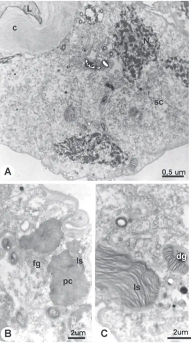
Newly emerged workers present a gland with cylindrical secretory cells that appear intact in all regions studied (Figs. 1A-C). The secretory space is enclosed by long microvilli (Figs. 1A, C). At 14 days of age, in the untreated workers (Group C1), the secretory cells appear slightly altered. The cytoplasm appears vacuolated, with disarrayed reticule and presence of concentric lamellar structures (Figs. 2A, B). The nuclei present an irregular shape and condensed chromatin (Fig. 2A). The lumen is filled by an homogeneous and electron dense secretion (Figs. 2A, B). These characteristics are observed at all glandular regions, but are more pronounced at the distal region (Fig. 2B). The characteristics of cellular degeneration are still more evident at all glandular regions of untreated workers with 25 days of age. These workers present an evidently altered
epithelium, with secretory cells of irregular shape (Fig. 3A). In addition to the concentric lamellar structures associated or not to electron dense granules that are dispersed throughout the cytoplasm (Fig. 3C), structures in a paracrystalline arrangement are also observed associated or not to membranous structures (Fig. 3B). The nucleus acquires a completely irregular shape showing a highly condensed chromatin and granular materials are found in the glandular lumen (Fig. 3A).
The treatment with juvenile hormone alters the sequence of this glandular cycle. Therefore, workers subjected to this treatment presented at 14 days of age a glandular epithelium with more alterations than in individuals of group C1 at this
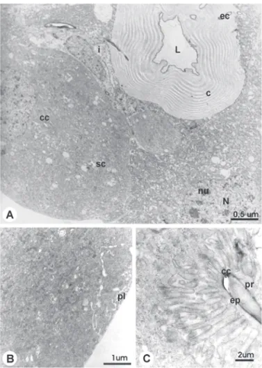
Figure 1. TEM micrographs showing the venom gland general organization of newly-emerged worker of A. mellifera.
A. General aspect of intima (i) cells convered by cuticle (c) around the lumen (L) and the excretory canaliculi (ec). Notice the secretory cells (sc) with collector canalicules (cc); N = nuclei; nu = nucleoli. B. Distal region with peripheral labyrinth (pl). C. Detail of collector canaliculi (cc) showing the epicuticle (ep) and the pro-cuticle (pr).
age. The cellular structures with characteristics of cellular degeneration are more evident (Figs. 4A-D). The cytoplasm appears completely vacuolated (Figs. 4A, C), with numerous concentric lamellar structures usually located around the terminal apparatus (Fig. 4B). At this age, in the treated group, we noted the presence of structures that were only observed in the C1 control group at 25 days of age, such as the presence of concentric lamellar structures associated to electron dense
granules (Fig. 4D) and structures with a paracrystalline arrangement at the distal region (Fig. 4C).
Workers treated with the solvent of the juvenile hormone, hexane (Group C2), presented at 14 days of age a proximal region with a fairly intact glandular epithelium (Fig. 5A), even more preserved than in group C1. At this region, the intracellular spaces are dilated, the rough endoplasmic reticule is well developed. and several regions of Golgi complexes can be observed in the cytoplasm (Fig. 5A). Nevertheless, the secretory cells of the distal region appear more altered, showing a great loss of cytoplasm (Fig. 5B).
At 25 days, the differences between the group treated with juvenile hormone and the groups C1 and C2 are more discreet, since at this age, independently of the treatment
Figure 3. TEM micrographs of the venom gland of 25 day-old workers of A. mellifera from the control without treatment (C1 group); A. Distal region general view, showing the secretory cell (sc) with nucleous (N) with irregular shape and condensed chromatin; L = lumen; c = cuticle. B. Magnification showing structures with para-cristalin (pc) associated to membranous lamellas (ls) and regions showing thin granulation (fg) which can be precursory of the crystal. C. Lamellar structures (ls) with
and region under consideration, the glandular epithelium appears completely degenerated. Figs. 6A and 6B show the characteristics observed in the group treated with juvenile
hormone and Figs. 6C through 6E show the characteristics observed in group C2, treated with hexane. In Fig. 6C it is possible to observe the presence of an homogeneous mass that represents the condensation of a secretory cells, probably in process of cellular death.
Discussion
Accompanying the ultrastructural modifications of the glandular cells of the untreated control group, it is possible to observe that cellular alterations occur according to the age of the worker and glandular region under consideration. The stage of greater development of the secretory apparatus of the cell, and therefore the stage of active secretion of the gland, must occur between emergence and 14 days of age, since at this age we found some alterations at the distal region of the gland, even though the proximal region showed intact cells. The fact that there was secretion inside the lumen shows that this secretion is still being produced or that it is still being transported into the reservoir. At 25 days, the cells have already lost their secretory characteristics, such as the presence of collecting canaliculi in the cytoplasm, presence of rough endoplasmic reticule and nuclei with disperse chromatin and regular shape.
The venom gland presents a single secretory cycle, which begins at the end of pupation and reaches its maximum around the 16th day of the worker’s adult life. From this point, the venom is stored inside the reservoir and the gland enters the degeneration process, which is completed around 30 days after emergence (Cruz-Landim & Kitajima 1966, Cruz-Landim et al. 1967). The present study confirms this result and shows an asynchrony in the cellular function between the glandular regions studied, since at 14 days the signs of cellular degeneration are more frequent in the distal region than in the other regions of the gland studied.
The venom is used by the workers for defense and, thus, must be available at the time when the task they execute exposes them to the greater dangers, that is, in the workers performing the tasks of guardians, which precedes the foraging task. Therefore, the secretion of the venom must precede the onset of this activity. On the other hand, the worker that stings looses its sting and the posterior part of the abdomen, leading to its death. Thus, there are no reasons that justify another secretory cycle under natural conditions. Nevertheless, it is interesting to point out that if the venom reservoir is emptied artificially without harming the sting, it could fill up again, thus indicating that the venom gland is able to secrete again (Abreu 1996). Therefore, in this case, as in others reported previously, the gland is capable of responding to external stimuli, altering its secretory cycle.
The treatment with juvenile hormone acts on the venom gland maintaining the pattern of cellular asynchrony, with the distal region of the gland of individuals of the treated group presenting at 14 days characteristics that were only observed at 25 days in the untreated C1 group. Abreu (2000), through histological and histochemical studies of the venom gland of workers, also observed that the glandular filament presents asynchrony. However, according to this author the
Figure 5. TEM micrographs of the venom gland of 14 day-old workers of A. mellifera from the control with the hexane treatment (C2 group). A. General aspect and details of the proximal region. Note secretory cells (sc) preserved, oppened intercellular spaces (is), rough endoplasmic reticulum (rer) and many Golgi regions (G). The nuclei (N) is with irregular shape, chromatin condensed (cr), and nucleolis (nu); B. Distal region general view, showing altered structure; l = lumen; c = cuticle; ec = excretory canaliculi; cc = collector canaliculi; sc = secretory cell.
at the distal end.
The juvenile hormone III is the only form found in the workers of A. mellifera (Fluri et al. 1982, Robinson & Ratnieks 1987, Robinson et al. 1989) and the titer of this proximal region degenerates first, which contradicts the
results presented here but agrees with observations in other glands (Cruz-Landim & Mello 1981, Silva & Silva de Moraes 1999), in which the glandular degeneration begins
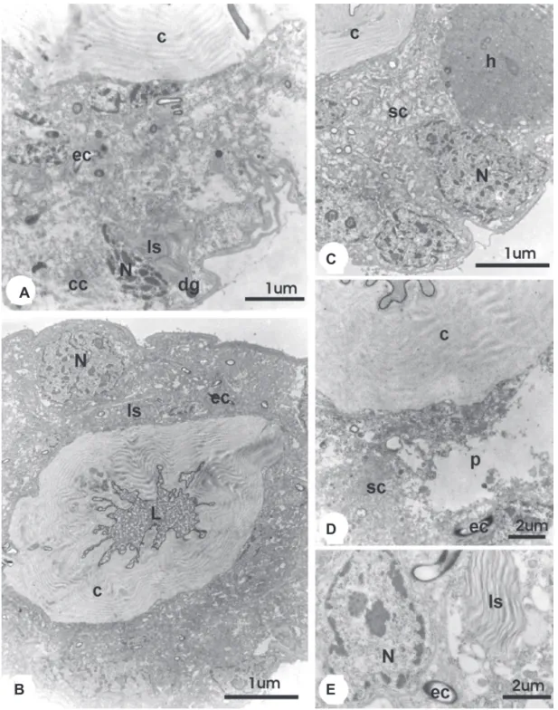
Figure 6. TEM micrographs of the venom gland of 25 day-old workers of A. mellifera from the group treated with juvenile hormone (HJ group) and the control with the hexane treatment (C2 group). A e B. Proximal region general view of the gland of workers treated with juvenile hormone, showing the presence of lamellar structure (ls) and nuclei (N) with irregular shape and condensed chromatin; c = cuticle; ec = excretory canaliculi; cc = collector canaliculi; dg = dense granules; L = lumen. C. Distal region general view of the gland of workers treated with hexane. Note the secretory cell (sc) with an homogeneous mass (h) and nuclei (N) with irregular shape and condensed chromatin; c = cuticle. D. Proximal region general view of the gland of workers treated with hexane, showing the secretory cell (sc) with cytoplasmic lost (p); c = cuticle; ec = excretory canaliculi. E. Aspect of cytoplsmatic desorganization of the gland of workers treated with hexane, showing concentric lamellar structures (ls) and condensation of the nuclei (N); ec = excretory canaliculi.
E A
B
hormone increases with the age of the worker, being that immediately after emergence, at the first day of adult life, these bees present approximately 5 pmol of this hormone for every 100 ml of haemolymph, but at 21 days of age they present about 20 pmol per 100 ml of haemolymph (Fluri et al. 1982, Robinson & Ratnieks 1987). Thus, the variations in the levels of juvenile hormone during adult life, mainly during the transition from performing activities inside the colony to foraging, affect the secretory cycle of the venom gland, as was shown through the ultrastructural analysis. When the workers present low levels of juvenile hormone, the cells of the venom gland present characteristics of active secretory cells. These characteristics were observed in newly emerged workers and in the untreated control group C1 at 14 days of age, although a few signs of degeneration were detected in the individuals of this group. Nevertheless, when the titer of juvenile hormone is high, the venom gland presents evident signs of degeneration. similar to the ones observed in workers of the untreated group C1 but at 25 days of age. These facts show that the anticipation of the secretory cycle and the degeneration of the venom gland promoted by the treatment with juvenile hormone is reflected on the glandular ultrastructure as a disarray of the secretory apparatus of the cells.
A similar effect was observed in other glands that are related to intra-colonial tasks. In nurse workers, the hypopharyngeal glands secrete substances that serve as food for the larvae. The treatment with juvenile hormone (Jaycox
et al. 1974, Rutz et al. 1976) on newly emerged workers causes the premature degeneration of these glands, a process that would normally occur when the workers ceases to perform tasks inside the colony. This result shows, once more, that the hormonal determinism of behavior is expressed through the maturation of the organs involved in the functions to be performed during each life stage.
In the present work, the treatment with hexane (group C2) produced at 14 days of age workers with secretory cells more preserved than group C1. In addition, hexane presented an effect that was similar to the one induced by the treatment with juvenile hormone, but with less intensity. A similar effect for the treatments for the hormone and its solvent hexane was verified by Nocelli et al. (2000) through morphological analyses of the venom gland of A. mellifera
and by Roat et al. (2004) through biochemical analysis by means of polyacrylamide gel electrophoresis (SDS-PAGE) on the band pattern of the glandular extracts.
We conclude that the treatment with juvenile hormone causes a precocious maturation or aging in the venom gland of
A. mellifera; thus, in this case, its effect is contrary to the one exerted during the larval stage when it’s topical application reverts the condition of the gland into a previous stage.
Acknowledgments
We wish to thank Antônio Sérgio Pascon for the collection of bees, and Antônio Teruyoshi Yabuki and Mônica Iamonte for technical support. We also thank CAPES for financial support.
References
Abdalla, F.C., L.F. Gracioli, H.C. Salles, C. Cruz-Landim & R.L.M.S. de Moraes. 2001. Effect of the application of juvenile hormone (JH) in honeybee worker larvae on the development of the Dufour’s and Koschewnikow’s glands. Sociobiology 37: 185-191.
Abreu, R.M.M. 1996. Efeitos de choques elétricos no comportamento das glândulas de veneno de operárias de Apis mellifera L. (Hymenoptera, Apidae). Dissertação de mestrado, Instituto de Biociências/UNESP, Rio Claro, 138p.
Abreu, R.M.M. 2000. Padrões citoquímicos do desenvolvimento das glândulas de veneno de operárias de Apis mellifera L. (Hymenoptera, Apidae). Tese de doutorado, Instituto de Biociências/UNESP, Rio Claro, 176p.
Abreu, R.M.M., R.L.M.S. de Moraes & O. Malaspina. 2000. Histological aspects and protein content of the venom gland of Apis mellifera L. workers: Effect of electrical shocks in summer and winter. J. Ven. Anim. Tox. 6: 87-98.
Akamatsu, Y., P.E. Dunn, F.J. Kezdy, K.J. Kramer, J.H. Law, D. Rubstein & L.L. Sanburg. 1975. Biochemical aspects of juvenile hormone action in insects, p.123-49. In R. Meints & E. Davies (eds.), Control mechanisms in development. Plenum Publishing Corp., New York, 149p.
Amabis, J.M., J.S. Morgante & L.C.G. Simões. 1981. (eds.) Textos de genética. São Paulo, EDUSP, 690p.
Bonetti, A.M., C. Cruz-Landim & W.E. Kerr. 1994. Sex determination in bees XXX. Effects of juvenile hormone on the development of tergal glands in Melipona. J. Apic. Res. 33: 11-14.
Cruz-Landim, C. & E.W. Kitajima. 1966. Ultraestrutura do aparelho venenífero de Apis (Hymenoptera, Apidae). Mem. Instituto Butantan 33: 701-710.
Cruz-Landim, C. & R.A. Mello. 1981. Desenvolvimento e envelhecimento de Scaptotrigona postica (Hymenoptera: Apidae). Aspectos histológicos e histoquímicos. Aciesp 31: 118.
Cruz-Landim, C., S. Baldissera & D. Beig. 1967. Degeneração da glândula de veneno em operárias de Apis durante o verão e inverno. Rev. Biol. 27: 355-361.
Fluri, P., M. Lüscher, H. Wille & L. Gerig. 1982. Changes in weight of the pharyngeal gland and haemolynph titres of juvenil hormone, protein and vitellogein in worker honey bees. J. Insect Physiol. 28: 61-68.
Huang, Z.Y., G.E. Robinson & D.W. Borst. 1994. Physiological correlates of division of labor among similarly aged honey-bees. J. Comp. Physiol. 174: 731-739.
Huang, Z.Y., G.E. Robinson, S.S. Tobe, K.J. Yagi, A. Strambi & B. Stay. 1991. Hormonal regulation of behavioural development in the honey bees is based on changes in the rate of juvenile hormone biosynthesis. J. Insect Physiol. 37: 733-741.
mellifera venom gland. I – Application on the larvae (Hymenoptera: Apidae). Sociobiology 40: 457-464.
Owen, M.D. & A.R. Bridges. 1976. Aging in the venom glands of queen and worker bees (Apis mellifera L.) Some morphological and chemical observations. Toxicon 14: 1-5.
Oliveira, V.T.P. & C. Cruz-Landim. 2001. Experimental control of the effect of extra doses of juvenile hormone on the bee development: the case of the wax glands of Apis mellifera (Hymenoptera, Apidae). Sociobiology 38: 513-521.
Rembold, H. 1976. The role determinatior in the honey bee, p. 21-34 In M. Lüscher (ed.), Phase and caste determination in insects: endocrine aspects. Oxford, Pergamon Press, 150p.
Roat, T.C., R.C.F. Nocelli, R.L.M. S. de Moraes & C. Cruz-Landim. 2004. Juvenile hormone effect on the venom gland secretory cycle in workers of Apis mellifera (Hymenoptera, Apidade). Sociobiology 43: 193-199.
Robinson, G.E. 1985. Effects of juvenile hormone analogue on honey bee foraging behavior and alarm pheromone production. J. Insect Physiol. 31: 277-82.
Robinson, G.E. 1987. Regulation of honey bee age polyethism by juvenile hormone. Behav. Ecol. Sociobiol. 20: 329-338.
Robinson, G.E. 1992. Regulation of division of labor in insects
societes. Annu. Rev. Entomol. 37: 637-665.
Robinson, G.E. & F.L.W. Ratnieks. 1987. Induction of premature honey bee (Hymenoptera; Apidae) flight by juvenile hormone analogs administered orally or topically. J. Econ. Entomol. 80: 784-787.
Robinson, G.E., R.E. Page, C. Strambi & A. Strambi. 1989. Hormonal regulation of behavioral integration in the honey bee colonies. Science 246: 109-12.
Sasagawa, H., M. Sasaki & I. Okada. 1989. Hormonal control of the division of labor in adult honeybees (Apis mellifera L.) I. Effect of methoprene on corpora allata and hypopharyngeal gland, and its a-glucosidae activity. Appl. Entomol. Zool. 24: 66-77.
Silva, E.C.M. & R.L.M.S. de Moraes. 1999. Silk gland degeneration of Apis mellifera (Hym., Apidae). Acta Microscopica 8: 93.
Sullivan, J.P., O. Jassini, S.E. Fahrback & G.E. Robinson. 2000. Juvenile hormone paces behavioral development in the adult worker honey bee. Horm. Behav. 37: 1-14.