ARTIGO ORIGINAL
/ ORIGINAL
ARTICLE
SEQUENTIAL SWALLOWS HAVE NO
INFLUENCE ON ESOPHAGEAL
CONTRACTIONS OF PATIENTS WITH
IRON DEFICIENCY ANEMIA
Roberto Oliveira DANTAS and Adriana Leonarda Martins MIRANDA
ABSTRACT – Background – An experimental study showed that thyropharyngeal, cricophar yngeal and cervical esophageal muscles of
rabbits with iron deficienc y anemia had morphological changes similar to those observed in muscular dystrophy, causing myastenic changes in muscles involved in swallowing. Our hypothesis is that patients with iron def iciency anemia may have a decrease in esophageal contractions with successive swallows. Patients and Method – We studied the esophageal motility of 12 women with iron deficienc y
anemia aged 31 to 50 years (median 36 years) with ser um iron from 11 to 40 µg/dL (median 21 µg/dL), and 13 asymptomatic women
aged 26 to 49 years (median 35 years) with serum iron over 60 µg/dL. We used the manometric method with continuous perfusion. The
esophageal contractions were measured at 3, 9 and 15 cm from the upper margin of a sleeve that straddled the lo wer esophageal sphincter. Each subject performed 10 swallows of a 2 mL bolus of water alter nated with 10 swallows of a 7 mL bolus, with an interval of 30 seconds between swallows. We measured the amplitude, duration, v elocity and area under the curve of contractions. Results – There was no difference between the swallows of a 2 mL or 7 mL bolus. The amplitude, duration and area under the curve were lower in patients with iron def iciency than in asymptomatic volunteers, mainly in the proximal and middle esophageal body. There was no difference in velocity. Sequential swallows did not change contraction amplitude, duration, velocity or area under cur ve in patients and volunteers. Conclusion – Although the power of esophageal contractions was decreased in patients with iron def iciency anemia, sequential swallows did not cause further impairment.
HEADINGS – Deglutition. Esophagogastric junction. Anemia, iron-def iciency.
INTRODUCTION
An experimental study showed that the thyrophar yngeal, cricopharyngeal and cervical esophageal muscles of rabbits with iron deficiency anemia had morphological changes similar to those observed in progressive muscular dystrophy, leading to the conclusion that iron deficienc y may cause
myastenic changes in muscles that per tain to swallowing(7).
In myastenia g ra vis the pharynx undergoes progressive
worsening of function upon repeated swallo w s(2).
Oculopharyngeal muscular dystrophy predominantly affects the striated pharyngeal muscles and the palpebral levator. The esophageal smooth muscle in the disease also shows dysfunction characterized by nonpropulsive, simultaneous,
retrograde and failed acti vity(1, 11).
Iron def iciency causes reduction of the constricting po wer of the pharyngeal muscle for propulsion of bolus into the
esophagus(7). In the study of the esophagus in patients with
iron deficiency anemia we found a decrease in esophageal
contractions(5, 6) and an increased esophageal transit duration(6).
Our hypothesis is that with the mor phological alterations of muscles involved in swallowing there is the possibility of a decrease in esophageal contraction with repeated swallows in patients with iron def iciency anemia.
PATIENTS AND METHODS
We studied 12 female patients aged 31 to 50 years
(median 36 years) with serum iron from 11 to 40 µg/dL (median
21 µg/dL). The nor mal range for serum iron in women is
Depar tament of Clinical Medicine, Faculty of Medicine of Ribeirão Preto, University of São Paulo, Ribeirão Preto, SP, Brazil.
49 to 151 µg/dL. The patients had hemoglobin levels below 11µg/dL and hematocrit below 30%. The duration of the symptoms attrib uted to anemia ranged from 2 months to 22 years (median 6 years). Two patients complained of dysphagia. The cause of anemia was hyper menor rhea. The patients did not have any systemic disease that could influence esophageal motility and none had been submitted to previous g astrointestinal surgical treatment. Endoscopic examination was perfor med in all patients, with normal results. None of the patients had malnutrition or was underweight.
As a control g roup we studied 13 asymptomatic female volunteers aged 26 to 49 years (median 35 years) with normal hematologic
evaluation and ser um iron over 60 µg/dL. Informed consent was
obtained from each volunteer and patient. The study was approved by the Human Research Committee of the University Hospital of Ribeirão Preto, SP, Brazil.
Esophageal manometr y was perfor med using an eight-lumen manometric catheter assembly incor porating a 6-cm sleeve device at its distal end. Side-hole recording orif ices were cut at the distal and proximal margins of the sleeve. Five additional side-hole recording orif ices were cut at 3 cm intervals along the assemb ly, starting 3 cm proximal to the sleeve (Arndorfer Specialties Inc, Greendale, WI, USA). The catheter assembly was connected to external pressure transducers, which in turn were connected to a PC Pol ygraph HR (Synectics Medical, Stockolm, Sweeden). The manometric signals were stored in a computer. During manometric recordings, a minimally compliant pneumohydraulic pump (JS Biomedicals Inc, Ca, USA) perfused distilled water at 0.5 mL/min through the slee ve and the side holes.
Each subject was studied after an over night fast. The catheter assembly was passed through the nose and positioned so that the
6-cm long sleeve straddled the lower esophageal sphincter (LES). The contractions in the esophageal body were registered by the side holes localized at 3, 9 and 15 cm from the upper mar gin of the sleeve. All volunteers and patients were studied in the supine position. They perfor med 10 swallows of a 2 mL bolus of water at roam temperature alter nated with 10 swallows of a 7 mL bolus of water. Using the computer Polygram Upper GI software version 6.4 (Gastrosoft, Inc) we measured the amplitude, duration, area under curve (AUC) and velocity of the peristaltic contractions.
For statistical analysis we used the unpaired t-test, calculating the one-tailed P value and anal ysis of variance. The swallows of the 7 mL bolus were analyzed. The Kolmogorov and Smirnov method showed that the data were sampled from populations that followed Gaussian distrib ution. The results are repor ted as mean ± SEM unless otherwise indicated.
RESULTS
There was no difference between the swallows of a 2 mL or 7 mL bolus.
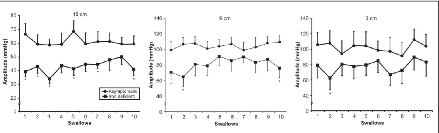
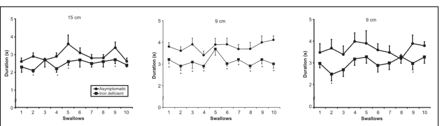
The contraction amplitude (Figure 1), duration (Figure 2) and AUC (F igure 3) were lower in patients with iron deficiency than in asymptomatic volunteers for most of the swallows, mainly in the proximal and middle esophageal body. There was no difference in peristaltic contraction velocity.
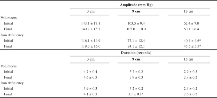
The contraction amplitude duration, AUC and velocity did not change signif icantl y with sequential swallows. There was no difference between the mean of the initial f ive s wallows and the mean of the f inal five swallows (Tables 1, 2) in volunteers or patients.
FIGURE 1 – Amplitude of esophageal contractions measured in 10 swallows of a 7 mL bolus of water at 3, 9 and 15 cm from the upper
margin of the sleeve in asymptomatic volunteers (n = 13) (z) and patients with iron def iciency anemia (n = 12) (). The results
DISCUSSION
Our results showed a decrease in the power of esophageal contraction, mostly in the middle and proximal esophagus, manifested in the amplitude, the duration and area under curve (amplitude x duration) of contractions in patients with iron deficiency. The comparison of the results of esophageal contractions between patients with iron deficincy and asymptomatic volunteers have been published
in a previous paper(6).
Studies of the effect of iron deficiency on the skeletal muscle metabolism of the rat found metabolic changes that are consistent with either a reduction in supply of oxygen to the muscle cell or
altered oxidati ve phosphor ylation by the mitochondria(9). The
decreased mitochondrial activity is likely to be due either to a reduction in the numbers of mitochondrial or to decreased activity of
the mitochondrial oxidative components(9). A leakage of mitochondria
in type I muscle fibers was found in rabbits with iron def icienc y(7).
In humans with iron deficiency anemia of moderate to severe degree
there was not evidence for a major mitochondrial oxidative defect(10).
Consequent to the description of alterations in muscles involved
in swallowing in animals with iron def iciency(7, 9), our hypothesis was
that the esophageal contraction, mainly in the proximal esophagus, where there is a proportion of striated muscle, might be impaired. Since it has also been suggested that myastenic changes are found in muscles of iron deficient animals, sequential swallows might show a progressive decrease in the power of esophageal contractions. The
FIGURE 2 – Duration of esophageal contractions measured in 10 swallows of a 7 mL bolus of water at 3, 9 and 15 cm from the upper mar gin
of the sleeve in asymptomatic volunteers (n = 13) (z) and patients with iron deficiency anemia (n = 12) (). The results are
shown as mean ± SEM. *P < 0.05
FIGURE 3 – Area under curve (AUC) of esophageal contractions measured in 10 swallows of a -7 mL bolus of water at 3, 9 and 15 cm from
the upper margin of the sleeve in asymptomatic volunteers (n = 13) (z) and patients with iron deficiency anemia (n = 12) ().
TABLE 1 – Amplitude and duration of esophageal contractions in asymptomatic volunteers (n = 13) and in patients with iron def iciency
anemia (n = 12) measured at 3, 9 and 15 cm from the upper mar gin of the sleeve, after 10 swallows of a 7 mL bolus of water. Swallows 1 to 5 are the initial ones and 6 to 10 are the f inal ones (mean ± SEM)
Amplitude (mm Hg)
3 cm 9 cm 15 cm
Volunteers
Initial 143.1 ± 17.1 103.5 ± 9.4 62.4 ± 7.0
Final 140.2 ± 15.3 105.0 ± 10.0 60.1 ± 6.4
Iron deficiency
Initial 116.1 ± 14.9 77.1 ± 12.4 40.4 ± 4.6*
Final 119.3 ± 16.0 84.1 ± 12.1 45.6 ± 5.3*
Duration (seconds)
3 cm 9 cm 15 cm
Volunteers
Initial 4.7 ± 0.4 3.7 ± 0.2 2.9 ± 0.3
Final 4.6 ± 0.3 3.9 ± 0.3 2.9 ± 0.2
Iron deficiency
Initial 3.9 ± 0.3 3.2 ± 0.2 2.4 ± 0.2
Final 4.1 ± 0.3 3.1 ± 0.1* 2.6 ± 0.2
P < 0.05 vs. volunteers
TABLE 2 – Area under curve and velocity of esophageal contractions in asymptomatic volunteers (n = 13) and in patients with iron def iciency
anemia (n = 12), measured at 3, 9 and 15 cm from the upper margin of the sleeve, after 10 swallows of a 7 mL bolus of water. Swallows 1 to 5 are the initial ones and 6 to 10 are the f inal ones (mean ± SEM)
Area under curve (mm Hg x sec)
3 cm 9 cm 15 cm
Volunteers
Initial 378.1 ± 66.6 206.3 ± 23.7 106.6 ± 18.8
Final 357.1 ± 57.9 210.5 ± 22.3 105.6 ± 18.0
Iron deficiency
Initial 250.4 ± 42.4 131.2 ± 24.5* 55.4 ± 8.9*
Final 276.8 ± 50.5 136.5 ± 22.0* 68.0 ± 10.3*
Velocity (cm/sec)
15 9 cm 9 3 cm
Volunteers
Initial 2.7 ± 0.2 3.1 ± 0.3
Final 2.6 ± 0.1 3.1 ± 0.3
Iron deficiency
Initial 2.8 ± 0.3 3.1 ± 0.3
Final 2.7 ± 0.3 3.2 ± 0.3
present results confirm the possibility that the esophagus of subjects with iron deficiency has an impaired contraction power, but did not confirm the possibility that sequential swallows cause progressive impairment of contractions.
The alterations observed in muscles involved in swallowing should affect the phar yngeal phase. A decrease in the power of pharyngeal
contractions(7) and a slower bolus penetration into the proximal
esophagus was detected in humans(6). The loss of the power of
pharyngeal contractions affects the opening of the upper esophageal
sphincter(3). In the present study, we detected a lower amplitude of
contractions in the proximal esophagus of patients compared to asymptomatic subjects.
Our results showed that the esophageal motility of patients with iron deficiency resembled the digestive motor impair ment seen in muscular dystrophy b ut not in myastenia g ravis.
In oculopharyngeal muscular dystrophy, muscle biopsies have demonstrated changes in levator, phar yngeal and vastus lateralis muscles, suggesting that the disorder may be a manifestation of
mitochondrial myopathy(2 ). It is a m yopathy affecting almost
exclusively the bulbar muscles and the levator muscle of the eyes. The major differential diagnoses are myastenia gravis and mitocondrial
myopathies. The disease also affects esophageal motility(1, 11), which
is associated with delayed esophageal isotope clearance(2).
Myastenia gravis is characterized by destr uction of acetylcholine receptors at neuromuscular junctions. The musculature controlled by
the cranial nerves is almost always involved. Diagnosis is confirmed by detecting acetylcholine receptor antibodies, which are present in
85% of the cases(2). The pharyngeal radio graphic examination shows
diffuse functional abnormalities with progressive worsening on repeated
swallows, with improvement after parenteral injection of neostigmine(8).
The comparison of the initial group of s wallows (1 to 5) with the final one (6 to 10) did not show signif icant differences. We previously described the same results in normal volunteers, patients
with Chagas' disease and patients with idiopathic achalasia(4). These
previous results and the present ones show that with an inter val of 30 seconds between sequential s wallows there is no change in amplitude, duration or velocity of peristaltic contractions. It is possible that in patients with iron deficiency sequential swallows performed within a shorter time inter val cause progressive impairment of contractions.
In conclusion, although there is impairment of contractions in patients with iron def iciency anemia, sequential swallows do not change the contractions, with the changes in esophageal motility showing some similarity to those of muscular dystrophy.
ACKNOWLEDGEMENTS
This work was supported in part by “Fundação de Apoio ao Ensino, Pesquisa e Assistência do Hospital das Clínicas da Faculdade de Medicina de Ribeirão Preto”, USP (FAEPA), and Fundação de Estudos e Projetos (FINEP), Pronex Grant number 42/97.
Dantas RO, Miranda ALM. Deglutições não modif icam as contrações esofágicas de pacientes com anemia ferropriva. Arq Gastroenterol 2004;41(1):27-32.
RESUMO – Racional – Estudo experimental encontrou que os músculos tirof aríngeo, cricofaríngeo e do esôfago cervical de coelhos com anemia ferropriva
têm alterações morfológicas semelhantes às encontradas na distrof ia muscular, provocando alterações miastênicas em músculos envolvidos com a deglutição. Nossa hipótese é de que pacientes com anemia ferropriva têm diminuição das contrações esofágicas com uma seqüência de de glutições sucessivas.
Objetivo – Avaliar as contrações esofágicas em pacientes com anemia ferropriva. Pacientes e Método – Estudou-se a motilidade do esôfago de 12
mulheres com anemia ferropriva, com idades entre 31 e 50 anos (mediana 36 anos) com fer ro sérico de 11 a 40 µg/dL (mediana 21 µg/dL), e 13 mulheres
assintomáticas, com idades entre 26 e 49 anos (mediana 35 anos) com ferro sérico acima de 60 µg/dL. Foi utilizado o método manométrico com perfusão
contínua. As contrações no esôf ago foram medidas a 3, 9 e 15 cm da margem superior de sensor longo (“sleeve”) colocado no esfíncter inferior do esôfago. Cada pessoa fez 10 deglutições de 2 mL de água, alternadas com 10 deglutições de 7 mL, com inter valo de 30 segundos entre as deglutições. Mediram-se a amplitude, duração, velocidade e área sob a curva das contrações. Resultados – Não houve diferença entre as deglutições dos volumes de 2 mL e 7 mL. A amplitude, duração e área sob a cur va foram menores nas pacientes do que nas voluntárias assintomáticas, principalmente em par tes proximal e média do esôf ago. Não houve diferença na velocidade. A seqüência de deglutições não modif icou a amplitude, duração, velocidade e área sob a curva das contrações nas pacientes e nas assintomáticas. Conclusão – Embora as contrações esofágicas estejam diminuídas em pacientes com def iciência de ferro, a seqüência de deglutições não provocou modif icações nessas contrações.
REFERENCES
1. Castell JA, Castell DO, Duranceau A, Topart P. Manometric characteristics of the pharynx, upper esophageal sphincter, esophagus, and lower esophageal sphincter in patients with oculophar yngeal muscular dystrophy. Dysphagia 1995;10:22-6.
2. Cook IJ. Disorders causing oropharyngeal dysphagia. In: Castell DO, Richter JE, editors. The esophagus. 3rd ed. Philadelphia: Lippincott Williams & Wilkins; 1999. p.165-84.
3. Cook IJ, Dodds WJ, Dantas RO, Massey B, Kern M, Lang IM, Brasseur JG, Hogan WJ . Opening mechanisms of the human upper esophageal sphincter. Am J Physiol 1989;257:G748-59.
4. Dantas RO. Effect of successi ve swallows on esophageal motility of normal volunteers, patients with Chagas’ disease, and patients with idiopathic achalasia. Neurogastroenterol Motil 2003;15:57-62.
5. Dantas RO, Villanova MG. Esophageal motility impairment in Plummer-Vinson syndrome. Correction by iron treatment. Dig Dis Sci 1993;38:968-71.
6. Miranda ALM, Dantas RO. Esophageal contractions and orophar yngeal and esophageal transits in patients with iron def iciency anemia. Am J Gastroenterol 2003;98:1000-4.
7. Okamura H, Tsutsumi S, Inaki S, Mori T. Esophageal web in Plummer-Vinson syndrome. Laryngoscope 1988;98:994-8.
8. Ott DJ. Radiology of the oropharynx and esophagus. In: Castell DO, Richter JE, editors. The esophagus. 3rd ed. Philadelphia: Lippincott Williams & Wilkins; 1999. p.45-87. 9. Thompson CH, Green YS, Ledingham JG, Radda GK, Rajagopalan B. The effect of iron def iciency on skeletal muscle metabolism of the rat. Acta Physiol Scand 1993;147:85-90.
10. Thompson CH, Kemp GJ, Taylor DJ, Radda GK, Rajagopalan B. No evidence of mitochondrial abnormality in sk eletal muscle of patients with iron-deficient anaemia. J Intern Med 1993;234:149-54.
11. Tiomny E, Khilkevic O, Korczyn AD, Kimmel R, Hallak A, Baron J, Blumen S, Asherov A, Gilat T. Esophageal smooth muscle dysfunction in oculopharyngeal muscular dystroph y. Dig Dis Sci 1996;41:1350-4.