Residence of liquids in the
infra-junctional portion of the
proximal stomach in patients with
gastroesophageal reflux disease
Departamento de Clínica Médica, Faculdade de Medicina de Ribeirão Preto, Universidade de São Paulo, Ribeirão Preto, SP, Brasil
C.L.A. Barbieri, L.E.A. Troncon, J.R.L. Herculano Jr., L.R.O. Aprile and R.O. Dantas
Abstract
Patients with gastroesophageal reflux disease may have disturbances of gastric motility, which could play a role in the pathophysiology of the disease. Recent studies have suggested that the gastric region just below the gastroesophageal junctionmay have a distinct physiologi-cal behavior. We determined whether patients with gastroesophageal reflux disease have abnormal residenceof food in the infra-junctional portion of the stomach after ingesting a liquid nutrient meal. Fasted adult patients with reflux disease (N = 11) and healthy volunteers (N = 10) ingested a liquid meal (320 ml; 437 kcal) labeled with 99mtechnetium-phytate and their total gastric emptying half-time and
regional emptying from the stomach infra-junctional region were determined. In 8 patients, episodes of postprandial acidic reflux to the esophagus were measured for 2 h using pH monitoring. There were no differences between reflux patients and controls regarding total gas-tric emptying time (median: 68 min; range: 39-123 min vs 65 min and
60-99 min, respectively; P > 0.50). Food residence in the infra-junctional area was similar for patients and controls: 23% (range: 20-30) vs 27% (range: 19-30%; P = 0.28) and emptying from this area
paralleled total gastric emptying (Rs = 0.79; P = 0.04). There was no correlation between residence of food in the infra-junctional area and episodes of gastroesophageal reflux (Rs = 0.06; P = 0.88). We conclude that it is unlikely that regional motor disturbances involving the infra-junctional region of the stomach play a relevant role in the pathogenesis of acidic gastroesophageal reflux.
Correspondence
L.E.A. Troncon
Departamento de Clínica Médica Hospital das Clínicas, FMRP, USP 14048-900 Ribeirão Preto, SP Brasil
Fax: +55-16-633-6695 E-mail: ledatron@fmrp.usp.br
Research supported by CNPq, PRONEX/FINEP, FAPESP, and FAEPA/HCFMRPUSP. C.L.A. Barbieri was the recipient of an
undergraduate “Introduction to Science” fellowship from CNPq.
Presented as a “poster of distinction” at the Digestive Diseases Week - 2004 (American Gastroenterological Association) and published in abstract form in
Gastroenterology 2004; 126 (Suppl 2): A-495 (Abstract T1705).
Received January 27, 2005 Accepted June 21, 2005
Key words
•Gastroesophageal reflux disease
•Gastric emptying •Gastric motility •Intragastric distribution •Proximal stomach
Introduction
Gastroesophageal reflux disease (GERD) is a common chronic disease, usually caused by increased episodes of acidic reflux from
Studies on animals and man have indi-cated that the stomach may play a central role in the origin of GERD. For example, gastric distension may elicit transient relax-ations of the lower esophageal sphincter (TRLES), which may be associated with increased episodes of gastroesophageal re-flux (5-7). Also, delayed gastric emptying, which might cause distension of the stom-ach, has been reported in substantial propor-tions of patients with GERD (8-12), although others have failed to confirm this finding (13-16). More recently, it has been shown that patients with GERD may have regional disturbances of gastric motility, such as in-creased (16-19) or dein-creased (20) retention of chyme in the proximal stomach, which may occur independently of gastric empty-ing rates.
Two recent studies exploring the chemi-cal characteristics of the intragastric con-tents collected from just below the gastro-esophageal junction have produced data sup-porting the view that this area may have a peculiar physiological behavior, distinct from more distal gastric sites (21,22). A study conducted on healthy volunteers reported that, after intragastric administration of ni-trates, nitric oxide concentrations over the segment just below the gastroesophageal junction were significantly higher than at all more distal gastric sites (21). In another study from the same group (22) carried out on dyspeptic patients, data from dual gastric and esophageal pH pull-trough recordings showed a regional variation in intragastric pH, with a “pocket” of more acidic contents just below the gastroesophageal junction (22). It was suggested that the contents of this region escaped the buffering effect of meals, remaining highly acidic in compari-son with contents collected from the body of the stomach (22).
We therefore hypothesized that disturbed motor performance of the infra-junctional (IJ) portion of the proximal stomach (the gastric area just below the lower esophageal
sphincter) of patients with GERD might lead to accelerated emptying of ingested meals. This would be associated with less buffered, more acidic contents, thus contributing to the pathogenesis of GERD. Hence, charac-terizing motor abnormalities involving the IJ portion of the proximal stomach might prompt the development of therapeutic strat-egies focused on this particular anatomical region. The objective of the present study was to determine the postprandial residence of fluids and the emptying rate of the IJ portion of the proximal stomach in GERD patients compared to healthy volunteers.
Material and Methods
Subjects
Eleven patients with GERD (4 males and 7 females, median age: 51 years, range: 19-62 years, median body mass index (BMI): 24.6 kg/m2, range: 19.38-29.82 kg/m2) and
10 healthy, non-symptomatic volunteers from the medical staff and other hospital employ-ees (8 males and 2 females) participated in the study after giving written informed con-sent. Median age in this group was 24.5 years (range: 18-45 years) and median BMI was 25.22 kg/m2 (range: 19.71-28.14 kg/
m2). There were no significant differences
(Student t-test) between patients and con-trols regarding age (P > 0.10) or BMI (P > 0.50). The local Ethics Committee approved the study protocol (Statement number 9326/ 00), which was carried out according to the Declaration of Helsinki for research in hu-man beings.
in those patients with typical GERD symp-toms and a normal endoscopy. On the basis of this investigation, 9 patients were found to have Savary-Miller (23) grade I erosive esophagitis, with no significant (<2 cm) hia-tus hernia, and 2 patients were found to have non-erosive esophagitis. The criteria for in-clusion in the GERD group therefore con-sisted of at least one cardinal GERD symp-tom (3) plus either erosive esophagitis at endoscopy (23) or abnormal results of the esophageal pH study (3,4). Criteria for in-clusion in the control group consisted of the absence of any active symptom or history of digestive complaints or diseases. None of the subjects included in the study were smok-ers, had evidence of other diseases or had previous abdominal operations. None of the healthy controls used any drugs. For the GERD patient group, all medications in use were discontinued at least one week before the tests.
Study protocol
Gastric emptying and the intragastric dis-tribution of a radio-labeled liquid nutrient meal were assessed in all patients and con-trol volunteers by external abdominal scin-tigraphy. In 8 of the GERD patients, a 2-h postprandial ambulatory esophageal pH re-cording using the same test meal was per-formed in another session carried out within 2-7 days after the first study. All tests were carried out in the morning, after an overnight fast.
Determination of gastric emptying and meal residence in the proximal stomach and in the infra-junctional region
Subjects ingested 320 ml of a liquid test meal containing 437 kcal (64 g carbohy-drate, 20 g protein, and 11 g fat, 800 mOsm/ kg) and prepared as described elsewhere (20). Before ingestion, the test meal was labeled with 18 MBq 99mtechnetium coupled
with phytate (Phytosyd; Sydma Medical Equipments and Reagents, Ribeirão Preto, SP, Brazil) as a non-absorbable carrier. Im-mediately after meal ingestion, images of the radioactivity in the anterior and posterior aspects of the abdomen were simultaneously collected (30 s of acquisition time) from subjects standing between the low energy, high sensitivity collimators of a dual-headed gamma camera (Sopha Vision, model DST, “Sophycamera”, Sopha Medical Vision America, Twinsburg, OH, USA). The cam-era was set up to record activity with a 20% window around the 140-keV photo peak of technetium. The acquisition apparatus was connected to an image processing system. Further serial images of the abdomen were then taken every 5 min during the first 20 min, and then at 10-min intervals up to 90 min. In the intervals between image acquisi-tions subjects sat comfortably in a chair near the camera.
poste-rior-to-anterior movement of the marker within the body.
For each region, “activity versus time” curves, expressed as percentages of activity in the total stomach immediately after the end of meal ingestion, were obtained. These curves permit the calculation of the follow-ing variables: gastric emptyfollow-ing half-time (T1/
2), the time (min) needed for the initial
activ-ity in the total stomach to fall by 50%; meal residence in the proximal stomach through-out gastric emptying: the ratio between the average retention of radioactivity in the ROIs corresponding to the proximal and the total stomach. Meal residence in the IJ area throughout gastric emptying was defined as the ratio between the average retention of radioactivity in the ROI corresponding to the IJ area and in both the proximal and total stomach. IJ area emptying half-time (T1/2)
was defined as the time (min) needed for the initial activity in this area to fall by 50%.
Determination of gastroesophageal reflux
Patients underwent conventional manom-etry (24) and immediately after the proce-dure a pH probe (Synetics Multi-use pH-Catheter; Synetics Medical, Stockholm, Sweden) with a single antimony electrode was introduced so that its tip was positioned 5 cm above the lower esophageal sphincter. The probe was connected to a pH meter (Digitrapper MkIII, Synetics Medical) and a basal recording was obtained for 45 min. The patient was then fed the same liquid test meal as used in the abdominal scintigraphy,
but without adding the radio marker. After ingestion, the patient sat comfortably and was instructed to stand up for one min every 10 min up to 2 h, in order to simulate the variations of body position observed during the isotope study.
The number of acidic reflux episodes during this study was counted after visual inspection of each individual recording us-ing a dedicated software (Polygram function testing for Windows, Synetics Medical). An acidic reflux episode was defined as a fall in esophageal pH to below the arbitrary limit of 4 pH units for at least 10 s. A further drop of at least 1 pH unit when esophageal pH was already below 4.0 was considered to be a new-reflux episode (20).
Statistical analysis
Data are reported as median and range and were analyzed by nonparametric tests sincethe data did not seem to have a normal distribution. Differences between controls and patients were analyzed by the Mann-Whitney test. Except for the analysis regard-ing gastric emptyregard-ing, the two-tailed test was utilized. The Spearman rank correlation co-efficient was employed for the assessment of the relationships between the various gas-tric motor variables, as well as between resi-dence of liquid in the IJ area and the number of postprandial acidic reflux episodes. Dif-ferences were considered to be statistically significant when P < 0.05.
Results
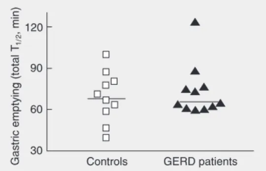
As shown in Figure 1, there was no sig-nificant difference between GERD patients and controls regarding total gastric empty-ing T1/2 values (median: 68 min; range:
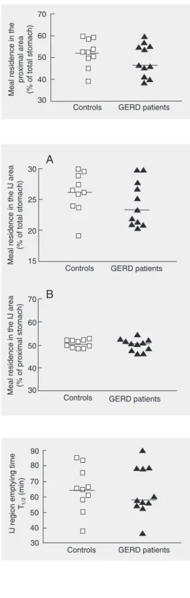
39-123 vs 65 min and 60-99 min, respectively). Data regarding residence of food in the proximal stomach are presented in Figure 2, which also shows that there was no signifi-cant difference between GERD patients and Figure 1. Gastric emptying
half-times (total T1/2) for patients with
the controls concerning this variable (46%; 38-60 vs 52%; 39-59%; P > 0.50).
Residence of food in the IJ area through-out gastric emptying in GERD patients was similar to that observed in controls, both when expressed as a fraction of total (23%; 20-30 vs 26.5%; 19-30%; P > 0.20) or proxi-mal stomach residence (50.6%; 46.1-54.4 vs 50.5%; 48.0-53.1%; P > 0.50). These data are presented in Figure 3A and B. Values for the emptying T1/2 of the IJ area of GERD
patients, although slightly lower, were also not significantly different from those ob-tained for the control group (58 min; 37-90 vs 64.5 min; 38-85 min; P > 0.50), as shown in Figure 4. Data in Table 1 shows that, in both groups, the motor behavior of the IJ area paralleled those of both the total and the proximal stomach since highly significant values for the correlation coefficient between the relevant variables were observed. The relationship between the rates of emptying of the IJ area and the total stomach is shown in Figure 5.
In GERD patients, there was no signifi-cant correlation between residence of food in the IJ area and the number of acidic gas-troesophageal reflux episodes (Rs = 0.06; P = 0.88), as shown in Figure 6.
Discussion
Our findings show that, after a liquid nutrient meal, the residence of chyme in the gastric region just below the lower esoph-ageal sphincter (IJ area) parallels the resi-dence rates of both the proximal and total stomach. Indeed, the median values for meal residence in the IJ area of both groups were numerically close to those corresponding to the size of the ROI for that area, when ex-pressed in proportion of either the proximal (~50%) or the total stomach (~25%). Also, no differences were found between GERD patients and healthy volunteers. Taking to-gether, these findings indicated that it is unlikely that an abnormal motor behavior of
Figure 2. Residence of food in the proximal stomach (reported as a fraction of that for the total stomach) throughout gastric emptying in patients with gastro-esophageal reflux disease (GERD) and control subjects, after a liquid nutrient meal. The horizontal bars indicate the me-dians for patients (N = 11) and controls (N = 10). P > 0.05 (Mann-Whitney test).
Figure 3. Residence of food in the infra-junctional (IJ) region of the proximal stomach (the area just below the lower esophageal sphincter) throughout gastric emptying in patients with gastro-esophageal reflux disease (GERD) and control subjects, after a liquid nutrient meal. Data are reported as fractions of both total (A) and proximal stomach (B) residence values. The hori-zontal bars indicate the medians for patients (N = 11) and con-trols (N = 10). A, P > 0.05; B, P > 0.05 (Mann-Whitney test).
Figure 4. Emptying half-time (T1/2) of the infra-junctional (IJ)
region of the proximal stomach (the area just below the lower esophageal sphincter) in pa-tients with gastroesophageal re-flux disease (GERD) and con-trol subjects, after a liquid nutri-ent meal. The horizontal bars in-dicate the medians for patients (N = 11) and controls (N = 10). P > 0.05 (Mann-Whitney test). the IJ region is involved in the pathogenesis
of acidic gastroesophageal reflux.
proximal stomach and may be associated with increased rates of both TRLES (1) and reflux episodes (4,5). Nevertheless, we were unable to find any evidence of rapid empty-ing from this region, or any significant cor-relation between residence of food in the IJ area and the number of acidic gastroesopha-geal reflux episodes.
Our patients probably are a rather repre-sentative sample of GERD, since this group included only patients with mild or no esoph-agitis and without hiatus hernia, but with pathological reflux, as demonstrated by 24-h esop24-hageal pH monitoring. T24-his kind of patient corresponds to the largest contingent in the clinical spectrum of GERD (1,4), and also represents a group in which TRLES originating in the stomach is regarded as a major pathogenic mechanism of gastroesoph-ageal reflux (1,4,26,27).
As far as the test meal employed in our study is concerned, we regard it as suitable for testing the proposed hypothesis of di-minished residence of liquids in the IJ re-gion, given its relatively high calorie content and osmolarity, and its ability to elicit a substantial number of postprandial episodes of acidic gastroesophageal reflux, as seen in a previous study with the same meal (20). Nevertheless, we cannot rule out the possi-bility that using a composite, mixed solid-liquid test meal, or more exclusively, a usual solid meal might produce different results.
Measurements of total and regional tric emptying and the number of acidic gas-troesophageal reflux episodes were per-formed on two different days. However, this is unlikely to be relevant for our negative findings, since the studies were performed under similar conditions, the interval be-tween the two studies was rather short and both scintigraphy (28) and esophageal pH monitoring (29) are quite reproducible tech-niques.
Another potential methodological limi-tation of the present study is the definition of a possibly too large ROI for the IJ region of Figure 6. Correlation between
values for residence of food in the infra-junctional (IJ) region of the proximal stomach (the area just below the lower esophageal sphincter) and the number of acidic gastroesophageal reflux episodes in patients with gastro-esophageal reflux disease, after a liquid nutrient meal. Rs = 0.06 and P > 0.05 (Spearman’s cor-relation).
Figure 5. Correlation between emptying rates from the infra-junctional (IJ) region of the proxi-mal stomach (the area just below the lower esophageal sphincter) and from the total stomach in patients with gastro-esophageal reflux disease, after a liquid nutrient meal. Data are reported for N = 11 patients. Rs = 0.71 and P = 0.018 (Spear-man’s correlation).
the proximal stomach might show a peculiar physiological behavior (21,22), we hypoth-esized that meal emptying from this area might be accelerated, leading to less buff-ered and more acidic contents (22). Since transfer of liquids from the proximal to the distal stomach appears to be dependent on gastric fundal pressure (25), abnormally rapid chyme emptying from the IJ area may be associated with increased pressure. This in turn may stretch the cardiac region of the
Table 1. Application of Spearman’s correlation coefficient to motor variables related to the infra-junctional (IJ) region of the stomach and those related to both the proximal and the total stomach in patients with gastroesophageal reflux disease (GERD) and in healthy volunteers (controls).
Subjects Emptying T1/2 - IJ vs Residence - IJ vs
total stomach proximal stomach
GERD (N = 11) Rs = 0.71 (0.018) Rs = 0.95 (<0.001) Controls (N = 10) Rs = 0.88 (0.001) Rs = 0.94 (0.0002)
Data in parentheses correspond to the relevant P values, for the hypothesis that the
correlation is significantly different from zero. T1/2 is the emptying half-time for each
the proximal stomach. This was done so that the longitudinal diameter of this ROI would correspond to 50% of that for the total stom-ach, in order to reduce the chance of missing the emergence of the esophagus into the stomach, which may vary from one indi-vidual to another. However, this relatively large size may have interfered with the dem-onstration of more subtle regional motor abnormalities regarding the IJ area. Never-theless, our findings of preserved emptying from the IJ area agree with those reported in a very recent study which used a different approach to define the IJ area (30). In this study, Sifrim et al. (30) used a radioactive marker to precisely define the gastroesopha-geal junction and a ROI as small as 10% of the total stomach around the marker corre-sponding to this particular gastric region and did not find any abnormal retention in this area.
The lack of a difference between GERD patients and controls for gastric emptying time in the present study differs from other studies showing delayed gastric emptying (8-12). Nevertheless, our findings agree with previous studies that either failed to demon-strate an abnormality in GERD, or found that only a small proportion of patients had delayed emptying (13-16,20). Indeed, close inspection of our data (Figure 1) shows that only one of the 11 GERD patients had an exceedingly high gastric emptying T1/2. Also,
we did not find differences between GERD patients and controls regarding meal resi-dence in the proximal stomach, which is in contrast to others who reported either
in-creased postprandial proximal residence of food (16-19) or diminished retention in the proximal stomach of the same liquid meal as used here (20). However, it is possible to assume that differences between our find-ings and other studies that demonstrated de-layed gastric emptying or postprandial intra-gastric food maldistribution in GERD are related to different criteria for patient inclu-sion or diversity in patient demographic and clinical characteristics. Also, it is difficult to rule out that the relatively small number of patients included in the present study may have contributed to the absence of differ-ence between the GERD and control groups, as well as to the discrepancy between our findings and those from other studies (13-20).
The present study did not demonstrate any differences between GERD patients and healthy volunteers regarding meal residence in the infra-junctional portion of the proxi-mal stomach. Our results therefore do not support the hypothesis that physiological motor abnormalities in the gastric area lo-cated just below the lower esophageal sphinc-ter play a relevant role in the pathogenesis of acidic gastroesophageal reflux.
Acknowledgments
The authors wish to thank Miss Marie Secaf for excellent technical assistance and Professor Daniel Sifrim, Leuven Catholic University, Leuven, Belgium, for his intel-lectual contribution to this study.
References
1. Galmiche JP & Janssens J (1995). The pathophysiology of gastro-oesophageal reflux disease: an overview. Scandinavian Journal of
Gastroenterology, 30 (Suppl 211): 7-18.
2. Koelz HR, Blum AL & The Swiss Esophagitis Study Group (1986). Cardinal symptoms of reflux esophagitis. Gastroenterology, 90 (Suppl 2): 1498 (Abstract).
3. Stacher G & Weiss W (1989). Diagnosis of gastro-esophageal reflux disease. Scandinavian Journal of Gastroenterology, 24 (Suppl 156):
21-24.
4. Richer JE & Castell DO (1982). Gastro-esophageal reflux. Annals of
Internal Medicine, 97: 93-103.
5. Holloway RH, Hongo M, Berger K et al. (1985). Gastric distention: A mechanism for post-prandial gastroesophageal reflux.
Gastroenter-ology, 89: 779-784.
of Physiology, 259 (Part 1): G380-G385.
7. Boulant J, Fioramonti J, Dapoigny M et al. (1994). Cholecystokinin and nitric oxide in transient lower esophageal sphincter relaxation to gastric distention in dogs. Gastroenterology, 107: 1059-1066. 8. McCallum RW, Berkowitz DM & Lerner E (1981). Gastric emptying
in patients with gastro-oesophageal reflux. Gastroenterology, 80: 285-291.
9. Maddern GJ, Chatterton BE, Collins PJ et al. (1985). Solid and liquid gastric emptying in patients with gastro-oesophageal reflux. British
Journal of Surgery, 72: 344-347.
10. Cunningham KM, Horowitz M, Riddell PS et al. (1991). Relations among autonomic nerve dysfunction, oesophageal motility, and gas-tric emptying in gastro-oesophageal reflux disease. Gut, 32: 1436-1440.
11. Scarpignato C & Farneze A (1992). Esophageal exposure to acid in GERD patients with and without delayed gastric emptying. Effect of cisapride. Hepatogastroenterology, 39: 91-92.
12. Benini L, Sembenini C, Castellani G et al. (1996). Gastric emptying and dyspeptic symptoms in patients with gastro-esophageal reflux.
American Journal of Gastroenterology, 91: 1351-1354.
13. Csendes A & Henriquez A (1978). Gastric emptying in patients with reflux oesophagitis or benign strictures of the esophagus secondary to reflux compared to controls. Scandinavian Journal of
Gastroen-terology, 13: 205-207.
14. Shay SS, Eggli D, McDonald C et al. (1987). Gastric emptying of solid food in patients with symptomatic gastroesophageal reflux.
Gastroenterology, 92: 459-465.
15. Keshavarzian A, Bushnell DL, Sontag S et al. (1991). Gastric empty-ing in patients with severe reflux esophagitis. American Journal of
Gastroenterology, 86: 738-742.
16. Stacher G, Lenglinger J, Bergmann H et al. (2000). Gastric empty-ing: a contributory factor in gastro-oesophageal reflux activity? Gut, 47: 661-666.
17. Penagini R, Hebbard G, Horowitz M et al. (1998). Motor function of proximal stomach and visceral perception in gastro-oesophageal reflux disease. Gut, 42: 251-257.
18. Tefera S, Gilja OH, Hatlebakk JG et al. (2001). Gastric accommoda-tion studied by ultrasonography in patients with reflux esophagitis.
Digestive Diseases and Sciences, 46: 618-625.
19. Tefera S, Gilja OH, Olafsdottir E et al. (2002). Intragastric
maldistri-bution of a liquid meal in patients with reflux oesophagitis assessed by three-dimensional ultrasonography. Gut, 50: 153-158.
20. Herculano Jr JRL, Troncon LEA, Aprile LRO et al. (2004). Dimin-ished retention of food in the proximal stomach correlates with increased acidic reflux in patients with gastroesophageal reflux disease and dyspeptic symptoms. Digestive Diseases and
Sci-ences, 49: 750-756.
21. Iijima K, Henry E, Moriya A et al. (2002). Dietary nitrate generates potentially mutagenic concentration of nitric oxide at the gastro-oesophageal junction. Gastroenterology, 122: 1248-1257. 22. Fletcher J, Wirz A, Young J et al. (2001). Unbuffered highly acidic
gastric juice exists at the gastro-esophageal junction after a meal.
Gastroenterology, 121: 775-783.
23. Savary M & Miller G (1978). The esophagus. In: Savary M & Miller G (Editors), Handbook and Atlas of Endoscopy. Gassman Verlag AG, Solothurn, Switzerland.
24. Dantas RO & Mamede RCM (1996). Esophageal motility in patients with esophageal caustic injury. American Journal of Gastroenterol-ogy, 91: 1157-1161.
25. Mayer EA (1994). The physiology of gastric storage and emptying. In: Johnson LR (Editor), Handbook of Physiology. Raven Press, New York, 929-976.
26. Mittal RK & McCallum RW (1988). Characteristics and frequency of transient relaxations of the lower esophageal sphincter in patients with reflux esophagitis. Gastroenterology, 95: 593-599.
27. Holloway RH, Kocyan P & Dent J (1991). Provocation of transient lower esophageal sphincter relaxations by meals in patients with symptomatic gastroesophageal reflux. Digestive Diseases and
Sci-ences, 36: 1434-1439.
28. Tosetti C, Paternicó A, Stanghellini V et al. (1998). Reproducibility of a solid and of a liquid caloric meal for gastric emptying studies.
Nuclear Medicine Communications, 19: 581-586.
29. Streets CG & DeMeester TR (2003). Ambulatory 24-hour esoph-ageal pH monitoring: why, when, and what to do. Journal of Clinical
Gastroenterology, 37: 14-22.