From the Department of Orthopaedics and Traumatology, Hospital das Clínicas, Faculty of Medicine, University of São Paulo.
THE USE OF BONE BRIDGES IN TRANSTIBIAL
AMPUTATIONS
Auro Mitsuo Okamoto, Roberto Guarniero, Rafael Ferreira Coelho, Fabricio Ferreira Coelho, and André Pedrinelli
RHCFAP/3014 OKAMOTO A M et al. - The use of bone bridges in transtibial amputations. Rev. Hosp. Clín. Fac. Med. S. Paulo
55 (4):121-128, 2000.
We sought to describe the bone bridge technique in adults, and present a variation for use in children, as well as to present its applicability as an option in elective transtibial amputations. This paper presents a prospective study of 15 transtibial amputations performed between 1992 and 1995 in which the bone bridge technique was employed. The patients’ ages ranged from 8 to 48 years, with an average of 22.5 years. This technique consisted of the preparation of a cylinder of periosteum extracted from the tibia and with cortical bone fragments attached to it to promote a tibiofibular synostosis on the distal extremity of the amputation stump. We noted that the cortical bone fragments were dispensable when the technique was employed in children, due to the increased osteogenic capacity of the periosteum. This led to a variation of the original technique, a bone bridge without the use of the cortical bone fragments.
Results: The average time spent with this procedure, without any significant variation between adults and children, was 171 minutes. The adaptation to the definitive prosthesis was accomplished between 20 and 576 days, with an average of 180 days. Revision of the procedure was necessary in 3 amputations.
Conclusions: This technique may be employed in transtibial amputations in which the final length of the stump lies next to the musculotendinous transition of the gastrocnemius muscle, as well as in the revision of amputation stumps in children, where the procedure has been shown to be effective in the prevention of lesions due to excessive bone growth.
DESCRIPTORS: Amputation. Lower limb. Operatory technique. Rehabilitation. Prospective studies.
Complications of transtibial ampu-tations are not rare. Prolonged prosthe-sis use may generate alterations in the amputation stump due to muscular at-rophy and mobility between the tibia and the fibula. The most frequently found alteration is the shortening of the distal diameter of the stump, which as-sumes a conical form and migrates to-wards the end of the prosthesis socket. This occurs by the approximation of the fibula behind the tibia due to socket compression, creating pressure points that may cause cutaneous ulcers and make prosthesis use impracticable. An-other common complication is the presence of pain at the amputation stump. Its main cause is the presence
of neuromas adhered to local scars. However, even on technically adequate amputations, the patient may have pain. Intraosseous circulatory alter-ations on extremities are also frequent in conventional amputations and result in a painful amputation stump at lower temperatures. Bone closure with bone bridge techniques, as we shall describe, promote conservation of pressure dis-persion, improving venous drainage of the bone and consequently, diminish-ing pain1.
Moreover, another complication of transtibial amputations, even though limited to pediatric patients, is the ex-cessive osseous overgrowth at the am-putation stump extremity, which causes skin lesions, allowing bacterial infec-tions to settle. Furthermore, bone pro-trusion, without skin laceration, hinders prosthesis use. Therefore, a revision of the amputation stump is necessary in children every two or three years due to excessive bone growth2. Techniques
like epiphysiodesis and the closure of the distal bone extremities with syn-thetic material have been proposed with acceptable results.
In 1949, in Hungary, Ertl2 proposed
amputation stump revisions in adult patients. In this technique, a perios-teum cylinder is built from the tibia, connecting it to the fibula, which re-sults in a synostosis or tibiofibular bone bridge, obtaining a much larger trans-versal area than in conventional transtibial amputations. Ertl called this procedure osteoperiosteoplasty, and
according to him, it results in a better distribution of forces on the amputation stump inside the prosthesis socket, de-creasing the possibility of cutaneous lesions. Ertl addressed the principles of physiological amputations (muscular modelling, individualized vascular ligature, and nerve section) as a funda-mental part of the technique, in order to produce a stump with adequate venous drainage, temperature similar to the rest of the body, absence of pain, and possessing contractile muscle3,4.
This technique allows the mainte-nance of the tibiofibular distance, an important factor for the support of the stump on the prosthesis socket, by im-peding the migration of the distal part of the fibula behind the tibia, with con-sequent chronic deformation of the amputation stump. In children, the pro-cedure is also shown to be efficient, impeding excessive bone growth on the distal extremity of the stump3.
PATIENTS AND METHODS
In the period between 1992 and 1995, 12 patients in our Orthesis Group underwent transtibial tion or revision of transtibial amputa-tion with the use of the bone bridge technique. Three out of these 12 pa-tients underwent bilateral procedures.
These patients were divided into 2 age groups: Group A comprising those aged over 14 years and who were op-erated on employing the original tech-nique described by Ertl and Group B comprising patients aged 14 who un-derwent the modified technique, herein
described as periosteoplasty.
Patient age ranged from 8 to 48 years, and the average patient age was 22 years, 6 months. In Group A, (av-erage age 32 years, 10 months) 4 am-putations (50%) were performed on male patients, and 4 amputations (50%) were performed on female pa-tients. Group B (average age 10 years, 8 months) 1 amputation (14.3%) was performed on a male patient, and 6 (85.7%) amputations were performed on female patients.
Regarding previous amputations, in Group A, 7 (87.5%) amputations were primary and 1 (12.5%) was an ampu-tation stump revision. Myelomeningo-cele sequelae (3 cases; 42.9%), fol-lowed by nervous lesions (2 cases, 28.6%), were the most frequent rea-sons for amputation in this group.
In Group B, 2 (28.6%) amputations were primary, and 5 (71.4%) were am-putation stump revisions. The most fre-quent indication for amputation was septicemia-induced vasculitis (3 cases;
42.9%), followed by myelomeningo-cele sequelae (2 cases; 28.6%).
Once amputation or stump revision was indicated, patients underwent pre-operative evaluation in order to estab-lish the adequate amputation level or the degree of shortening to be attained in the revision cases. The causes that led to the amputation were considered in the primary amputation cases, while in the stump revision cases the stump alterations that made prosthesis use impracticable were evaluated. Addi-tionally, the osseus level of the ampu-tation was also established in accor-dance with the regions of the tibia that presented with cortex integrity.
Criteria for selection of the patients to undergo the technique were: - Indication of transtibial amputation
due to foot deformity or functional incapacity without involvement of the leg;
- Indication of lower limb amputation stump revision in cases in which the revision stump, after the revision,
Table 1 - Amputation causes in group “A”.
Amputation cause Frequency Proportion(%)
Myelomeningocele sequelae 3 42.86
Trauma 1 14.29
Neurofibromatosis 2 14.29
Nervous lesion 1 28.57
Deep mycosis 1 14.29
Total 8 100
Table 2 - Amputation causes in Group “B”.
Amputation cause Frequency Proportion(%)
Myelomeningocele sequelae 2 28.57
Septic vasculitis 3 42.86
Trauma 1 14.29
Vacular lesion 1 14.29
!
would have a final length near themusculotendinous transition of the gastrocnemius muscle;
- Indication of amputation revision in children with transtibial amputation, due to excessive bone growth in the distal extremity of the stump in cases in which the revision also resulted in a transtibial amputation stump; - All patients with vascular alterations,
malignant osseus neoplasia, articular deformity with flexion of the knee exceeding 20 degrees, or signs of ac-tive infection were excluded.
After the determination of the ideal osseous level, patients in Group A un-derwent the bone bridge technique. These patients were anesthetized with a spinal block. The osseus level was marked on the crista tibialis at the level of the musculotendinous transition of the gastrocnemius muscle. Two equally-sized cutaneous flaps were then marked (the first one anteromedial and the other posterolateral) from the marked osseus level to the most distal level with skin integrity.
Foot resection was performed en bloc, 8 cm distally to the marked
am-putation level. In the amam-putation revi-sion cases, the distal extremity of the stump was removed en bloc at the most
distal region of the tibia where its anatomy was preserved. Skin flap dis-section followed at the subfascial plane, with exposure of the leg muscles. The anterior and lateral com-partments of the leg were isolated as one muscle group to the marked am-putation level.
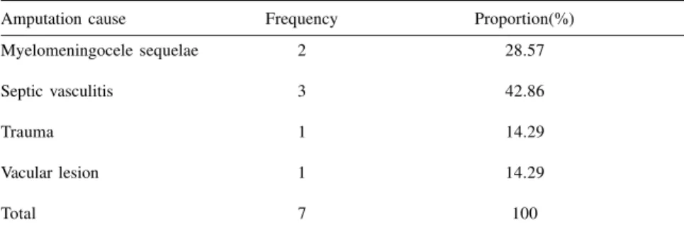
Osteotomy of the fibula was per-formed 1 cm proximal to the marked level of the tibia. The periosteum of the anteromedial face of the fibula was de-limited and detached proximally with the use of an osteotome, with frag-ments of cortical bone adhered to it (Fig. 1). The same procedure was done on the periosteum of the posterior and anterolateral faces of the tibia, so that two periosteum strips with cortical
bone fragments were obtained, both 8 cm in length. Subsequently, tibial sec-tion was accomplished at the definitive level. The osseous extremity was rounded with a rasp.
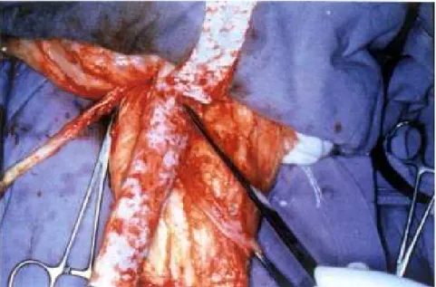
The fibular and the posterior and anterior tibial arteries, as well as their corresponding veins, were then identi-fied and ligated, while the tibial and the deep and superficial fibular nerves were sectioned and placed deep in the muscle group.
The posterior osteoperiosteal strip was taken to the distal extremity of the fibula and therein fixed through holes in the fibula, made with a 2.5 mm drill. The anteromedial osteoperiosteal strip was taken to the fibula, covering the medullar layers of the tibia and fibula and positioning the bone fragments against the fragments of the other strip. The borders of the strips were then su-tured to each other forming a cylinder of periosteum filled with bone grafts, connecting the extremities of the fibula and the tibia (Fig. 2).
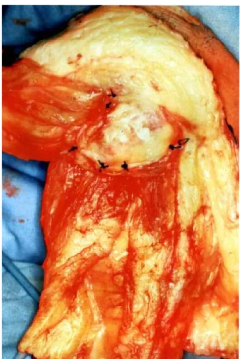
Next, myoplasty was done, suturing the muscular group composed of the posterior compartment to the muscular group formed by the anterior and lat-eral compartments (Fig. 3). The mus-cular groups were also sutured to the
osteoperiosteal cylinder. The skin flap layers were then sutured, followed by a compressive plaster bandage.
The patients of Group B underwent periosteoplasty. They were given gen-eral anesthesia, while antiseptic proce-dures and marking of the osseus level were made as described for Group A. Foot resection (or distal extremity of the stump in the revision cases) was performed 4 to 6 cm distal to the marked level, and the detachment of the periosteum was done without the tibial cortex bone fragments. There-fore, the resulting cylinder (bone bridge) did not contain bone fragments. Fastening to the fibula was done by su-turing the periosteum directly, which, due to its thickness, obviates drilling in the distal extremity of the bone. The other steps were analogous to the tech-nique described for Group A.
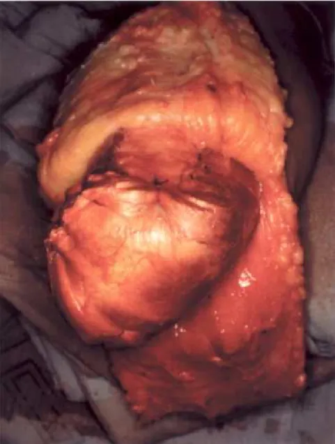
The prosthesis indication was made after the radiologic establishment of the bone bridge (Fig. 4) and the stabi-lization of stump dimensions.
RESULTS
"
when the orthopedic prosthesis could be used painlessly and when the bone bridge was consolidated. Results were considered “bad” when bone spiculae formed and/or patients presented with pain at the amputation stump, thus mak-ing prosthesis use impossible. Accord-ing to these criteria, the obtained results where “good” in 14 (93%) operations and “bad” in only one.
In Group A, the average duration of
the surgery was 190 minutes, ranging from 105 to 320 minutes (SD = 65.65). Adaptation to the orthopedic prosthesis occurred on the average 223 days after surgery, ranging from 36 to a maximum of 576 days (SD = 170.45).
In Group B, the average length of the surgery was 150 minutes, ranging between 120 and 180 minutes (DP = 19.24). Comparison between the two groups did not show any statistical
#
necessary for the treatment of tibial anddeep fibular nerve neuromas, which sig-nificantly reduced the pain. One case of stump ulcer was verified in a patient from Group B, and credited to an error in the manufacture of the socket of the orthopedic prosthesis. The ulcer was closed with surgical treatment and did not occur again after the manufacture of a new socket. Additionally, one case in Group B had osseous spiculae. Once the patient underwent revision of the opera-tion, it was found that the periosteoplasty had been performed with inadequate technique, with the
pe-riosteum tearing during the operation. The spiculae were removed, but the manufacture of a new periosteoplasty was not possible due to the short length of the stump. The patient was able to continue to use a prosthesis after this correction.
DISCUSSION
Delayed changes in the amputation stump are common reasons that patients seek medical treatment after prosthesis adaptation. Following a classic
transtibial amputation, rehabilitation, and prosthesis adaptation, the patient usually returns to everyday life and sel-dom makes regular follow-up visits. Meanwhile, the stump undergoes pro-gressive alterations in its form, resulting in chronic stump pain (because of a pos-terior position of the fibula relative to the tibia) with cutaneous lesions and muscular atrophy. The distal migration of the stump inside the socket occurs because of the retraction of the in-terosseous membrane and compression of the lateral walls of the socket, there-fore supporting body weight on its dis-tal extremity.
The use of poorly adjusted sockets may result in chronic lymphedema. This occurs when there is space be-tween the stump and the end of the socket, caused by the critical distal drainage of the stump, by the orthos-tatic position, and by the lack of muscles capable of pumping blood to-wards the inferior vena cava. The edema thus formed may become chronic, frequently associated with Figure 4 - Radiologic establishment of the bone bridge.
$
skin metaplasia, keratinization, and secondary infection.
The absence of axial load on the bone and muscular disinsertion are also important factors contributing to the establishment of progressive osseous demineralization (analogous to underuse osteoporosis of persons who undergo prolonged immobilization), which could be one of the causes of pain at the amputation stump.
The opening of the bone marrow reduces intraosseous venous pressure, as has been stated before. This results in venous stasis due to reduction of the osseous venous drainage capacity and local hypothermia. This phenomenon takes place both in patients with vas-cular disease and in trauma patients, as well as in other causes of amputation.
Revision of the transtibial amputa-tion stump in children is usually due to excessive osseous growth. This causes deformity and generates a point of in-creased mechanical pressure on the distal part of the stump, resulting in lo-cal ischemia and rupture of the skin, resulting in osseous exposition and consequently infection.
The solution for the aforemen-tioned problems might be the manufac-ture of a new prosthesis socket, and this is effectively done for amputation stumps with less than a year of devel-opment. However, in older cases, pain and skin alterations may not improve with this procedure, and surgical treat-ment may be required, which invari-ably results in a shorter final length. For this reason, the development of techniques that stabilize stump form and impede the development of the aforementioned alterations is highly desirable.
The technique created by Ertl, in addition to preventing the fibula from moving in relation to the tibia and main-taining the vascularization of the sec-tioned bone extremities, impedes the exposition of the tibial and fibular bone marrow. Occlusion of the bone marrow
also maintains intraosseous pressure, which is essential in preserving the venous drainage of the bone1. The bone
bridge, on the other hand, protects the vasculonervous pedicle, preventing the fibula from moving and forcing it to work with the tibia as one bone. This ensures the maintenance of the cylindric form of the amputation stump. Like-wise, the existence of bone tissue on the stump perpendicular to the axis of the limb creates a surface that can be loaded, preventing demineralization through the generation of forces in the axial length of the limb. In children, the treatment of the periosteum prevents the creation of osseous protrusions and skin ulceration. Additionally, the association of myoplasty to the bone bridge de-scribed by Ertl allows an osseous inser-tion for the muscle group, thus prevent-ing its atrophy.
Our Prosthesis and Orthesis Group has elected as the ideal level for transtibial amputation the musculoten-dinous transition of the gastrocnemius muscle, as stated by Murdoch7 and
Burgess8 (1971). Amputations made
distally to this point have an elevated incidence of trophic lesions due to the lack of a muscular cushion, whereas amputations performed proximally to the transition results in greater energy expenditure in perambulation6,7.
There-fore, once the transtibial amputation has been indicated, it must be per-formed at the most distal level that has healing potential. However, for those cases that presented with a viable pe-riosteum at the distal third of the tibia, we began to use the Ertl bone bridge technique, since 6 to 8 cm of the dis-tal part of the tibia would be removed in the amputation regardless of the technique used. This would retain the benefits of the ERTL technique with-out shortening of the final stump length.
Criteria for patient selection were established to encompass the concepts of physiological amputation and of
stump size maintenance. In order to construct the periosteum cylinder, 6 to 7 cm of distal periosteum of the tibia had to be in good condition. This por-tion of the tibia should be removed, determining the final stump length. While performing the technique, it was demonstrated that the loss of osseous length equals diameter of the cylinder, which in adults should be around 5 or 6 cm. Thus it was observed that if the transection were to be performed at the level of the musculotendinous transi-tion of the gastrocnemius, it was nec-essary that, in adults, the periosteum presented itself in good condition to 2 or 3 cm proximal to the tibiotarsal ar-ticulation. Consequently, the selected adult patients had an indication for transtibial involving only the foot. Pa-tients with an indication for amputation involving the leg proximal to the mal-leoli were excluded from the selection. Patients with vascular disease, malig-nant osseous neoplasias (because of the risk of compromising the surgical safety margin), flexion deformity of the knee or hip above 20 degrees (which could limit prosthesis use), or bacterial infection were also excluded9, 10.
%
is considered to be the most difficultsurgical step of this technique, due to the possibility of its rupture.
As has been stated before, there was a difference of 40 minutes in surgery length between the two groups (190 minutes in adults, 150 minutes in chil-dren), however, this was not shown to be statistically significant (p=0.06), in contrast to what was expected. One pos-sible explanation could be the small sample size, associated with a large variation inside each group. Another explanation is that this surgery may not depend so much on the manufacture of the bone bridge, but on other procedures for obtaining the physiological stump.
Regarding the time elapsed be-tween the operations and the respective prosthesis adaptations, it has been ob-served that the standard deviation is
very close to the average, indicating large variation on the registered times. In conclusion, the operative techniques are similar in time elapsed between the operations and the respective prothesis adaptations. This result is also differ-ent from what was expected, since the greater activity of the child’s periteum would, in theory, establish os-seous consolidation and consequent faster prosthesis adaptation.
CONCLUSION
Technical advances regarding the manufacture of orthopedic prosthesis could hinder the development of new amputation techniques. However, we believe that stump quality is more im-portant than prosthesis quality,
espe-cially in scenarios in which good qual-ity prostheses are not economically ac-cessible to the majority of the popula-tion.
It is fundamental to observe, how-ever, that the technique has precise in-dications, requiring an adequate patient selection to be employed with good re-sults. Based on the data shown, we conclude that transtibial amputation according to the Ertl bone bridge tech-nique, associated with the construction of the physiological stump, can be ap-plied to primary amputations in adult patients. Periosteoplasty, on the other hand, can be applied to pediatric pa-tients in the revision of transtibial stumps indicated by excessive osseous growth, preventing the appearance of secondary skin lesions.
RESUMO RHCFAP/3014
OKAMOTO AM e col. – O uso da técnica da osteoperiosteoplastia em amputações transtibiais. Rev. Hosp. Clín. Fac. Med. S. Paulo 55
(4):121-128, 2000.
Objetivo: Descrever a técnica da
osteoperiosteoplastia em adultos e apresentar a variação para a utilização em crianças, procurando demonstrar sua aplicabilidade como opção nas am-putações transtibiais eletivas.
Casuística e métodos: O artigo
apresenta um estudo prospectivo de 15 amputações transtibiais realizadas en-tre 1992 e 1995 em que se utilizou a técnica da osteoperiosteoplastia. A ida-de dos pacientes variou ida-de 8 a 48 anos, com média de 22,53 anos. A técnica
consistiu na confecção de um cilindro de periósteo retirado da tíbia, conten-do fragmentos de osso cortical presos ao mesmo, para promover uma sinos-tose tibiofibular na extremidade distal do coto de amputação. Observou-se que os fragmentos de osso cortical eram dispensáveis quando a técnica foi empregada em crianças, pela maior capacidade osteogênica do periósteo. Isto levou a uma variação da técnica original, que consiste numa periosteoplastia sem a utilização de fragmentos de osso cortical.
Resultados: O tempo médio
des-pendido com a técnica, sem variação significativa entre adultos e crianças, foi de 171 minutos. A adaptação da prótese definitiva dos pacientes foi
ob-tida entre 20 e 576 dias, com média de 180 dias. Revisões da técnica empre-gada foram necessárias em três ampu-tações (20%).
Conclusão: A técnica encontra
aplicação nas amputações transtibiais em que o comprimento final do coto seja próximo da transição musculo-tendínea do gastrocnêmio e nas revi-sões de cotos de amputação de crian-ças onde a técnica mostrou ser efetiva na prevenção das lesões decorrentes do excessivo crescimento ósseo.
&
REFERENCES
1. LOON HE - Biological and biomechanical principles in amputation surgery. In: INTERNATIONAL PROSTHETICS COURSE, 2nd.
Copenhagen, 1960. Proceedings. Committee on Prosthesis, Braces, and Technical Aids. Copenhagen, 1960. p. 41-58. 2. MARQUADT E & CORREL J - Amputations and prosthesis for
lower limb amputee. Int Orthop 1984; 8: 139-144.
3. ERTL J - Über amputationsstumpfe. Chirurg 1949; 20: 218-224. Apud FRIEDMANN, LW10 - An overview and tissue techniques.
4. PINTO MAGS, FILHO NA, GUEDES JPB et al. - Ponte óssea na amputação transtibial. Rev Bras Ortop 1998; 33:7. p. 525-31. 5. DEDERICH R - Stump correction by muscle plastic procedure. In:
INTERNATIONAL PROSTHETICS COURSE, 2nd. Copenhagen,
1960. Proceedings.Committee on Prosthesis, Braces, and Technical Aids. Copenhagen, 1960. p. 59-61
6. GONZALES EG, CORCORAN P J & REYES RL - Energy expenditure in below knee amputees: correlation with stump length. Arch Phys Med Rehabil 1974; 55:111-115.
7. MURDOCH G - Levels of amputation and limiting factors. Ann R Coll Surg Engl 1967; 40: 204-216.
8. BURGUESS EM, ROMANO RL, ZETTL JH et al. – Amputations of the leg for peripheral vascular insufficiency. J Bone Joint Surg 1971; 53-A:874-8.
9. WEISS M Apud FRIEDMANN LW - An overview and tissue techniques. In: THOMAS CC - The surgical treatment of the amputee. Springfield. 1978; p. 153-8 (separata).
10. FRIEDMANN LW - An overview and tissue techniques. In: THOMAS CC - The surgical treatment of the amputee. Springfield, 1978. p. 153-8 (separata).