382 Rev. odonto ciênc. 2010;25(4):382-385
Received: January 23, 2010 Accepted: September 26, 2010
Conflict of Interest Statement: The authors state that there are no financial and personal conflicts of interest that could have inappropriately influenced their work.
Copyright: © 2010 Mollica et al.; licensee EDIPUCRS. This is an Open Access article distributed under the terms of the Creative Commons Attribution-Noncommercial-No Derivative Works 3.0 Unported License.
Original Article
Temperature variation in pulp chamber during
dental bleaching in presence or absence of
light activation
Variação da temperatura na câmara pulpar durante o
clareamento dental na presença ou ausência de fotoativação
Fernanda Brandão Mollica a
Daniel Maranha da Rocha a
Alessandro Caldas Travassos a
Marcia Carneiro Valera a
Maria Amélia Maximo de Araujo a
a Department of Restorative Dentistry, Sao Jose dos Campos Faculty of Dentistry, State Paulista University, Sao Jose dos Campos, SP, Brazil
Correspondence:
Rua Jose Ferreira, 92, Jd Aquarius Sao Jose dos Campos, SP – Brazil 12246-004.
E-mail: [email protected]
Abstract
Purpose: In addition to the chemical damage due to bleaching gels penetration into the pulp during pulp vitality dental bleaching, another possible aggressive factor could be the heat generated by the exothermal oxidation reaction of the bleaching gel, which may also be aggravated by the use of light activation. This study assessed the temperature variation in the pulp chamber in human teeth, using three different bleaching gels with or without LED light activation.
Methods: Thirty human pre-molars were cut longitudinally to obtain buccal and lingual halves. The 60 specimens were divided into 3 groups, and the bleaching gel used varied as follows: 35% hydrogen peroxide (WHP); 37% carbamide peroxide (W) and 38% hydrogen peroxide (OX). Half of the specimens were submitted to bleaching with light activation and, the other half, without light activation. The light source used was the light emitting diode appliance (LED, 3-Light, Clean Line), and the intrapulpal temperatures were measured by using a digital thermometer. Data were analyzed by ANOVA and Tukey’s tests (alpha=5%).
Results: The intrapulpal temperatures (in oC) were as follows: without light activation (WHP=
0.68b; W= 0.40b; OX= 0.48b); with light activation (WHP= 2.35a; W= 1.60a; OX= 1.80a ).
Conclusion: LED light activation of bleaching gels increased the temperature in the pulp chamber, but did not achieve the critical temperature value of 5.5 oC.
Key words: Bleaching; activation; temperature; light; peroxide
Resumo
Objetivo: Além da agressão química devido à penetração dos géis clareadores na polpa durante o clareamento de dentes com vitalidade pulpar, outro possível fator agressor pode ser o calor gerado pela reação de oxidação exotérmica do gel clareador, que pode também ser agravada pela fotoativação. Este estudo avaliou a variação da temperatura na câmara pulpar de dentes humanos, utilizando-se três diferentes géis clareadores, com ou sem fotoativação.
Metodologia: Trinta pré-molares humanos foram cortados longitudinalmente para obtenção de duas metades: vestibular e lingual. Os 60 espécimes foram divididos em 3 grupos e os agentes clareadores utilizados variaram como segue: peróxido de hidrogênio 35% (WHP), peróxido de carbamida 37% (W) e peróxido de hidrogênio 38% (OX). Metade dos espécimes foi submetida ao clareamento com fotoativação e, a outra metade, sem fotoativação. A fonte de luz utilizada foi o aparelho à base de diodo emissor de luz (LED, 3-Light, Clean Line) e as temperaturas foram medidas por um termômetro digital. Os dados foram analisados por análise de variância e teste de Tukey (alfa=5%).
Resultados: Os resultados de temperatura foram: sem fotoativação (WHP= 0.68b; W= 0.40b; OX= 0.48b); com fotoativação (WHP= 2.35a; W= 1.60a; OX= 1.80a).
Conclusão: A fotoativação dos géis clareadores com LED contribuiu para um maior aumento de temperatura na câmara pulpar, mas não se atingiu a temperatura crítica de 5,5oC.
Rev. odonto ciênc. 2010;25(4):382-385 383
Mollica et al.
Introduction
Dental bleaching has become increasingly outstanding among the esthetic procedures routinely performed by dentists. It is very important to point out that a smile with a
healthy appearance signiicantly increases the patient’s self-esteem and conidence and projects an image of health. In
addition, studies have proved that the satisfaction with smile
is very important in personal and professional’s life (1).
The most frequently used substances for bleaching vital
teeth are 10% to 37% carbamide peroxide and 1.5% to 38% hydrogen peroxide, with the most concentrated products reserved for professional use in the dental ofice (2). The great advantage of in-ofice whitening is the time economy
because this high concentration bleaching gel acts faster
being able to alter the shade from 8 to12 tones (3). The
use of light to enhance the effects of the bleaching gel is recent, and a study have pointed out that this procedure can
accelerate dental bleaching process (4), but more recent publications indicate that the beneit of the additional use of light is limited (5).
The light activation units most used at present are the quartz-halogen-tungsten (QHT), argon lasers, plasma arc and light emitting diodes (LEDs), which have the advantage of promoting photo-chemical reactions and later selective and
controlled heating of only the gel, instead of the entire dental
structure (5). Michida et al. (6) found that light activation with LED promoted less temperature variation in the pulp
chamber when compared to halogen light and Nd:YAG laser.
A frequent concern as regards the use of peroxides at high
concentrations is their penetrability into dental tissues. Benetti et al. (7) demonstrated that signiicant amounts of hydrogen peroxide were found in the pulp chamber in teeth bleached with carbamide peroxide at high concentrations. Hanks et al. (8) afirmed that hydrogen peroxide diffusion
is higher as the bleaching gel concentration and the time it remains in contact with the tooth increases. According to
Leonard et al. (9), clinically hypersensitivity is a result of reversible reactions and it can be observed in both in in-ofice and home bleaching.
In addition to the chemical action due to bleaching gels penetration into the pulp chamber during dental bleaching with pulp vitality, another possible aggressive factor could
be the heat generated by the exothermal oxidation reaction of the bleaching gel (7), which may also be aggravated by the use of light activation. According to Brännström (10,11) the heat causes the liquid in the dentinal tubules to expand, and can lead to vascular injury and tissue necrosis. In view of this, the aim of this study was to make an in vitro
assessment of the temperature variation in the pulp chamber in human teeth, using three different bleaching gels with or
without LED light activation.
Methods
Thirty healthy human premolars, extracted due to periodontal
problems, were cleaned with periodontal curettes to remove
the periodontal ligament remains. The selected teeth were
stored in distilled water at 4ºC, exchanged periodically until the time of use, not exceeding a period of 6 months (ISO 11405). Next, the root apexes were included in chemically activated acrylic resin (Jet, Clássico, Sao Paulo, Sao Paulo, Brazil) with the use of a silicone mold (Silibor, Clássico, Sao Paulo, Sao Paulo, Brazil) to enable the teeth to be cut longitudinally, in the mesio-distal direction, just in the
middle of the occlusal surface, using a cutting machine
under refrigeration (Labcut 1010 Extec, Sao Paulo, Sao Paulo, Brazil), producing a buccal and a lingual segments
to facilitate the access to the pulp chamber.
The dental thickness of the buccal and lingual faces was standardized by using a thickness meter, at 2 mm for each
segment by wearing down the lateral, buccal or lingual wall of the pulp chamber with a high speed spherical carbide burr
(JET, Beavers Dental, Morrisburg, ON, Canada). Each root
portion was embedded in acrylic resin.
The 60 specimens obtained were randomly divided into 3 groups (n=20), according to the bleaching gel used: Group WHP: 35% hydrogen peroxide (Whiteness HP 35%, FGM, Joinville, SC, Brazil), Group W: 37% carbamide peroxide (Whiteness Super 37%, FGM, Joinville, SC, Brazil) and Group OX: 38% hydrogen peroxide (Opalescence Xtra Boost, Ultradent, South Jordan, Utah, USA). The bleaching
gel was applied over the entire enamel area, according to the
manufacturer’s instructions.
In each of the groups, half of the specimens were submitted to bleaching with light activation and the other half, without
light activation. The light source LED 3-Light (500 mW/cm2, Clean Line, Taubate, SP, Brazil) was applied for 30 s, keeping a distance of 5 mm to the gel surface. Whitening procedures
were performed by the same operator.
To measure the temperature variation during the whitening
procedure, a digital thermometer MT 507 (Minipa, Sao Paulo, Sao Paulo, Brazil) was used. Room temperature was standardized by an air conditioner unit set at 23oC±1oC.
Before the experiment began, the specimens were placed in a container with water at room temperature (23oC±1oC) so that
they would not dry out, what could inluence on temperature
variation. Thus, the specimens were removed from the container one by one before the whitening procedure began,
and lightly dried with an air jet. Next, the pulp chamber was illed with a thermal paste (Implastec, Votorantim Indústria Brasileira, Sao Paulo, SP, Brazil) to improve thermal
conductance, and the thermocouple tip was positioned in the pulp chamber as closely as possible to the buccal surface.
The thermocouple wire was ixed to the tooth with wax.
The temperature inside the pulp chamber was measured
before the bleaching gel application (T0); during light activation of the bleaching gel (T1) and during the color change of the bleaching gel until achieving a clear color (T2). So, it would be possible to verify any increase in temperature
384 Rev. odonto ciênc. 2010;25(4):382-385
Temperature in pulp chamber during dental bleaching
The temperature variation was calculated by the following
equation: maximum temperature value during T1 or T2–T0.
Data followed a normal distribution and were analyzed by
using a two-way ANOVA and Tukey’s test for comparison of means between groups (Mini-Tab 14.12, 2004). A level of signiicance of 0.05 was set for all tests.
Results
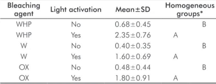
There was a statistically signiicant difference between acti-vation and no actiacti-vation groups (P<0.001). LED light acti-vation of bleaching gels increased the temperature (Table 1).
Table 1. Comparison of mean of temperature variation (ºC) of
the experimental groups of bleaching agent with and without light activation.
Bleaching
agent Light activation Mean±SD
Homogeneous groups*
WHP No 0.68±0.45 B
WHP Yes 2.35±0.76 A
W No 0.40±0.35 B
W Yes 1.60±0.69 A
OX No 0.48±0.44 B
OX Yes 1.80±0.91 A
* Means followed by the same letter do not differ statistically (Tukey’s test, P>0.05).
Discussion
This study showed that LED light activation of bleaching
gels contributed to greater variation of temperature increase in the pulp chamber, but did not achieve the critical value
of 5.5oC. It must be pointed out that in vitro study models, such as the used in this study, are not capable of reproducing
the pulp blood low, which is capable of dissipating the heat applied (12). So, it should be considered that, in vivo, the temperature rise presents lower values than those found
in this study (3). The temperature values measured in this
study cannot be directly applied for temperature variations
in vivo.
In the present study, the premolar surface thickness up to the pulp chamber was standardized at 2 mm. According to
Wetter et al. (4), the size of teeth interferes in temperature increase, since in their study the temperature was lower in
the molars than in the incisors and pre molars. Furthermore, Sulieman et al. (3) found out that the smaller teeth, such as
the lateral incisors, presented a higher temperature increase when compared with the canines, possibly due to the
difference in dentinal thickness between these teeth. In-ofice bleaching products are based on hydrogen peroxide applied as a paste or gel on the tooth surface (13). In the bleaching process with hydrogen peroxide, the bleaching
gel may or may not be indicated in association with a light
and/or heat source. The purpose of the energy supplied is to accelerate hydrogen peroxide degradation and prove an enhanced effect in a shorter clinical time (14).
Although eficient, there have been reports of post-operative
sensitivity by patients, as regards whitening procedures,
mainly those performed in-office (15). Bowles and
Thompson (16) showed that pulp enzymes were inhibited by hydrogen peroxide, especially when the more concentrated agents were used. Tjan and Dunn (13) and Greenwall (17) related that sensitivity could be related to pulp
inlamma-tion induced by heat, which could cause irreversible pulp
damage if a temperature increase higher than 5.5ºC is generated (18).
In the present study, the condition with activation always presented higher temperature variations for all the bleaching gels used. Also, when the type of bleaching gel was assessed, the mean values with activation differed statistically from the
condition without activation. These results were expected as when light is projected onto a bleaching gel, a small fraction is absorbed and its energy is converted into heat. So, the use
of light can have a thermal catalytic and a photolytic effect.
The irst one happens as the release of hydroxyl-radicals from peroxide is accelerated by temperature rise, and the last one is explained through direct excitation of peroxide by light (5).
Although the potential increase in eficacy of bleaching
gels by light activation is still not well documented, neither the application of light nor the application of heat seem to
increase the decomposition rate of peroxide (19). The use of halogen, LED and plasma arc light were equally eficient to promote hydrogen peroxide decomposition when compared with groups without light activation (20-22). The best results
were associated with a longer contact time of the bleaching gel with the tooth, and mainly with a greater number of times
that the gel was changed (20). Moreover, some side effects
of light activation, such as the temperature increase in the
pulp and the increased penetration of peroxide into the pulp chamber should be emphasized (5).
In the present study, the use of light activation signiicantly
increases temperature in the pulp chamber, but no group
achieved the critical temperature of 5.5oC. These indings
are in agreement with those by Zanin et al. (23), who used LEDs for activating the bleaching gel with temperature rise of only 2ºC. Although the present study did not show critical
temperature variations, it is important to be aware of the
possible variations that may occur in the dental pulp. Even small temperature rises can cause an inlammatory process
in the pulp tissue, which may be reversible or irreversible depending on the temperature reached and on the duration
that the tooth was submitted to heating (17). Application of 35% hydrogen peroxide with heat showed pulp alterations,
including obliteration of odontoblasts, hemorrhage,
inlammation and internal dentin resorption (24). Clinical
data revealed sporadic and reversible reactions both with
in-ofice and home techniques (9).
Although there are defense mechanisms against the irritation
of bleaching gels, Leonard et al. (22) emphasized the need
for care in the use of bleaching gels. It should be pointed out
that even with a temperature rise lower than 5.5 ºC, there is
Rev. odonto ciênc. 2010;25(4):382-385 385
Mollica et al.
when associated with this temperature rise in vivo. Thus, not
using external sources to activate hydrogen peroxide would be a safer procedure to keep pulp tissue health.
A variety of light sources that greatly differ in their properties are available to be used for light activation
of bleaching products. In this study, it was used a LED appliance with a power output of 500 mW/cm2 to promote the light activation of bleaching agents. Previous studies
have shown some advantages of this type of light source,
such as less temperature increase in the pulp chamber (6,25). This can be explained by the emission of light by LEDs used with systems for light-activated bleaching, which is
within the blue range and does not extend as far into the infrared spectral range as QHT or plasma arc lamps do. However, thermal pulp damage from LED-systems cannot be absolutely excluded and has to be considered especially when more powerful LEDs are used for a longer period of time (5).
It is still questionable whether the light activation results
in better tooth whitening in comparison with non-activated bleaching procedures. So, the application of light should be done carefully and following the manufacturer’s
recommendations in order to avoid undesired pulp responses.
References
Goldstein RE. In-office bleaching: where we came from, where we 1.
are today. J Am Dent Assoc 1997;128(Suppl):11S-15S.
Miserendino L, Neiburger E, Pick R. Current status of laser in 2.
dentistry. Int Dent J 1987;56:254-7.
Sulieman M, Addy M, Rees JS. Surface and intra pulpal temperature 3.
rises during tooth bleaching: an in vitro study. Br Dent J 2005;199: 37-40.
Wetter NU, Walverde DA, Kato IT, Eduardo CP. Bleaching efficacy 4.
of bleaching gels activated by xenon lamp and 960-nm diode radiation. Photomed Laser Surg 2004;22:489-93.
Buchalla W, Attin T. External bleaching therapy with activation by 5.
heat, light or laser – A systematic review. Dent Mater 2007;23: 586-96.
Michida SMA, Passos SP, Marimoto ARK, Valera MC, Araujo MAM. 6.
Intrapulpal temperature variation during bleaching with various activation mechanisms. J Appl Oral Sci 2009;17:436-9.
Benetti AR, Valera MC, Mancini MN, Miranda CB, Balducci I. In vitro 7.
penetration of bleaching agents into the pulp chamber. Int Endod J 2004; 37:120-4.
Hanks CT, Fat JC, Wataha JC, Corcoran JF. Cytotoxicity and dentin 8.
permeability of carbamide peroxide and hydrogen peroxide vital bleaching materials in vitro. J Dent Res 1993;72:931-8.
Leonard RH, Bentley C, Eagle JC, Garland GE, Knight MC, Phillips 9.
C. Nightguard vital bleaching: a long-term study on efficacy, shade retention, side effects, and patients’ perceptions. J Esthet Restor Dent 2001;13:357-69.
Bränström M. Dentinal and pulpal response III: Application of an air 10.
stream to exposed dentin. Long observation period. Acta Odontol Scand 1960;18:235-52.
Bränström M. Dentinal and pulpal response IV: Some experiments with 11.
heat and pressure illustrating the movement of odontoblasts into the dentinal tubules. Oral Surg Oral Med Oral Pathol 1962;15:203-12. Goodis HE, White JM, Andrews J, Watanabe LG. Measurement of 12.
temperature generated by visible light cure lamps: an in vitro model. Dent Mater 1998;5:230-4.
Tjan AHL, Dunn JR. Temperature rise produced by various light 13.
generations through dentin barriers. J Prosthet Dent 1998;59: 433-8.
Guimarães JGA. Analise térmica da câmara pulpar em dentes 14.
humanos submetidos ao clareamento com laser diodo – estudo in vitro [thesis]. São Paulo (SP): Faculdade de Odontologia da USP; 2004.
Glickman GN, Frys H, Baker FL. Adverse response to vital bleaching. 15.
J Endodont 1992;18:351-4.
Bowles WH, Thompson LR. Vital bleaching: the effects of heat 16.
and hydrogen peroxide on pulpal enzymes. J Endodont 1986;12: 108-12.
Greenwall L. Bleaching techniques in restorative dentistry. London 17.
(UK): Martin Dunitz; 2001.
Zach L, Cohen G. Pulp response to externally applied heat. Oral 18.
Surg 1965;19:515-30.
Hein DK, Ploeger BJ, Hartup JK, Wagstaff RS, Palmer TM, Hansen 19.
LD. In-office vital tooth bleaching – what do lights add? Compend Contin Educ Dent 2003;24:340-52.
Clinical Research Associates. New generation on in-office vital tooth 20.
bleach, Part 2. CRA Newsletter 2003;27:1-2.
Papathanasiou A, Kastali S, Perry RD, Kugel G. Clinical evaluation 21.
of a 35% hydrogen peroxide in-office whitening system. Compend Contin Educ Dent 2002;23:335-48.
Leonard RH, Sharma A, Haywood VB. Use of different concentrations 22.
of carbamide peroxide for bleaching teeth: an in vitro study. Quintessence Int 1998;29:503-7.
Zanin F, Brugnera Junior A, Campos DHS, Zanin VOZ. Clareamento 23.
dental com Laser e LED. RGO 2003;51:143-6.
Nathanson D. Vital tooth bleaching: Sensitivity and pulpal 24.
considerations. J Am Dent Assoc 1997;128(Suppl):41S-44S. Carrasco TG, Carrasco-Guerisoli LD, Fröner IC. In vitro study of the 25.