Psychology & Neuroscience, 2011, 4, 1, 7 - 9 DOI: 10.3922/j.psns.2011.1.002
PSYCHOLOGY
NEUROSCIENCE
Single-pass measurement of the optical quality of the opossum eye
Eduardo Oswaldo-Cruz
1, Cristovam W. Picanço-Diniz
2and Luiz Carlos L. Silveira
21 - Universidade Federal do Rio de Janeiro, Rio de Janeiro, RJ, Brazil
2 - Universidade Federal do Pará, Belém, PA, Brazil
Abstract
This paper reports the results that are part of a series of experiments designed to evaluate aspects of the spatial resolution of the visual system of the opossum, Didelphis marsupialis aurita. This nocturnal marsupial presents a well-developed eye, displaying features that relect specialization for operation at low levels of luminosity. The species was shown to be slightly myopic, a feature that may prove to be valuable because of the increased depth of ield. Opossum visual acuity has been previously evaluated by means of determining the Contrast Sensitivity Function (CSF). The results indicate rather poor visual acuity compared with other nocturnal animals. In this paper, we describe the results obtained for the optical quality of the opossum’s eye using a single-pass method. The results suggest that the opossum’s optical system is capable of forming images that can be resolved when separated by an angular distance on the order of 6 minutes of arc. Keywords: eye optics, eye optical quality, single-pass method, contrast sensitivity, opossum eye.
Received 8 June 2011; received in revised form 30 June 2011; accepted 30 June 2011. Available on line 30 June 2011
Eduardo Oswaldo-Cruz, Universidade Federal do Rio de Janeiro, Instituto de Biofísica Carlos Chagas Filho, Rio de Janeiro, Brazil. Cristovam W. Picanço-Diniz and Luiz Carlos L. Silveira, Universidade Federal do Pará, Instituto de Ciências Biológicas, Belém, Brazil. Luiz Carlos L. Silveira, Universidade Federal do Pará, Núcleo de Medicina Tropical, Belém, Brazil.
Correspondence regarding this article should be directed to:
Dr. Luiz Carlos L. Silveira, Universidade Federal do Pará, Núcleo de Medicina Tropical, Av. Generalíssimo Deodoro, no
92 (Umarizal), 66055-240 Belém, Pará, Brazil. Phone: +5591-32016819. Fax: +5591-32410032. E-mail: [email protected]
Introduction
The visual system provides information about the spatial and temporal distribution of the radiation of a
restricted portion of the electromagnetic spectrum from a wide extent of the space that surrounds an animal. The
analysis of spatial luminosity distribution plays a leading role in the organization of the behavior of the animal in its ecological niche. In higher vertebrates, spatial analysis
is performed on the image of the external world formed
over the photoreceptor layer by the refractive elements of the eye. Information provided by the selective activation of these elements is integrated at the peripheral level by
a complex network of neural elements that constitute the neural portion of the retina. These preliminary data are
then conveyed by means of the nerve ibers that constitute
the optic nerve to higher centers where further analysis occurs, resulting in perception.
Acuity is a measure of the ability to discriminate between separate points in space and for a particular
species is determined by three major factors. The irst
factor concerns the optical quality of the image formed
by the eye. The second factor concerns the ability of the
neural retina to code and analyze the activity that arises from receptor activation. The third factor is sensitivity.
In some species, the ability to resolve ine detail varies
with the intensity of the stimuli.
During several years of study, various morphological and functional aspects of the visual system of the opossum, Didelphis marsupialis aurita, a nocturnal marsupial with
a wide geographical distribution, were analyzed. This species offers unique opportunities for neurobiological
research. For example, its particular mode of reproduction
renders possible the study of the early development of
the organization of the visual pathway. Marsupials have many features in common with primitive eutherian
mammals; therefore, better knowledge of their nervous
system may offer important clues to understanding the
organization of the brains of higher mammals.
Several topics of the organization of the opossum
visual system have been studied, indicating that
marsupials have many features in common with higher mammals. To understand the optical and neural factors that determine the spatial resolution of this species, we performed a series of experiments designed to evaluate
the limitations set by optical and neural elements that
comprise opossum eye optics (Oswaldo Cruz, Hokoç, & Sousa, 1979), eye refractive state (Picanço-Diniz, Silveira, & Oswaldo-Cruz, 1983), retinal ganglion
cell density distribution (Hokoç & Oswaldo Cruz,
Oswaldo-Cruz, Picanço-Diniz and Silveira 8
Picanço-Diniz, & Oswaldo-Cruz, 1982). In this paper,
we describe the results obtained for the optical quality of the opossum’s eye using a single-pass method. The results were previously published in a symposium book with a very limited number of printed copies (Oswaldo
Cruz, Picanço-Diniz, & Silveira, 1982).
Materials, methods and results
We estimated the quality of the image formed at the
retinal level by the refractive system of the opossum’s eye,
evaluating the Line Spread Function of its dioptric system.
We used a “single-pass” method introduced by Hisako
Ikeda and colleagues (Ikeda & Wright, 1973) to study the optical quality of the retinal image of the eye of the cat.
One adult specimen of Didelphis marsupialis aurita
(WIED, 1826) that weighed 1300 g was used. The animal was anesthetized by an intraperitoneal injection of the barbiturate sodium pentobarbital, 25 mg/kg,
and mounted on a head holder. Eye movements were
prevented by continuous intravenous infusion of the neuromuscular blocking agent pancuronium bromide (Pavulon, 10 mg/kg/h; Organon, São Paulo, Brazil). The animal was artiicially ventilated. Cycloplegia, midriasis, and exophthalmia were induced by the
topical application of atropine and phenilephrine. Body
temperature was maintained by a thermostatically controlled electric blanket. Electrocardiographic
monitoring was conducted throughout the experiment. All photographic recordings of the fundus occuli were performed under anesthesia.
After an intravenous injection of a 10% solution of luoresceine, the retinal vessels of both animal
eyes were photographed using a fundus camera (Carl Zeiss Jena, Jena, East Germany). The negatives were developed using a gamma value of approximately 1. The negatives were then placed in an apparatus that consisted of a high-intensity light source, condenser,
and photographic objective. A rotary front surface mirror driven by a triangular waveform generator
displaced the enlarged image across a slit in front of
a photodetector. The proile of the smallest capillary vessels was then measured by means of the output of a
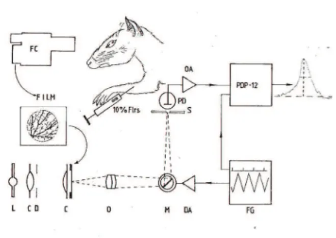
photomultiplier coupled to a PDP-12 digital computer (Digital Equipment Corporation, Maynard, MA, USA) and interactive software that allowed the averaging of the photomultiplier output (Zin & Oswaldo Cruz, 1981). Figure 1 is a semi-diagrammatic representation of the
procedure used to obtain these data. We determined the half-width at mid-height of the luminosity proiles
of the smallest vessels (n > 20) present in the fundus occuli photographic recordings. The results were in
the range of 6-12 minutes of arc and suggest that the optical system is capable of forming images that can be resolved when separated by an angular distance on the order of 6 minutes of arc. Therefore, the optical
quality of the image formed by the refractive system of the opossum’s eye is almost as good as that formed by
the eyes of cats and humans (i.e., 1-2 minutes of arc;
Campbell & Gubisch, 1966; Ikeda & Wright, 1973;
Ikeda, Wright, Young, & Nuza, 1973; Vos, Walraven,
& Van Meeteren, 1976; Westheimer, 1986; Williams, Brainard, McMahon, & Navarro, 1994).
Discussion
This analysis of the quality of the retinal image does not take into consideration the refractive state of the eye (Oswaldo-Cruz et al., 1979; Picanço-Diniz et al., 1983). Knowledge of the optical properties of the eye of the
opossum was obtained by computing the schematic eye for this species using Gaussian simpliication
(Oswaldo-Cruz et al., 1979). The results suggest that this species shows some degree of myopia (-1.97 D), contrasting with the hyperopia usually reported in small eyes when
slit retinoscopy is used. The results obtained by Gaussian calculation are valid for para-axial rays in
aberration-free systems. The small eye of this species (AP diameter approximately 10 mm), displaying high light-gathering properties, presents chromatic and spherical
aberrations as expected (unpublished observations). In an aberration-free system, resolution is a function of both the wavelength of light and aperture of the system, but in practice this limit is not attained because of the
Figure 1. Semi-diagrammatic representation of the experimental procedures used for the determination of the optical quality of the retinal image. A Zeiss Jena fundus camera was used to
photograph the retinal vessels following an intravenous injection of a 10% solution of luoresceine. After developing (gamma
approximately 1) the negatives, they were placed in an apparatus that consisted of a high-intensity light source, condenser, and
photographic objective. A rotary front surface mirror driven by
a triangular waveform generator displaced the enlarged image
across a slit in front of a photodetector. After ampliication, the
output of the sensor was averaged using an interactive program in
a PDP-12 digital computer. The luminance proile of the smallest
vessels was displayed and photographed. Measurements to determine the half-width at mid-height were performed on prints
Optical quality of the opossum eye 9
limitations introduced by the imperfection of refractive
components. Picanço-Diniz et al. (1983) used visually
evoked cortical potentials to study 25 animals and
estimated that this species shows an average myopia of -2.21 D, giving strong experimental support to the
myopic refractive state of the opossum’s eye.
Silveira et al. (1982) used visually evoked cortical potentials to estimate the contrast sensitivity and visual acuity of the opossum in a study of 11 animals. They found that average visual acuity in this sample was
1.25 cycles per degree, indicating that the opossum has
lower visual acuity than primates, felines, and squirrels
but higher visual acuity than albino and pigmented rats. The acuity obtained in the mesopic range (2.4 cd/m2)
was not improved when stimuli of considerably higher luminance (500-1000 cd/m2) were used, suggesting that the retina of this species does not present a higher-resolution system that operates at photopic levels.
Low contrast sensitivity, poor spatial resolution, and the
absence of a dual system that operates at different luminosity
levels offer further evidence that this species is endowed with a visual apparatus specialized to operate at low luminosity.
Acknowledgements
EOC, CWPD, and LCLS have received continuous support from CNPq, CAPES, and FINEP during their careers.
References
Campbell, F.W., & Gubish, R.W. (1966). Optical quality of human
eye. Journal of Physiology (London), 186, 558-578.
Hokoç, J.N., & Oswaldo Cruz, E. (1979). A regional specialization
in the opossum’s retina: quantitative analysis of the ganglion cell
layer. Journal of Comparative Neurology, 183, 385-395.
Ikeda, H., & Wright, M.J. (1973) Optical quality of the cat’s eye and
human eye. Journal of Physiology (London), 232, 34P-35P. Ikeda, H., Wright, M.J., Young, S., & Nuza, J. (1973). Relation
between refractive error and the spread of the image on the cat’s
retina. Vision Research, 13, 867-871.
Oswaldo-Cruz, E., Hokoç, J.N., & Sousa, A.P.B. (1979). A schematic eye for the opossum. Vision Research, 19, 263-278.
Oswaldo-Cruz, E., Picanço-Diniz, C.W., & Silveira, L.C.L. (1982).
Optical and neural factors involved in spatial resolution by the
visual system of the opossum Didelphis marsupialis. Proceedings of the Third Japan-Brazil Symposium on Science and Technology,
(pp. 147-161). Tokyo.
Picanço-Diniz, C.W., Silveira, L.C.L., & Oswaldo-Cruz, E. (1983). Electrophysiological determination of the refractive state of the eye of the opossum. Vision Research, 23, 867-872.
Silveira, L.C.L., Picanço-Diniz, C.W., & Oswaldo-Cruz, E. (1982). Contrast sensitivity function and visual acuity of the opossum. Vision Research, 22, 1371-1377.
Vos, J.J., Walraven, J., & Van Meeteren, A. (1976). Light proiles of
the foveal image of a point source. Vision Research, 16, 215-219.
Westheimer, G. (1986). The eye as an optical instrument. In K.R.
Boff, L. Kaufman, & J.P. Thomas (Eds.), Sensory processes and perception (series title: Handbook of perception and human performance, vol. 1) (pp. 4/1-4/20). New York, N.Y.: Wiley. Williams, D.R., Brainard, D.H., McMahon, M.J., & Navarro, R.
(1994). Double-pass and interferometric measures of the optical
quality of the eye. Journal of the Optical Society of America: A. Optics, image science, and vision, 11, 3123-3135.