O
RIGINALA
RTICLE Revista Brasileira de FisioterapiaEffects of low-level helium-neon laser on
induced wound healing in rats
Os efeitos do laser hélio-neônio de baixa intensidade na cicatrização de lesões
cutâneas induzidas em ratos
Viviane L. Busnardo1, Maria L. P. Biondo-Simões2
Abstract
Objective: To evaluate the effects of low-level helium-neon (HeNe) laser on cutaneous wound healing in rats. Methods:Sixty Wistar rats were divided into control group and experimental group. A sutured longitudinal, dorsal-medial incision was made, with simple separate stitches. The experimental group was irradiated daily in three areas of the wound with HeNe laser (5mW maximum continuous power, 632.8 nm wavelength, 4 J/cm2 energy density and 0.015 cm2 laser beam area) for 36 seconds. The areas were evaluated on
the third, seventh and fourteenth days postoperative. Histological sections were stained with hematoxylin-eosin to determine the type of inflammatory reaction according to the protocol by Vizzotto et al. (2003)* and with Picrosirius to identify types I and III collagen and the collagen maturation index (CMI). Immunohistochemical detection was employed with anti-CD45-LCA to recognize the inflammatory cells. Results: Both groups had the same inflammatory pattern. The experimental group had fewer inflammatory cells at the three evaluation times (p<0.001) with faster reduction in the number of leukocytes. The experimental group had greater total collagen density on the third day (p=0.001), with more type III collagen (p=0.001) and more type I collagen (p=0.001). There was no significant difference in the CMI.Conclusion: Low-level laser irradiation does not change the quality of the inflammatory response, but it does reduce its intensity; it increases collagen deposition in the early stages of the healing process and does not interfere with collagen maturation.
Key words: wound healing; low-level laser therapy; helium-neon laser; collagen.
* Vizzotto Jr AO, Noronha L, Scheffel LHD, Campos ACL. Influência da cisplatina administrada no pré e pós-operatório sobre a cicatrização de anastomoses colônicas em
ratos. J Bras Patol Med Lab. 2003;39(2):143-9.
Resumo
Objetivo: Avaliar os efeitos do laser de baixa potência hélio e neônio (HeNe) na cicatrização de feridas cutâneas de ratos. Métodos:
Sessenta ratos Wistar foram divididos em grupos controle e experimento. Utilizou-se ferida incisional, longitudinal, dorso-mediana, suturada com pontos separados simples. No grupo experimento, as feridas foram irradiadas diariamente com aparelho de laser de HeNe com potência contínua máxima de 5mW, comprimento de onda de 632,8 nm, visível com densidade de energia de 4J/cm2, área
de raio do laser de 0,015cm2, durante 36 segundos, em três pontos da lesão. As feridas foram avaliadas no 3º, no 7º e no 14º dia de
pós-operatório. Cortes histológicos foram corados com hematoxilina-eosina (H&E) e avaliados segundo protocolo de Vizzotto et al. (2003)* para identificar o tipo de reação inflamatória e com Picrosirius para identificar os colágenos I e III e o índice de maturidade da cicatriz (IMaC). Utilizou-se imunoistoquímica com anti-CD45-LCA para o reconhecimento das células inflamatórias. Resultados: Ambos os grupos mostraram o mesmo padrão inflamatório. No grupo experimento, observaram-se menos células inflamatórias nos três tempos estudados (p<0,001), com diminuição mais rápida do número de leucócitos. Verificou-se que as do grupo experimento tinham maior densidade de colágeno total no 3º dia (p=0,001), com mais colágeno III (p=0,001) e mais colágeno I (p=0,001). Não houve diferença significativa no IMaC. Conclusão: A irradiação com laser de baixa intensidade não modifica a qualidade da reação inflamatória, mas diminui a intensidade dela; aumenta a deposição do colágeno no início do processo cicatricial e não interfere na maturação da cicatriz.
Palavras-chave: cicatrização de feridas; laserterapia de baixa intensidade; laser de gases Hélio e Neônio; colágeno.
* Vizzotto Jr AO, Noronha L, Scheffel LHD, Campos ACL. Influência da cisplatina administrada no pré e pós-operatório sobre a cicatrização de anastomoses colônicas em
ratos. J Bras Patol Med Lab. 2003;39(2):143-9.
Received: 21/08/2008 – Revised: 10/02/2009 – Accepted: 30/06/2009
1 Graduate Program in Clinical Surgery, Pontifícia Universidade Católica do Paraná (PUCPR), Curitiba (PR), Brazil 2 Department of Surgery, Universidade Federal do Paraná (UFPR), Curitiba (PR), Brazil
Correspondence to: Viviane L. Busnardo, Universidade Positivo, Rua Prof. Pedro Viriato Parigot de Souza, 5.300, CEP 81280-330, Curitiba (PR), Brazil, e-mail: [email protected]
Introduction
he process of tissue healing is very complex and involves several biological efects, such as vascular and cell changes, epithelial proliferation, ibroblast proliferation, synthesis and deposition of collagen, production of elastin and
proteogly-cans, revascularization and wound contraction1,2. he
incor-poration of laser as a therapeutic tool in the biomedical ield has been investigated since 1960 but, in spite of the numerous studies on the efects of laser therapy, it is diicult to justify physical variables such as: application technique, dosages,
depth, modes and duration of exposure3. It has been observed
that photostimulation inluences macrophage production of
growth factors, which increases cell proliferation4-6. In 1976,
Mester et al.7 reported that low-level helium-neon (HeNe) laser
could aid the healing of mechanical injuries. Since then, it was shown that this laser has several efects on live tissue, efects known as laser biostimulation.
Laser photobiomodulation has been increasingly used with
the purpose of improving the quality of wound healing8. he
therapeutic efects of laser on the diferent biological types are broad and include trophic-regenerative, anti-inlammatory
and analgesic efects9,10. It has also been demonstrated that
tissue regeneration becomes more efective when treated
with low-level laser11-16. here are reports that laser irradiation
stimulates the release of ibroblast growth factor (FGF) and the
replication of these cells17,18. Irradiation with HeNe laser would
accelerate the healing process, with a better weave of collagen
ibers11,19 and greater collagen deposition,16,20,21 combined with
faster reepithelialization and neovascularization13,17,19,22.
In situations of deicient healing, such as ischemia, diabe-tes and pressure ulcers, irradiation with low-level laser could
be an alterative for the recovery21,23,24. he objective of the
pres-ent study was to evaluate the efects of low-level helium-neon (HeNe) laser on the healing process of skin wounds in rats.
Methods
he presented study was conducted in accordance with Federal Law no. 6638 and the recommendations of the Colégio Brasileiro de Experimentação Animal (COBEA), an entity as-sociated with the International Council for Laboratory Animal Science. his project was approved by the Animal Research Eth-ics Committee of Universidade Católica do Paraná (PUC-PR), under the protocol no. 181.06/CEUA-PUC-PR.
Sixty Wistar male rats (Rattus norvegicus albinus, Rodentia
mammalia) from the vivarium of PUC-PR were used in the
study. he mean weight was 397.84±32.74 g and the age ranged
from 100 to 150 days. Over the course of the study, the animals
remained in the vivarium and were divided into groups of ive
per box. he room temperature was kept at 22±2 ºC with a light/
dark cycle of 12 hours and ambient relative humidity and noise volume. All of the boxes were placed on shelves equally distant from the source of light. he animals had free access to water
and speciic food for the species (NUVILAB, NUVITAL®
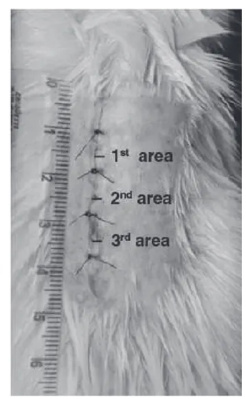
). he animals were anesthetized with an intramuscular injec-tion (0.1 ml/100g of weight) of ketamine (50 mg) and xylazine 2% (20 mg), the dorsal region was shaved and cleaned with pov-idone-iodine and the surgical ield was delimited with a sterile fenestrated drape. Next, a longitudinal dorsal-medial incision was made through the skin and subcutaneous tissue, starting below an imaginary line that corresponds to the insertion of the front paws. he wound measured approximately four cen-timeters and it was closed with four simple separate stitches one centimeter apart using monoilament nylon thread 4.0. he laser was applied to the three areas between the stitches (Figure 1). After the surgery, the animals received a single intra-muscular injection of diclofenac potassium (10 mg/kg) for
an-algesic purposes25. After the recovery from the anesthesia, the
rats were adequately marked and randomly distributed into two distinct groups, with 30 animals each: experimental group (EG) treated with laser and untreated control group (CG).
Each group was subdivided into three subgroups with the same number of animals to perform the time evaluation on postoperative days 3, 7 and 14. hese subgroups were de-nominated: EG day 3, EG day 7 and EG day 14 and CG day 3, CG day 7 and CG day 14. For the treatment, the device laser
He-Ne Plasmax IV, LHN 9709 (KLD Biossistemas®
) was used. he wounds were treated with HeNe laser at energy density of
4 J/cm2 over a 3 cm2 area, resulting in a calculated use of the
laser of 12 seconds per area of the wound. hus, the HeNe laser was applied at the maximal continuous energy level of 5 mW,
with wavelength of 632.8 nm and laser beam area of 0.015 cm2.
Twenty-four hours after the incision procedure, the wounds of the EG animals received treatment with HeNe laser. One subgroup was irradiated for 3 days, another for 7 days and the last for 14 days. For the administration of the laser, the animals were sedated with intraperitoneal propophol (10 mg/Kg). he animals from the CG received the same sedation. After
mac-roscopic analysis on the 3rd, 7th and 14th days, ten animals from
each group were selected by draw and euthanized with an intraperitoneal injection of sodium thiopental (120 mg/Kg) in the left iliac fossa.
In the sampled animals, skin segments (5x4cm) containing the wound in the center were resected. About half a centime-ter in the cranial and caudal extremities of the segment was rejected, and the three remaining centimeters of the wound were used for the study. he segments were ixed in bufered formalin at 10% for 24 hours and later submitted to routine
Figure 1. Appearance of the wound at the end of surgery, showing the laser application areas.
Parameters Intensity
Acute Moderate Mild Absent
Polymorphonuclear cells -3 -2 -1 0
Edema -3 -2 -1 0
Congestion -3 -2 -1 0 Monomorphonuclear cells 3 2 1 0 Granulation Tissue 3 2 1 0
Fibrosis 3 2 1 0
Table 1. Methods for quantification of histological findings in sections stained with hematoxylin-eosin (H&E).
Inflammatory Process Final Score
Acute -9 to -3
Subacute -2.9 to 3
Chronic 3.1 to 9
Table 2. Characteristics of the phases of inflammatory process according to the final score.
histological procedure. hrough the hematoxylin-eosin (H&E) staining, the general morphological evaluation of the wound was obtained, and the inlammatory pattern was recognized. Ten ields were viewed at 400x magniication, according to
the guidelines described by Vizzotto et al.26 (Table 1). For the
cell count, the following scale was adopted: no cell=0; up to 50 cells=1; 50 to 100 cells=2 and more than 100 cells=3, positive for monomorphonuclear cells and negative for
polymorphonu-clear cells26. After the attribution of the indices, their total was
calculated so that each group of animals had a inal score for
classiication into three phases of the inlammatory process26
(Table 2).
he histological sections stained with Picrosirius (
Picrosir-ius-red F3BA) under microscopy and polarized light allowed the identiication of the collagen density in the wounds and the fractions of type I and III collagen. With this technique, the thicker and highly birefringent type I collagen ibers ap-pear orange and red in color, and the iner, dispersed and less
birefringent type III collagen ibers appear green in color25. he
images were captured on a Sony® CCD 101/Trinitron® system,
digitized by an Oculus TCX® capture board and analyzed by the
Image Plus4.5 for Windows application. For each slide, three ields were read at 200X magniication over the wound area, and the mean was calculated.
he collagen maturation index (CMI27) was deined as the
ratio of the percentage of type I collagen to the percentage of type III collagen. his index varies from zero (percentage of type I collagen=0 and percentage of type III collagen=100) to ininite (percentage of type I collagen=100 and percentage of type III collagen=0). Values higher than one indicate that the percentage of type I collagen is greater than the percentage of
type III collagen and show the state of collagen maturation28.
he tissue segments used for anti-CD45-LCA immunos-taining (LCA – Leukocyte Common Antigen, Clone 136-4B5 IV
WS - IgG1, FK-Biotec®) at 1:100 dilution were submitted to
anti-gen retrieval in bain-marie in 10 mmol/L citrate bufer (pH 6.0) for further immunohistochemical processing. To guarantee the veracity of the staining, controls were made. For external posi-tive control, a lymph node fragment was used. he dermis and the epithelium were used as internal negative control. For the analysis, a count was taken of the positive cells stained brown for LCA per ield of magniication, in a total of ten ields.
he descriptive results obtained from the study were ex-pressed as means and standard deviations. Two-way ANOVA was used to determine the efect of the group and of the evalu-ation day on the study variables. he normality condition of the variables was assessed by the Shapiro-Wilk test, and the homogeneity of the variances, by Levene’s test. In the cases in which there was a signiicant interaction between the factors,
Student’s t test was used for intragroup analysis and ANOVA
Median Score
-5 -3 -2 -1 0 1 2 3 4 5
3 days 7 days 14 days
Control Experimental -4
p=0.579
p=0.912
p=0.661
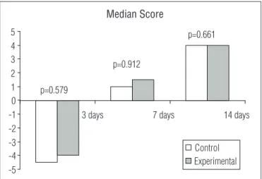
Figure 2. Graphic representation of the inflammatory process in the control and experimental groups at the three evaluation times, according to the score by Vizzotto et al.26
Figure 3. Histological appearance of the wounds on the fourteenth day postoperative.
Experimental Group (EG); B) Control Group (CG). Presence of inflammatory polymor-phonuclear cells (“) and fibroplasia (∆) (H&E stain – 100x magnification).
was used for within-day analysis. For the multiple compari-sons, the Least Signiicant Diference (LSD) test was used. For the analysis of the CMI tables, Fischer’s test was used. he level of signiicance adopted was 0.05.
Results
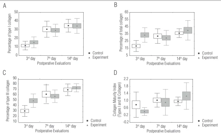
In the histological sections of the wounds on day 3 for both groups, the inlammation showed characteristics of an acute process evidenced by negative scores. For the between-group comparisons, on day 7 the inlammation had changed and had characteristics of a subacute process, and on day 14, it had char-acteristics of a chronic process evidenced by positive scores (Figures 2 and 3). With regard to the analysis of type I percent-age of collpercent-agen, there was no signiicant diference between the groups in the three moments analyzed (p=0.527; Figure 4A). here was higher type III collagen density in the wounds of EG day 3 (p=0.001). However, in the between-group comparisons of day 7 and 14, the diferences were not signiicant (Figure 4B). Furthermore, the between-group comparisons of the to-tal collagen showed higher density in the wounds of EG day 3
(p=0.001), without signiicant diferences in the comparisons of day 7 and 14 (Figure 4C).
Regarding the CMI, greater maturation was found in the wounds of CG day 3 when compared to EG day 3 (p=0.026; Fig-ure 4D). However, in the dichotomized analysis separating the number of wounds with indices up to 1 and those with indices higher than 1, there was no diference in frequency between the groups. A diference between groups was found on day 3 (p=0.474), day 7 (p=1) and day 14 (p=1) (Figure 5). he immuno-histochemical process showed a lower count of inlammatory cells for the EG group when compared to the CG group on the three assessment moments (p<0.001), with an earlier decrease in their number in the EG group (Figures 6 and 7).
Discussion
Wound healing can be improved when the main events that enable it are stimulated, e.g. nutrition, cell proliferation
and control of inlammation and infection11,12,29. In several
stu-dies with animal models, low-level laser has been identiied as an adjuvant for healing because it improves revasculariza-tion, generation of energy in form of adenosine triphosphate (ATP) for the repair cells, ibroblast proliferation and
inhi-bition of the chemical mediators of inlammation10,24. Given
the main factors involved in the healing process, the present study aimed to identify the inluence of laser on the general aspects of this process. he factors for analysis were chosen to observe the resolution of the inlammatory process and the collagen synthesis. he results conirm that the low-level
laser with 4 J/cm2 energy density promotes type III collagen
deposition on postoperative day 3.
Carvalho et al.11 conducted a morphometric analysis of the
percentage of collagen ibers by color density of the wounds of
48 rats. he authors used HeNe laser (4 J/cm2) for 36 seconds in
the EG and assessed the results on postoperative days 3, 7 and 14 and found a signiicant increase in the percentage of collagen from day 3 to day 7. When day 14 was compared to day 7, a decel-eration in the percentage of collagen was observed. he authors attributed this deceleration to the state of maturation of these wounds because of reports of decrease in cell proliferation and ibroblast number and size in the remodeling/maturation.
Pugliese et al.12 studied the efects of low-level laser
(GaA-lAs laser at diferent energy densities) on the standardized skin wounds of 62 Wistar rats. In the morphometric analysis, the authors observed a greater expression of the collagen ibers, however, without statistical signiicance. he best results were
found in the groups irradiated with 4 J/cm2. In the evaluation
of the inlammatory response, the irradiated wounds had an earlier reduction in edema, congestion and inlammatory cells.
Control Experiment Postperative Evaluations 0 10 20 30 40 50
3rd day 7th day 14th day 5
15 25 35 45 55 65 10 20 30 40 50 60 70 80 90 -0.2 0.2 0.6 1.0 1.4 1.8 2.2 Control Experiment Postperative Evaluations
3rd day 7th day 14th day
Control Experiment Postperative Evaluations
3rd day 7th day 14th day
Control Experiment Postperative Evaluations
3rd day 7th day 14th day
A
B
D
C
P er ce nt ag e of ty pe I co lla ge n P er ce nt ag e of to ta l c ol la ge n P er ce nt ag e of ty pe II I c ol la ge n C ol la ge n M at ur ity In de x (T yp es I an d III C ol la ge n)Figure 4. Mean percentages of the areas of the histological sections examined and represented by collagen in the two groups at each evaluation and the collagen maturation index.
Figure 6. Photomicrographs of histological sections showing the leukocytes marked by anti-CD45 (*) on the third-day evaluation (400x magnification).
1 Postoperative Evaluation
2 8 1 2 2 3
3rd day 7th day 14th day
Control INFLAMMATORY CELLS
Means
Experiment
Figure 7. Mean number of inflammatory cells (per site) marked with anti-CD45 in ten sites.
Figure 5. Photomicrograph of the histological sections of the wound areas of the EG and CG, stained with Sirius Red under a polarized light, (200x magnification) on the third-day evaluation.
Regarding the improvement in healing, the authors attributed to the laser a signiicant increase in the collagen deposition that can happen by induction of the cell proliferation or even by an increase in protein synthesis and release, with the added possibility of both processes occurring simultaneously.
Araújo et al.19 found more activated ibroblasts, more
col-lagen and more incorporation of 3H-proline in the wounds of rats treated with low-level laser and evaluated on days 8, 15
and 22 post-wounding. Carvalho et al.21 reported that this
ef-fect can be observed in the wounds of diabetic rats, showing that low-level laser therapy could be useful in adverse
situa-tions. Medrado et al.20, however, observed a slight increase in
collagen density.
Collagen maturation was evaluated using the CMI. he Picrosirius-stained sections allowed the assessment of the type of collagen and to quantify each type in the samples. Several authors have compared diferent staining methods for collagen analysis and concluded that the Picrosirius pigment is the most selective and the easiest to use and interpret. It is also speciic for the study of tissue collagen because the amount of adhered pigment is proportional to the amount of protein, allowing the
quantiication of the protein10,27,28.
Despite a higher density of type III collagen on day 3, the results of the CMI in the dichotomized analysis showed that the collagen maturation was similar throughout the duration of the study. In fact, the type I collagen density tended to be gre-ater in the EG, but it was not statistically signiicant. A larger sample could clarify this question by conirming or rejecting this tendency. he same can be stated about the wounds of the EG, which had a tendency for higher but non-signiicant maturation indices on assessment days 7 and 14. On day 14, a deceleration in the healing process was observed, a inding
reported in a previous study5 and that may be explained by the
decrease in the proliferation of cellular elements in this phase. he immunohistochemical method showed that the number of inlammatory cells was lower in the wounds of the EG, a fact that determines the signiicant reduction in the inlammatory process.
hese indings were veriied by other authors12,16,20who also
re-ported a reduction in inlammatory edema in the groups irradiated with low-level laser. According to their indings, the positive efects of laser on tissue healing are due to the increase in cell proliferation and to the signiicant reduction in inlammatory cells.
Despite its use in medical practice since the 1970s, laser is still an object of study. he present research found an increase in type III collagen density on postoperative day 3, an increase in the percentage of total collagen, a reduction in inlamma-tory cells and an earlier resolution of the inlammainlamma-tory phase in the irradiated wounds. hese facts justify the use of the laser in wound healing. It is worth noting that the pattern of inlam-matory response remained the same in both groups (acute on day 3, subacute on day 7 and chronic on day 14), but with lower intensity in the EG. If we consider that the maintenance of the inlammatory process delays ibroplasia, it would be expected that the collagen density would be higher in the EG, a fact that was not observed during the healing process. here was only an initial higher density in the wounds of the EG.
Other beneicial efects of low-level laser on the healing
pro-cess have been reported. Reis et al.16 used GaAlAs laser (4 J/cm2,
9mW, wavelength = 670nm) for 3 to 5 days and found increased collagen density and a better extracellular matrix distribution (p<0.05). he authors also reported a greater amount and
ac-tivity of ibroblasts. Houreld and Abrahamse24 showed that the
use of low-level HeNe laser stimulated interleukin-6 expression of the interleukin-6, cell proliferation and migration in diabetic
subjects, thus improving the healing process24. Silveira, Streck
and Pinho15 demonstrated that low-level laser can activate
mitochondrial enzymes, such as cytochrome c oxidase, and improve wound healing.
Although many clinical studies have been favorable to the use of conventional laser, the mechanism by which it ac-celerates the process of healing has not been fully elucidated. here are still some hypotheses to be tested, e.g. whether laser enhances the transcription of ribonucleic acid (RNA) while reducing the inlammatory phase and anticipating ibroplasia, and whether the laser-induced increase in oxygen and ATP is determined by an early angiogenesis.
To ensure the efects of low-level laser therapy on the hea-ling process, there is still a need for consensus on the standards for the physical variables: application times and techniques, energy densities, output powers and wavelengths. he com-parison of the results of several authors has been hindered by their use of diferent methodologies. he use of HeNe laser (632.8nm), applied with diferent densities can lead to dife-rent cellular responses, and this may preclude comparisons.
Hawkins and Abrahamse30 applied doses of 0.5, 2.5, 5, 10 and
16 J/cm2 to human skin ibroblasts on two consecutive days
and found that 5 J/cm2 stimulated mitochondrial activity, cell
proliferation and ibroblast migration. However, higher doses decreased cell viability and proliferation and damaged the cell
membrane and DNA30.
Conclusion
Irradiation with low-level HeNe laser in the conditions of this experiment did not change the quality of the inlamma-tory response, but decreased its intensity. Collagen deposition increased at the beginning of the healing process and the ma-turation of the wound was not afected.
1. Mandelbaum SH, Di Santis EP, Mandelbaum MHS. Cicatrização: conceitos atuais e recursos auxiliares – parte II. An Bras Dermatol. 2003;78(5):525-40.
2. Thomas DW, O’Neill ID, Harding KG, Sheperd JP. Cutaneous wound healing: a current perspective. J Oral Maxilofac Surg. 1995;53(4):442-7.
3. England S. Introduction to mid laser therapy. Physiotherapy. 1988;74(3): 100-3.
4. Young S, Bolton P, Dyson M, Harvey W, Diamantopoulos C. Macrophage responsiveness to light therapy. Lasers Surg Med. 1989;9(5):497-505.
5. Yu HS, Chang KL, Yu CL, Chen JW, Chen GS. Low-energy helium neon laser irradiation stimulates interleukin-1 alpha and interleukin-8 release from cultured human keratinocytes. J Invest Dermatol. 1996;107(4):593-6.
6. Skinner SM, Gage JP, Wilce PA, Shaw RM. A preliminary study of the effects of laser radiation on collagen metabolism in cell culture. Aust Dent J. 1996;41(3):188-92.
7. Mester E, Nagylucskay S, Döklen A, Tisza S. Laser stimulation of wound healing. Acta Chir Acad Sci Hung. 1976;17(1):49-55.
8. David R, Nissan M, Cohen I, Soudry M. Effect of low-power He-Ne laser on fracture healing in rats. Lasers Surg Med. 1996;19(4):458-64.
9. Ortiz MCS, Carrinho PM, Santos AAS, Gonçalves RC, Parizzoto NA. Laser de baixa intensidade: princípios e generalidades – parte 2. Fisioter Bras. 2001;2(6):337-52.
10. Woodruff LD, Bounkeo JM, Brannon WM, Dawes Jr KS, Barham CD, Waddel DL, et al. The efficacy of laser therapy in wound repair: a meta-analysis of the literature. Photomed Laser Surg. 2004;22(3):241-7.
11. Carvalho PTC, Mazzer N, Siqueira JF, Ferreira VJ, Silva IS. Análise de fibras colágenas através da morfometria computadorizada em feridas cutâneas de ratos submetidos à irradiação do laser HeNe. Fisoter Bras. 2003;4(4):253-8.
12. Pugliese LS, Medrado AP, Reis SR, Andrade Z de A. The influence of low-level laser therapy on biomodulation of collagen and elastic fibers. Pesqui Odontol Bras. 2003;17(4):307-13.
13. Hawkins D, Houreld N, Abrahamse H. Low level laser therapy (LLLT) as an effective therapeutic modality for delayed wound healing. Ann NY Acad Sci. 2005;1056:486-93.
14. Rocha JCT. Terapia laser, cicatrização tecidual e angiogênese. Rev Bras Promoção Saúde. 2004;17(1):44-8.
15. Silveira PC, Streck EL, Pinho RA. Evaluation of mitochondrial respiratory chain activity in wound healing by low-level laser therapy. J Photochem Photobiol B. 2007;86(3):279-82.
16. Reis SR, Medrado AP, Marchionni AM, Figueira C, Fracassi LD, Knop LA. Effect of 670-nm laser therapy and dexamethasone on tissue repair: a histological and ultrastructural study. Photomed Laser Surg. 2008;26(4):307-13.
17. Rocha Jr AM, Andrade LCF, Oliveira RG, Aarestrup FM, Farias RE. Modulação da proliferação fibroblástica e da resposta inflamatória pela terapia a laser de baixa intensidade no processo de reparo tecidual. An Bras Dermatol. 2006;81(2):150-6.
18. Ribeiro MS, Da Silva D de F, De Araujo CE, De Oliveira SF, Pelegrini CM, Zorn TM, et al. Effects of low-intensity polarized visible laser radiation on skin burns: a light microscopy study. J Clin Laser Med Surg. 2004;22(1):59-66.
19. de Araujo CE, Ribeiro MS, Favaro R, Zezell DM, Zorn TM. Ultrastructural and autoradiographical analysis show a faster skin repair in He-Ne laser-treated wounds. J Photochem Photobiol B. 2007;86(2):87-96.
20. Medrado AR, Pugliese LS, Reis SR, Andrade ZA. Influence of low level laser therapy on wound healing and its biological action upon myofibroblasts. Lasers Surg Med. 2003;32(3):239-44.
21. Carvalho PT, Mazzer N, dos Reis FA, Belchior AC, Silva IS. Analysis of the influence of low-power HeNe laser on the healing of skin wounds in diabetic and non-diabetic rats. Acta Cir Bras. 2006;21(3):177-83.
22. Corazza AV, Jorge J, Kurachi C, Bagnato VS. Photobiomodulation on the angiogenesis of skin wounds in rats using different light sources. Photomed Laser Surg. 2007;25(2):102-6.
23. Prado RP, Liebano RE, Hochman B, Pinfildi CE, Ferreira LM. Experimental model for low level laser therapy on ischemic random skin flap in rats. Acta Cir Bras. 2006;21(4):258-62.
24. Houreld N, Abrahamse H. Irradiation with a 632.8 nm helium-neon laser with 5 J/cm2 stimulates proliferation and expression of interleukin-6 in diabetic wounded fibroblast cells. Diabetes Technol Ther. 2007;9(5):451-9.
25. Junqueira LC, Bignolas G, Brentani RR. Picrosirius staining plus polarization microscopy, a specific method for collagen detection in tissue sections. Histochem J. 1979;11(4):447-55.
26. Vizzotto Jr AO, Noronha L, Scheffel LHD, Campos ACL. Influência da cisplatina administrada no pré e pós-operatório sobre a cicatrização de anastomoses colônicas em ratos. J Bras Patol Med Lab. 2003;39(2): 143-9.
27. Coelho ICML. Infuência da desnutrição intra-uterina na cicatrização da parede abdominal de ratos lactentes avaliada mediante estudo tensiométrico e da morfometria do colágeno [tese]. Curitiba (PR): Universidade Federal do Paraná; 2003.
28. Alves MRA. Influência da nicotina, durante a gestação e lactação, na cicatrização da parede abdominal de ratos lactentes: estudo tensiométrico, morfométrico e imunohistoquímico [tese]. Curitiba (PR): Universidade Federal do Paraná; 2006.
29. Reddy GK. Photobiological basis and clinical role of low-intensity lasers in biology and medicine. J Clin Laser Med Surg. 2004;22(2):141-50.
30. Hawkins DH, Abrahamse H. The role of laser fluence in cell viability, proliferation, and membrane integrity of wounded human skin fibroblasts following helium-neon laser irradiation. Lasers Surg Med. 2006;38(1):74-83.