Quim. Nova, Vol. 37, No. 4, 584-588, 2014
Artigo
http://dx.doi.org/10.5935/0100-4042.20140098
*e-mail: [email protected]
#Centro para el Desarrollo de la Nanociencia y la Nanotecnología, CEDENNA
SYNTHESIS, CHARACTERIZATION AND COMPUTATIONAL STUDIES OF (E)-2-{[(2-AMINOPYRIDIN-3-YL) IMINO]-METHYL}-4,6-DI-TERT-BUTYLPHENOL
Alexander Carreñoa, Andrés Vegaa,#, Ximena Zarateb, Eduardo Schottb, Manuel Gacitúac, Ninnette Valenzuelac, Marcelo Preited, Juan M. Manríquezc and Ivonne Chávezc,*
aDepartamento de Ciencias Químicas, Facultad de Ciencias Exactas, Universidad Andrés Bello, República 275, Santiago, Chile bLaboratorio de Bionanotecnología, Departamento de Ciencias Químico-Biológicas, Universidad Bernardo O´Higgins, General Gana 1780, Santiago, Chile
cDepartamento de Química Inorgánica, Facultad de Química, Pontiicia Universidad Católica de Chile, Avenida Vicuña Mackenna 4860, Santiago, Chile
dDepartamento de Química Orgánica, Facultad de Química, Pontiicia Universidad Católica de Chile, Avenida Vicuña Mackenna 4860, Santiago, Chile
Recebido em 04/06/2013; aceito em 09/12/2013; publicado na web em 10/04/2014
(E)-2-{[(2-Aminopyridin-3-yl)imino]-methyl}-4,6-di-tert-butyl-phenol (3), a ligand containing an intramolecular hydrogen bond, was prepared according to a previous literature report, with modiications, and was characterized by UV-vis, FTIR, 1H-NMR, 13C-NMR,
HHCOSY, TOCSY and cyclic voltammetry. Computational analyses at the level of DFT and TD-DFT were performed to study its electronic and molecular structures. The results of these analyses elucidated the behaviors of the UV-vis and electrochemical data. Analysis of the transitions in the computed spectrum showed that the most important band is primarily composed of a HOMO→LUMO transition, designated as an intraligand (IL) charge transfer.
Keywords: intramolecular hydrogen bond; Schiff base; DFT.
INTRODUCTION
Schiff base ligands derived from salicylaldehyde containing OH…N type hydrogen bonding have been studied due to the versatility of their electronic and steric properties.1-3 Different metal complexes with Schiff base ligands have been synthesized and studied because of their ability to form stable complexes.4 These complexes have also been used in analytical chemistry for the spectrophotometric deter-mination of heavy metals.5,6 Furthermore, they are used as catalysts in chemical and photochemical reactions as well as many other applications. Schiff bases with an -OH group in the ortho position relative to an imino group -C=N- have generated much interest due to the existence of an intramolecular hydrogen bond between the OH and the nitrogen atom.7,8 Bio-inspired compounds that contain an intra-molecular hydrogen bond have been utilized as dyes in photoelectro-chemical cells.9-11 A detailed study of such compounds demonstrated that their electrochemical activity is similar to the activity observed for redox processes in biological systems.12 This research emphasizes the importance of an intramolecular hydrogen bond in such systems, which has been the subject of much investigation.
With this in mind, we aimed to investigate the possible use of an aminopyridine, (E )-2-{[(2-aminopyridin-3-yl)imino]-methyl}-4,6--di-tert-butyl-phenol (3, Scheme 1),containing a Schiff base and possessing an intramolecular hydrogen bond as a substituent group. A complete study of its electronic properties was performed to un-derstand the role of the hydrogen bond.
The synthesis and characterization of compound 3 is reported. Compound 3 was obtained from a modified template pathway (Scheme 1), based on a synthesis described by Kleij et al.13,14 A study of its electronic properties and a computational study were performed, to understand its molecular properties.
EXPERIMENTAL
General procedures
NMR spectra were recorded on a Bruker AVANCE 400 MHz NMR spectrometer at 25ºC. Samples were dissolved in deuterated chloroform and acetone, using tetramethylsilane as an internal refe-rence. The NMR spectra were processed with TOPSIN 2.1 program. IR spectra of all compounds were recorded on a Perkin-Elmer 1310 Bruker FT-IR spectrophotometer in KBr discs and were recorded in the range of 250-4000 cm-1. UV-vis spectra were performed using a vis-NIR scanning spectrophotometer Shimadzu Model UV-3101 PC.
For the electrochemical experiments, the working solution con-tained 9.81 x 10⁻³ mol L-1 of compound 3 with 0.1 mol L-1 tetrabu-tylammoniumhexaluorophosphate (used as a supporting electrolyte, 99% Aldrich) and TBAPF6 in CH3CN (solvent, p.a. Aldrich). Before each experiment, the solution was purged with high purity argon, and an argon atmosphere was maintained over the solution during the entire experiment. A polycrystalline non-annealed platinum disc (diameter 2 mm) was used as the working electrode. Platinum gauze, separated from the cell main compartment by a piece of ine sintered glass, was used as the counter electrode. All potentials in this paper
Scheme 1. General reaction scheme of
Synthesis, characterization and computational studies of (E)-2-{[(2-aminopyridin-3-yl)imino]-methyl}-4,6-di-tert-butylphenol 585
Vol. 37, No. 4
utilize an Ag/AgCl electrode in tetramethylammonium chloride as a reference to match the potential of a saturated calomel electrode (SCE) at room temperature. All electrochemical experiments were performed at room temperature on a CHI900B bipotentiostat inter-faced with a PC running CHI 9.12 software for experimental control and data acquisition.
Theoretical computations were performed using density func-tional theory (DFT) with the B3LYP hybrid exchange/correlation (XC) functional, which includes the non-local exchange term, with three parameters of Becke and the correlation term of Lee-Yang-Parr.15,16 Gaussian basis set 6-311+G (2d,p) was employed.17 Vibrational frequencies were calculated to verify that the stationary state corresponds to a minimum value. Geometric optimizations for the compound were performed. Polarizable continuum model (PCM) was used to simulate the solvent effect using dichlorome-thane as solvent.18–20 In this model, the solvent is characterized for its dielectric constant as well as other parameters. All calculations were made using Gaussian 03.21
Syntheses
(E)-2-{[(2-Aminopyridin-3-yl)imino]-methyl}-4,6-di-tert-butyl-phenol (3) was prepared by condensation of 1,2-diaminopyridine (1) with 3,5-di-tert-butyl-2-ol-benzaldehyde (2) in 20 mL of methanol (see Scheme 1). The reaction was stirred for 24 h at room temperature and then iltered, and the precipitate was washed with methanol and diethyl ether. The yellow product was puriied by crystallization, and the product was dried under vacuum. Yellow crystals suitable for X-Ray diffraction were obtained by slow evaporation of the solvent. Yield 70%. UV/VIS: (dichloromethane, c=4.21x10⁻⁵ mol L-1) λ (ε) = 238 (33939.73 mol-1 dm3 cm-1), 279 (23569.23 mol-1 dm3 cm-1), 374 (22419.68 mol-1 dm3 cm-1). (Ethanol, c=4.21x10⁻⁵ mol L-1) λ (ε)= 204 (53751.39 mol-1 dm3 cm-1), 237 (20556.46 mol-1 dm3 cm-1), 373 (12925.49 mol-1 dm3 cm-1 ). (Acetonitrile, c=4.21x10⁻⁵ mol L-1) λ (ε)= 196 (75455.99 mol-1 dm3 cm-1), 236 (21702.93 mol-1 dm3 cm-1), 370 (12978.52 mol-1 dm3 cm-1).
FTIR (KBr, cm-1): υ
OH 3469, υNH2 3264 and 3136, υC=N 1609, υC-C 1590. 1H-NMR (400 MHz, CDCl
3, ppm): δ = 1.28 [s; 9H; -C(CH3)3], 1.47 [s; 9H; -C(CH3)3], 4.80 [bs; 2H; -NH2], 6.72 [dd: J=7.6; 5.0 Hz; 1H; H2], 7.24 [m; 1H; H3], 7.26 [d: J=2.0 Hz; 1H; H5], 7.48 [d: J=2.0 Hz; 1H; H6], 8.01 [dd: J=5.0; 1.6 Hz; 1H; H1], 8.63 [s; 1H; H4], 13.03 [s; 1H; OH]. 1H-NMR (400 MHz, Acetone-d
6, ppm): δ = 1.37 [s; 9H; -C(CH3)3], 1.48 [s; 9H; -C(CH3)3], 5.48 [bs; 2H; -NH2], 6.70 [dd: J=7.6; 5.0 Hz; 1H; H2], 7.48 [dd: J=7.6; 1.5 Hz; 1H; H3], 7.49 [d: J=2.5 Hz; 1H; H5], 7.54 [d: J=2.0 Hz; 1H; H6], 7.95 [dd: J=4.9; 1.5 Hz; 1H; H1], 8.88 [s; 1H; H4], 13.30 [s; OH]. 13C-NMR (400 MHz, CDCl
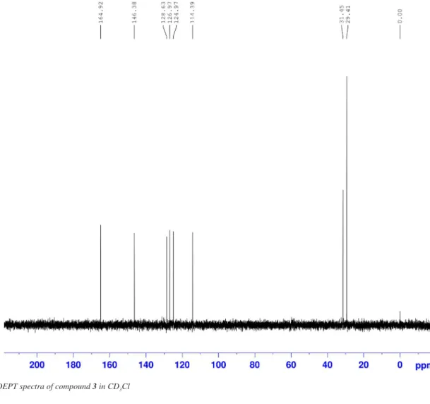
3, ppm): δ = 29.42 (-C(CH3)3), 31.44 (-C(CH3)3), 34.22 (-C(CH3)3), 35.13 (-C(CH3)3), 114.39 (C10), 118.40 (C11), 124.95 (C12), 126.96 (C4), 128.62 (C5), 130.71 (C9), 137.15 (C7), 141.08 (C1), 146.39 (C3), 153.32 (C2), 157.98 (C8), 164.91 (C6). Anal. Calcd for C20H27N3O: C, 73.84; H, 8.32; N, 12.92. Found C, 73.74; H, 11.93; N, 13.16.
RESULTS AND DISCUSSION
Compound 3 was a yellow solid obtained in 70% yield. The compound was characterized by FT-IR, and the spectrum exhibits several bands in the range from 2500-4000 cm-1 (Figure 1S in the Supplementary Material). The main absorption frequencies show strong symmetric and asymmetric bands due to an O-H band at 3469 cm-1 and two -NH
2 bands at 3264 and 3136 cm-1. The absorp-tions near 1609 and 1590 cm-1 were assigned to the stretching of the υC=N and υC-C bonds, respectively, and have been previously reported for a similar compound.22,23
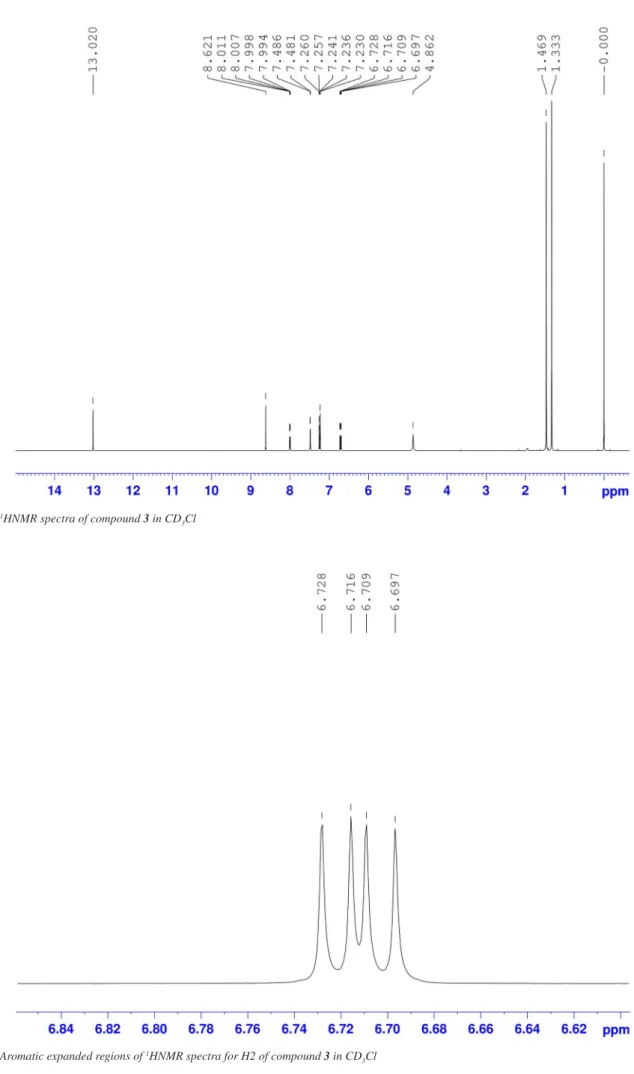
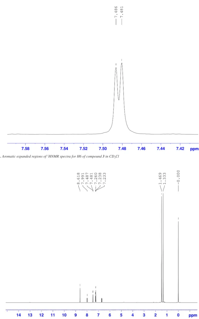
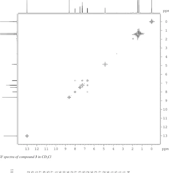
1D and 2D NMR spectra were obtained in CD3Cl or acetone-d6 solutions, and the numbering of protons and carbons are provided in the Supplementary Material (Figure 2S and 3S). The 1HNMR spectrum (CDCl3, Figures 4S-8S in the S.M.) exhibits a strong peak at approximately 13.03 ppm for –OH, and the amino proton at 4.80 ppm has been previously reported for similar compounds.24,25 After D2O exchange, (Figure 9S in the S.M.) these two signals disappear from the spectrum. Signals assigned to the tert-butyl group appear at 1.47 and 1.28 ppm, and the aromatic protons of both rings appear at 7.50-6.70 ppm and 8.65-8.00 ppm (Figure 10S in the S.M.).
In the pyridine ring, H2 appears at 6.72 ppm, H3 at 7.24 ppm and H1 at 8.01 ppm, while H5 appears at 7.26 ppm and H6 at 7.48 ppm, as shown in Figure 1.
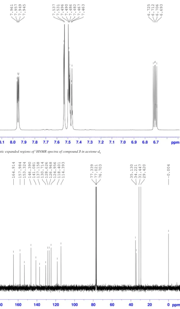
For the assignment of H3 and H5, which appear between 7.10-7.60 ppm and overlap with the CD3Cl signal, a spectrum in acetone-d6 was obtained (Figure 11S in the S.M.). The protons of the pyridine ring appear at 7.95 ppm (H1), 6.70 ppm (H2) and 7.48 ppm (H3), and the signals in the phenol ring appear at 7.49 ppm (H5) and 7.54 ppm (H6) (Figure 12S in the S.M.). To complete the assignment, a 1D TOCSY spectrum allowed us to identify the resonances for all protons (Figure 2). When irradiating at H1 (Figure 2A), an enhancement was observed for H2 and H3. In the same manner, when H2 (Figure 2D) was irradiated, a spectrum showing enhancement for H1 and H2 was obtained. When H5 was irradiated instead (Figure 2B), enhancements in H6, and both -C(CH3)3 signals were observed. When H6 was irradiated (Figure 2C), it was impossible to avoid irradiating H3 as well, so enhance-ments for the H4, H1, H5, H6, H3, H2 and -C(CH3)3 signals were observed. These experiments allowed us to assign all signals to their respective protons.
13CNMR broad-band decoupled and DEPT spectra in CDCl 3 solution allowed for total peak assignment (Figure 13S and 14S in the S.M.). A signal commonly assigned to the Schiff base carbon (C6) was observed at 169.91 ppm, consistent with previous literature reports.4-6 Other signals corresponding to the phenol ring appeared at 157.98 (C8) ppm, and those for the pyridine ring at 153.32 (C2) ppm and 141.08 (C1) ppm.
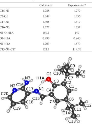
A crystal structure of compound 3 has been previously reported by Carreño et al., showing an intramolecular hydrogen bond with an OH…N distance of 2.621 Å. The dihedral angle between the aromatic rings is 33o, which is almost planar.26 According to the optimized calculated geometry shown in Figure 3, the molecule adopts a co-planar conformation with an angle between the rings of 39o. Table 1
Figure 1.Expanded aromatic region of the 1HNMR of 3 in CD
Carreño et al.
586 Quim. Nova
presents the important parameters of the experimental and theoretical data, which agree well.
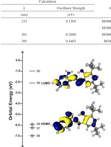
The UV-vis spectra in dichloromethane, ethanol and acetonitrile showed absorption bands centered at 375 nm and 190 nm (see Table 2), which were assigned to n→p* (C=N) and p→p* transitions.27,28 Time-dependent density functional theory (TD-DFT) calculations were conducted further elucidate the UV-vis observed transitions. Figure 4 shows an overlay of the calculated and experimental UV-vis spec-tra. The composition of the calculated transitions is shown in Table 3. The TD-DFT calculation shows that the absorption band located experimentally at 238 nm (33939.73 mol-1 dm3 cm-1) is theoreti-cally found at 233 nm. This calculated transition is composed of a
HOMO→LUMO+3 and a HOMO→LUMO+4 transition. The bands located experimentally at 281 nm (23569.23 mol-1 dm3 cm-1) and at 374 nm (22419.68 mol-1 dm3 cm-1) are theoretically calculated at 281 nm and 385nm, respectively. Those calculated excitations are associ-ated with a HOMO→LUMO+1 and a HOMO→LUMO transition, respectively. Experimentally, there is no signal shift in the UV-Vis spectra when the solvent polarity changes.
To gain additional insight into the nature of the O-H...N hydrogen bond, topological analysis was performed with the DGrid package in the framework of the atoms in molecules (AIM) theory, which were performed for B3LYP/BASE densities.29,30 In this system, a bond criti-cal point (BCP) was found between the H and N atoms and shows a charge density value of 0.0526 a.u., which is the order of magnitude of the charge density in the hydrogen bonds.31 In addition, as natural bond orbital (NBO) theory32 studies the role of orbital interactions by considering the interactions between occupied donor and empty acceptor orbitals, the second-order perturbative energy analysis es-timates of donor-acceptor interactions in the natural bond orbital (NBO) basis showed an orbital occupancy donated by the nitrogen LP (lone pair) to the BD* of the hydrogen. This interaction stabilizes the system by 40.28 kcal mol-1.
Compound 3 exhibits no luminescent properties. Compounds with an imine N=CH- bond have been previously reported as not showing emission properties.23-36
The frontier molecular orbitals for compound 3 are plotted in Figure 5. The HOMO is composed mainly of amino and phenol groups, which indicates that these groups are involved in the oxida-tion process.37 The LUMO is composed of the imine group, which is the region that the electron will be attracted to in the reduction process. The isosurfaces provide some suggestions regarding the experimental results obtained from electrochemical studies.38 The observed behavior agrees with the electrochemical properties shown
Figure 2. 1D TOCSY experiment for 3 in acetone-d6, irradiating at 3182.67
Hz (A), 3014.60 Hz (B), 2998.01 Hz (C) and 2684.80 Hz (D)
Table 1. Selected bond lengths (Å) and angles (˚) for compound 3
Calculated Experimental*
C15-N1 1.288 1.279
C3-O1 1.349 1.356
C17-N1 1.406 1.417
C16-N3 1.372 1.357
N1-O-H1A 150.1 149
O1-H1A 0.990 0.840
N1-H1A 1.709 1.870
C15-N1-C17 121.1 119.76
Figure 3. DFT optimized structure of compound 3
Table 2. Principal absorption bands of compound 3 in dichloromethane, ethanol and acetonitrile, with λ in nm and ε1 (in mol-1 dm3 cm-1) in parentheses.
Solvents λ(ε) λ(ε) λ(ε) Dichlorometane 238 (33939.73) 279 (23569.23) 374 (22419.68) Ethanol 204 (53751.39) 237 (20556.46) 373 (12925.49) Acetonitrile 196 (75455.99) 236 (20556.46) 370 (12978.52)
Figure 4. Experimental (---) and calculated (___) (B3LYP/6-311+G(2d,p))
Synthesis, characterization and computational studies of (E)-2-{[(2-aminopyridin-3-yl)imino]-methyl}-4,6-di-tert-butylphenol 587
Vol. 37, No. 4
for aminophenol compounds reported by Salavagione et al.39 When the electrochemical study was performed with compound 3, the CV proiles at 200 mV s-1 showed several signals (see Figure 6), which were solved by working in the window potential. Compound 3 showed no electrochemical reversible peak potentials, whereas an important irreversible oxidation and an irreversible reduction process were observed.
The voltammetric pattern of compound 3 shows a peak at -1.78 V, which is attributed to the reduction process. This value corresponds
to an irreversible electron transfer.40 The irreversible reduction peak is attributed to an intramolecular reductive coupling of the imine group, which involves a self-protonation reaction. In those reactions, the phenolic hydroxyl group acts as a proton donor, and its reductive value depends on the solvent used and on the structure of the Schiff base.41 Furthermore, one peak is observed at 1.27 V, which is attributed to the oxidation of the NH2 and OH groups.42-46
CONCLUSIONS
We synthesized and fully characterized a pyridine Schiff base ligand and showed the presence of an intramolecular hydrogen bond. UV-vis spectra in different solvents showed no band-shift, conirming the stability of the hydrogen bond.
Theoretical geometrical parameters showed good agreement with previously reported experimental data. Additionally, the UV-vis bands were predicted by means of TD-DFT calculations. The stability of the hydrogen bond was also conirmed by the absorption bands, as the irst band is located experimentally at 374 nm. Theoretically, this band is centered at 385 nm and is associated with a HOMO LUMO transition with OH group involvement; this transition is described as an intraligand charge transfer.
The voltammetric peak at -1.78 V is attributed to the reduc-tion of the imine group, and involves a self-protonareduc-tion reacreduc-tion, as shown previously. This result is consistent with the theoretical study, which shows the importance of the intramolecular hydrogen bond. Additionally, a peak at 1.27 V is observed and attributed to the oxidation of amino and hydroxyl groups. The use of compound 3 as a ligand in d-transition metal complexes will be the subject of further research.
SUPPLEMENTARY MATERIAL
Available at http://quimicanova.sbq.org.br, in the form of a PDF ile, with free access. The following data, is found: FTIR spectrum (Figure 1S), numbering used in proton and carbon assignments (Figure 2S and 3S), 1HNMR spectra and aromatic expanded regions in CD3Cl (Figure 4S to 8S), D2O exchanged 1HNMR spectra, in CD3Cl (Figure 9S), HHCOSY spectra in CD3Cl (Figure 10S), 1HNMR spectra and aromatic expanded region, in acetone-d6 (Figure 11S and 12S), 13CNMR and DEPT spectra,in CD
3Cl (Figure 13S and 14S). ACKNOWLEDGMENT
We are grateful to Dr. Maria A. del Valle (PUC) for access to her instrumental facilities, Dr. Claudio Lopez A. (PUC) for the NMR spectra, and B.A. Alfonso Inzunza G. for his help with the English translation. We thank “Proyecto RC120001 de la Iniciativa Cientíica Milenio del Ministerio de Economía, Fomento y Turismo” and UNAB-DI-28-10/I for inancial support. A. Carreño acknowl-edges Universidad Andrés Bello for his doctoral fellowship. A.Vega Table 3. Calculated and experimental transitions of compound 3 in dichloromethane
Calculation
Assignment
Experimental
λ Oscillator Strength λ Extinction coeficient
(nm) (eV) (nm) (mol-1 dm3 cm -1)
233 0.1505 HOMO → LUMO+3 238 33939.73
HOMO → LUMO+4
281 0.2680 HOMO → LUMO+1 279 23569.23
385 0.4403 HOMO → LUMO 374 22419.68
Figure 5. Energy level diagram of the frontier molecular orbitals for
com-pound 3
Figure 6. CV profiles of compound 3 at 200 mVs-1. Interface: Pt/ 1.0 x 10⁻⁵mol
L-1, 1.0 x 10⁻⁴ mol L-1 TBAPF
Carreño et al.
588 Quim. Nova
is a member of “Financiamiento Basal para Centros Cientíicos y Tecnológicos de Excelencia FB0807”. E. Schott thanks Fondecyt 1130707.
REFERENCES
1. Costamagna, J.; Vargas, J.; Latorre, R.; Alvarado, A.; Mena, G.; Coord. Chem. Rev. 1992, 119, 67.
2. Gupta, K.C.; Sutar, A.K.; Coord. Chem. Rev. 2008, 252, 1420.
3. Prakash, A.; Adhikari, D.; Int. J. Chem. 2011, 3, 1891.
4. Holbach, M. ; Zheng, X.; Burd, C.; Jones, C.W.; Weck, M.; J. Org. Chem. 2006, 71, 2903.
5. Kormaly, E.; Kylic, E.; Talanta 2002, 58, 793.
6. Fakhari, A.R.; Khorrami, A.R.; Naeimi, H.; Talanta 2005, 66, 813. 7. Benisvy, L.; Blake, A.J.; Collison, D.; Davies, E.S.; Garner, D.;
Mc-Innes, E.J.L.; McMaster, J.; Whittaker, G.; Wilson, C.; Chem. Commun.
2001, 1824.
8. Benisvy, L.; Bill, E.; Blake, A.J.; Collison, D.; Davies, E.S.; Garner, C.D.; Guindy, Ch. I.; McInnes, E.J.L.; McArdle, G.; McMaster, J.; Wilson, C.; Wolowska, J.; Dalton Trans. 2004, 3647.
9. Kodis, G.; Terazono, Y.; Liddell, P.; Andreasson, J.; Garg, V.; Ham-bourger, M.; Moore, T.; Moore, A.; Gust, D.; J. Am. Chem. Soc. 2006,
128, 1818.
10. Brune, A.; Jeong, G.; Liddell, P.A.; Sotomura, T.; Moore, T.A.; Moore, A.L.; Gust, D.; Langmuir 2004, 20, 8366.
11. Moore, G.F.; Hambourger, M.; Gervaldo, M.; Poluektov, O.G.; Rajh, T.; Gust, D.; Moore, T.A.; Moore, A.L.; J. Am. Chem. Soc. 2008, 130,
10466.
12. Markle, T.F.; Rhile, I.J.; DiPasquale, A.G.; Mayer, J.M.; PNAS 2008, 15,
8185.
13. Kleij, A.W.; Kuil, M.; Tooke, D.M.; Lutz, M.; Spek, A.L.; Reek, J.N.H.;
Chem. Eur. J. 2005, 11, 4743.
14. Haak, R.M.; Wezenberg, S.J.; Kleij, A.W.; Chem. Commun. 2010, 46,
2713.
15. Becke, A.D.; J. Chem. Phys. 1993, 98, 5648.
16. Lee, C.; Yang, W.; Parr, R.G.; Phys. Rev. 1998, B37, 785.
17. Ditchield, R.; Hehre, W.J.; Pople, J.A.; J. Chem. Phys. 1971, 54, 724.
18. Cossi, M.; Scalmani, G.; Rega, N.; Barone, V.; J. Chem. Phys. 2002,
117, 43.
19. Cammi, R.; Mennucci, B.; Tomasi, J.; J. Phys. Chem. A 2000, 104, 5631. 20. Cossi, M.; Barone, V.; J. Chem. Phys. 2001, 115, 4708.
21. Frisch, M.J.; Trucks, G.W.; Schlegel, H.B.; Scuseria, G.E.; Robb, M.A.; Cheeseman, J.R.; Montgomery, J.A.; Vreven, T.; Kudin, K.N.; Burant, J.C.; Millam, J.M.; Iyengar, S.S.; Tomasi, J.; Barone, V.; Mennucci, B.; Cossi, M.; Scalmani, G.; Rega, N.; Petersson, G.A.; Nakatsuji, H.; Hada, M.; Ehara, M.; Toyota, K.; Fukuda, R.; Hasegawa, J.; Ishida, M.; Nakajima, T.; Honda, Y.; Kitao, O.; Nakai, H.; Klene, M.; Li, X.; Knox, J.E.; Hratchian, H.P.; Cross, J.B.; Adamo, C.; Jaramillo, J.; Gomperts, R.; Stratmann, R.E.; Yazyev, O.; Austin, A.J.; Cammi, R.; Pomelli, C.; Ochterski, J.W.; Ayala, P.Y.; Morokuma, K.; Voth, G.A.; Salvador, P.; Dannenberg, J.J.; Zakrzewski, V.G.; Dapprich, S.; Daniels, A.D.; Strain, M.C.; Farkas, O.; Malick, D.K.; Rabuck, A.D.; Raghavachari, K.;
Fores-man, J.B.; Ortiz, J.V.; Cui, Q.; Baboul, A.G.; Clifford, S.; Cioslowski, J.; Stefanov, B.B.; Liu, G.; Liashenko, A.; Piskorz, P.; Komaromi, I.; Mar-tin, R.L.; Fox, D.J.; Keith, T.; Al-Laham, M.A.; Peng, C.Y.; Nanayak-kara, A.; Challacombe, M.; Gill, P.M.W.; Johnson, B.; Chen, W.; Wong, M.W.; Gonzalez, C.; Pople, J.A.; Gaussian 03; Computer Program for Computational Chemistry; Gaussian, Inc.; Pittsburgh, USA, 2003. 22. Ambroziak, K.; Rozwadowski, Z.; Dziembowskaa, T.; Bieg, B.; J. Mol.
Struct. 2002, 615, 109.
23. Majerz, I.; Pawlukojc, A.; Sobczyk, L.; Dziembowska, T.; Grech, E.; Szady-Chelmieniecka, A.; J. Mol. Struct. 2000, 552, 243.
24. Waldeck, D.H.; Chem. Rev. 1991, 415.
25. Yildiz, M.; Kilic, Z.; Hokelek, T.; J. Mol. Struct. 1998, 441, 1. 26. Carreño, A.; Ladeira, S.; Castel, A.; Vega, A.; Chávez, I.; Acta
Crystallogr. 2012, E68, o2507.
27. Tozzo, E.; Romera, S.; dos Santos, M.P.; Muraro, M.; Santos, R.H.; Liao, L.M.; Vizotto, L.; Dockal, E.R.; J. Mol. Struct. 2008, 876, 110. 28. Brescian-Pahor, N.; Calligaris, M.; Delise, P.; Dodie, G.; Nardin, G.;
Randaccio, L.; J. Chem. Soc., Dalton Trans. 1976, 23, 2478. 29. Kohout, M.; Programm DGrid; version 4.6; Radebeul, Germany, 2010. 30. Bader, R.F.W. In Atoms in Molecules: A Quantum Theory; Bader,
R.F.W., eds.; Claredon Press: Oxford, UK, 1990. 31. Koch, U.; Popelier, P.L.A.; J. Phys. Chem. 1995, 99, 9747. 32. Foster, J.P.; Weinhold, F.; J. Am. Chem. Soc. 1980, 102, 7211.
33. Rau, H.; Luddecke, E.; J. Am. Chem. Soc. 1982, 104, 1616.
34. Karatsu, T.; Kitamura, A.; Zeng, H.; Arai, T.; Sakuragi, H.; Tokumara, K.; Bull. Chem. Soc. Jpn. 1995, 68, 920.
35. Gopal, R.V.; Reddy, A.M.; Rao, V.J.; J. Org. Chem. 1995, 60, 7966.
36. Boere, R.T.; Roemmele, T.L.; Coord. Chem. Rev. 2000, 210, 369. 37. Carreño A.; Gacitúa M.; Linares C.; McLeod Carey D.; Pizarro N.;
Preite M.; Manríquez J.M.; Arratia-Perez R.; Vega A.; Chávez I.; Resumos do XVI Simpósio Brasilero de Química Teórica, Ouro Preto,
Brasil, 2011.
38. Korth, H.G.; de Heer, M.I.; Mulder, P.; J. Phys. Chem. A2002, 106,
8779.
39. Salavagione, H.J.; Arias, J.; Garces, P.; Morallon. E.; Barbero, C.; Vazquez, J.L.; J. Electroanal. Chem. 2004, 565, 375.
40. Isse, A.; Gennaro, A.; Vianello, E.; Electrochim. Acta 1997, 42, 13.
41. Zolezzi, S.; Spodine, E.; Decinti, A.; Polyhedron 2002, 21, 55. 42. Yamada, K.; Teshima, K.; Kobayashi, N.; Hirohashi, R.; J. Electroanal.
Chem. 1995, 394, 71.
43. Lapuente, R.; Cases, F.; Garces, P.; Morallon, E.; Vazquez, J.L.; J. Elec-troanal. Chem. 1998, 451, 163.
44. Salavagione, H.J.; Arias-Pardilla, J.; Perez, J.M.; Vazquez, J.L.; Mo-rallon, E.; Miras, M.C.; Barbero, C; J. Electroanal. Chem. 2005, 576,
139.
45. Carreño A.; Gacitúa M.; Valenzuela N.; Manríquez J.M.; Vega A.; Chávez I.; Resumos da 8th Workshop in Computational Chemistry and Molecular Spectroscopy, Punta de Tralca, Chile, 2012.
46. Carreño A.; Preite M.; Manríquez J.M.; Chávez I.; Vega A.; Resumos da 2nd International Conference on Materials Science, Valdivia, Chile,
Quim. Nova, Vol. 37, No. 4, S1-S7, 2014
Material Suplementar
*e-mail: [email protected]
#Centro para el Desarrollo de la Nanociencia y la Nanotecnología, CEDENNA
SYNTHESIS, CHARACTERIZATION AND COMPUTATIONAL STUDIES OF (E)-2-{[(2-AMINOPYRIDIN-3-YL) IMINO]-METHYL}-4,6-DI-TERT-BUTYLPHENOL
Alexander Carreñoa, Andrés Vegaa,#, Ximena Zarateb, Eduardo Schottb, Manuel Gacitúac, Ninnette Valenzuelac, Marcelo Preited, Juan M. Manríquezc and Ivonne Chávezc,*
aDepartamento de Ciencias Químicas, Facultad de Ciencias Exactas, Universidad Andrés Bello, República 275, Santiago, Chile bLaboratorio de Bionanotecnología, Departamento de Ciencias Químico-Biológicas, Universidad Bernardo O´Higgins, General Gana 1780, Santiago, Chile
cDepartamento de Química Inorgánica, Facultad de Química, Pontiicia Universidad Católica de Chile, Avenida Vicuña Mackenna 4860, Santiago, Chile
dDepartamento de Química Orgánica, Facultad de Química, Pontiicia Universidad Católica de Chile, Avenida Vicuña Mackenna 4860, Santiago, Chile
Figure 3S. FT-IR spectra of compound 3
Carreño et al.
S2 Quim. Nova
Figure 5S.Aromatic expanded regions of 1HNMR spectra for H2 of compound 3 in CD
3Cl
Figure 4S.1HNMR spectra of compound 3 in CD
Synthesis, characterization and computational studies of (E)-2-{[(2-aminopyridin-3-yl)imino]-methyl}-4,6-di-tert-butylphenol S3
Vol. 37, No. 4
Figure 7S.Aromatic expanded regions of 1HNMR spectra for H1 of compound 3 in CD
3Cl
Figure 6S.Aromatic expanded regions of 1HNMR spectra for H5 and H3 of compound 3 in CD
Carreño et al.
S4 Quim. Nova
Figure 9S.D2O exchange
1HNMR spectra of compound 3 in CD 3Cl
Figure 8S.Aromatic expanded regions of 1HNMR spectra for H6 of compound 3 in CD
Synthesis, characterization and computational studies of (E)-2-{[(2-aminopyridin-3-yl)imino]-methyl}-4,6-di-tert-butylphenol S5
Vol. 37, No. 4
Figure 11S.1HNMR spectra of compound 3 in acetone-d
6
Carreño et al.
S6 Quim. Nova
Figure 12S.Aromatic expanded regions of 1HNMR spectra of compound 3 in acetone-d
6
Figure 13S. 13CNMR spectra of compound 3 in CD
Synthesis, characterization and computational studies of (E)-2-{[(2-aminopyridin-3-yl)imino]-methyl}-4,6-di-tert-butylphenol S7
Vol. 37, No. 4