R E S E A R C H
Open Access
Metabolic syndrome, diabetes and
inadequate lifestyle in first-degree relatives
of acute myocardial infarction survivors
younger than 45 years old
Maria Helane C. Gurgel
1,2, Renan M. Montenegro Junior
2,5*, Clarisse M. Melo Ponte
2, Tamara Cristina S. Sousa
3,
Paulo Goberlanio B. Silva
2, Lucia de Sousa Belém
4, Frederico Luis Braz Furtado
2, Lívia A. de Araújo Batista
2,
Alexandre C. Pereira
1and Raul D. Santos
1Abstract
Background:A premature myocardial infarction (PMI) is usually associated with a familial component. This study evaluated cardiovascular risk factors in first-degree relatives (FDR) of patients with PMI not presenting the familial hypercholesterolemia phenotype.
Methods:A cross-sectional study comprising FDR of non-familial hypercholesterolemia patients who suffered a myocardial infarction <45-years age matched for age and sex with individuals without family history of cardiovascular disease. Subjects were evaluated for presence of the metabolic syndrome and its components, lifestyle, statin therapy, and laboratory parameters.
Results:The sample was composed of 166 FDR of 103 PMI patients and 111 controls. The prevalence of smoking (29.5 vs. 6.3%;p< 0.001), prediabetes (40.4 vs. 27%; p < 0.001), diabetes (19.9 vs. 1.8%; p < 0.001), metabolic syndrome (64.7 vs. 36%; p < 0.001), and dyslipidemia (84.2 vs. 31.2%;p= 0.001) was greater in FDR. There was no difference on the prevalence of abdominal obesity between groups. In addition, FDR presented higher triglycerides (179.0 ± 71.0 vs. 140. 0 ± 74.0 mg/dL;p= 0.002), LDL-cholesterol (122.0 ± 36.0 vs. 113.0 ± 35 mg/dL;p= 0.031), non-HDL-cholesterol (157.0 ± 53.0 vs. 141.0 ± 41.0 mg/dL;p= 0.004), and lower HDL-cholesterol (39.0 ± 10.0 vs. 48.0 ± 14.0 mg/dL; p< 0.001) than controls. Thyrotropin levels (2.4 ± 1.6 vs. 1.9 ± 1.0 mUI/L;p= 0.002) were higher in FDR. The risk factor pattern was like the one of index cases. Only 5.9% (n= 10) of FDR were in use of statins.
Conclusions:FDR of non-familial hypercholesterolemia patients with PMI presented an elevated prevalence of metabolic abnormalities, inadequate lifestyle and were undertreated for dyslipidemia.
Keywords:Myocardial infarction, Risk factors, Metabolic syndrome, Family history, Dyslipidemia, Thyroid hormones/ metabolism
* Correspondence:renanmmjr@gmail.com 2Federal University of Ceará, Fortaleza, Brazil
5Professor Costa Mendes, 1608, Zip-code: 60416-200. Rodolfo Teófilo,
Fortaleza, Ceará, Brazil
Full list of author information is available at the end of the article
Background
The occurrence of an acute myocardial infarction (AMI) before the age of 45 years is unusual. At the Framing-ham study, the 10-year incidence of AMI was 12.9:1000 in men aged 30 to 34 years and 5.2:1000 in women aged 35 to 44 years. In comparison with older individuals, the incidence of AMI is approximately 8-fold smaller in the younger population [1].
Premature coronary artery disease (CAD) is strongly as-sociated with a familial component. Many studies have shown high proportions of cardiovascular risk factors as well as subclinical coronary atherosclerosis in first-degree relatives (FDR) of individuals with a premature myocardial infarction (PMI) [1, 2]. The importance of a positive family history as an independent biomarker for premature CAD is now clearly established [3]. Family history of CAD is usually defined as a coronary event occurring in a FDR, before ages 55 and 65 years in male and females, respect-ively [4]. However, there is little information regarding lifestyle factors that predispose to cardiovascular disease in FDR of AMI survivors younger than 45 years of age.
Familial hypercholesterolemia has been associated with PMI and there has been a renewed interest on this dis-ease due to the development of newer treatments to re-duce LDL-cholesterol (LDL-C) [5], however its presence explains only a minority of PMI cases [6]. Therefore, the aim of this study was to evaluate CAD risk factors in FDR of Brazilian patients who suffered a myocardial in-farction before age 45 years who did not present the fa-milial hypercholesterolemia phenotype.
Methods
Study design and subjects
This is a cross-sectional, single-center study. We con-secutively selected index cases aged <45 years, from both sexes, diagnosed with an AMI according to the Ameri-can Heart Association criteria [7]. All included patients presented CAD confirmed by angiography, and were screened between September 2011 and January 2015, at the cardiometabolism outpatient unit of the Hospital do Coração Dr. Carlos Alberto Studart Gomes (Messejana hospital), a tertiary cardiology hospital in Fortaleza, Brazil. Subjects with diagnosis of thyroid dysfunction; with suspicion of familial hypercholesterolemia defined as LDL-C > 190 mg/dL or the presence of cutaneous or tendinous xanthomas; using corticosteroids, immunosu-pressants, or illicit drugs; as well as pregnant or lactating women were excluded.
We invited the FDR of all index subjects to participate in the study through letter or telephone contact to per-form a clinical and laboratory evaluation. Asymptomatic sex and age volunteers without family history or prema-ture CAD were used as controls.
Clinical evaluation
All index patients, FDR and asymptomatic volunteers were interviewed and examined by the same physician (MHG). The following risk factors were assessed: (1) smoking: current smokers and ex-smokers who had quit smoking for less than 3 years were considered smokers [8]; (2) dyslipidemia: classified as isolated hypercholes-terolemia (LDL-cholesterol − LDL-C≥160 mg/dL), iso-lated hypertriglyceridemia (triglycerides– TG≥150 mg/ dL), mixed hyperlipidemia (LDL-C≥160 mg/dL and TG ≥150 mg/dL), and low HDL-cholesterol (HDL-C) values (isolated, males <40 mg/dL and females <50 mg/dL; or associated with high levels of LDL-C or TG) [9]; (3) hypertension: patients using antihypertensive medication or with a history of systolic blood pressure > 140 mmHg and/or diastolic blood pressure > 90 mmHg previously assessed at least during 3 different occasions [10]; (4) ab-normal glycemic status: presence of diabetes mellitus (glycated hemoglobin −HbA1c ≥6.5%, or fasting plasma glucose≥126 mg/dL, or 2-h plasma glucose≥200 mg/dL during an oral glucose tolerance test, or a random plasma glucose ≥200 mg/dL in patients with classical symptoms of hyperglycemia or hyperglycemic crisis), or individuals at high risk for developing diabetes (impaired fasting plasma glucose value between 100 and 125 mg/ dL), or impaired glucose tolerance: 2-h plasma glucose in 75-g oral glucose tolerance test value of 140 to 199 mg/dL, or HbA1c 5.7 to 6.4%), according to the American Diabetes Association [11]; (5) excess body weight: overweight (body mass index − BMI ≥25.0 to 29.9 kg/m2) and obesity (BMI ≥30.0 kg/m2) [12]; (6) sedentarism: physical activity of less than 150 min per week [13]; (7) metabolic syndrome: presence of at least three criteria defined by the International Diabetes Fed-eration; abdominal obesity defined as waist circumfer-ence > 90 cm in males, >80 cm in females or BMI ≥25 kg/m2; fasting TG levels ≥150 mg/dL; HDL-C < 40 mg/dL in males and <50 mg/dL in females; elevated blood pressure defined as values ≥130/85 mmHg or current use of antihypertensive drugs; impaired fasting glucose defined as fasting plasma glucose ≥110 mg/dL or use of antidiabetic medications or previous history of type 2 diabetes [14].
Anthropometric assessment
Hard coronary heart disease risk estimation
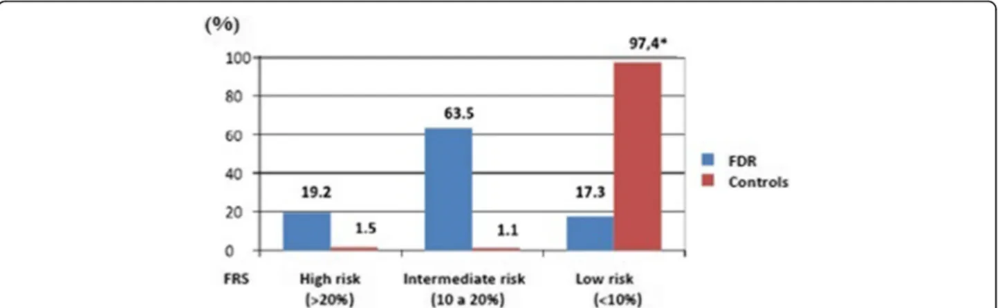
The 10-year hard coronary heart disease risk estimation was calculated using Framingham risk score equations [15] in FDR and controls. Subject were stratified as high risk (>20%); intermediate risk (10 to 20%); and low risk (<10%).
Laboratory evaluation
The following laboratory blood tests were performed in study subjects: fasting blood glucose, total plasma choles-terol, HDL-C, LDL-C, TG and thyroid stimulant hormone (TSH). Fasting plasma glucose was evaluated using the en-zymatic colorimetric hexokinase method. TG and total plasma cholesterol were measured using an enzymatic col-orimetric method with cholesterol esterase oxidase and glycerol phosphate oxidase, respectively. HDL-C was assayed using the enzymatic colorimetric method with polyethylene glycol. LDL-C was calculated using the Frie-dewald formula for TG levels <400 mg/dL. Non-HDL-C was calculated by the formula: Total cholesterol - HDL-C. All analyses were performed in the hospital’s central la-boratory using Roche Diagnostic commercial kits by the multichannel automatic analyzer Roche Cobas™6000.
Statistical analysis
Continuous variables are expressed as mean ± standard deviation (SD) and normality was tested, using the Kolmogorov-Smirnov test, followed by analysis with Stu-dent’s t test (parametric data) or Mann-Whitney’s test (nonparametric data). Categorical data are expressed as absolute frequency and percentages, and were tested by the chi-square test for bivariate analysis. Results were considered significant whenp< 0.05.
Results
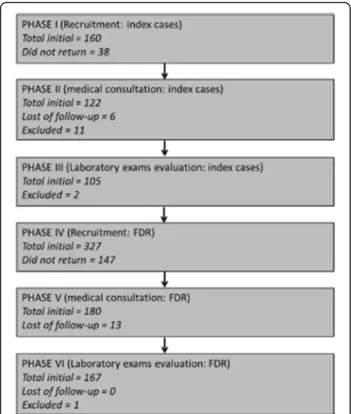
Figure 1 shows patient recruitment algorithm. A total of 103 index cases and 327 FDR were invited. The reasons for non-inclusion were: no response to invitation (n= 147) and lack of laboratory sample collection (n = 14). Hypothyroidism was an exclusion criteria for one par-ticipant. A total of 166 asymptomatic volunteers were enrolled (aged between 18 to 70 years) in the FDR group. The mean age of FDR and controls were 43.6 ± 12.6. and 41.5 ± 6.5 years (p= 0.148) respectively. There was a predominance of female sex in both groups 63.2% (n= 105) and 68.4 (n= 76) respectively in FDR and con-trol groups (p= 0.278).
Table 1 shows clinical and laboratory characteristics of index cases. There was an elevated prevalence of obesity, sedentarism, smoking, diabetes and hypertension. More than 80% of index cases fulfilled the criteria for the meta-bolic syndrome. The most frequent dyslipidemia patterns were low HDL-C levels and hypertriglyceridemia despite the use of statins by 90% of patients. Of importance 42%
of index cases presented a history of early CAD in the family and only 18.6% (n= 19) had LDL-C < 70 mg/dL. Table 2 shows the comparison of clinical and laboratory characteristics between FDR and controls according to sex. The prevalence of dyslipidemia, mainly low HDL-C levels and hypertriglyceridemia, smoking, pre-diabetes, diabetes, and the metabolic syndrome was higher in FDR individuals than in controls (all parameters p< 0.001). However, there were no differences concerning the preva-lence of hypertension, abdominal obesity and overweight.
Fig. 1Study subject inclusion and exclusion algorithm
Table 1Clinical and laboratory characteristics of 103 patients <45 years with myocardial infarction
Parameters (n= 103)
Age (years) 39.6 ± 5.7
Male sex n (%) 56.0 (54.4)
Overweight n (%) 86.0 (86.0)
Abdominal obesity n (%) 87.0 (88.8)
Dyslipidemia n (%) 96.0 (93.2)
Hypertension n (%) 44.0 (42.7)
Sedentarism n (%) 87.0 (84.3)
Smoking n (%) 59 (57.3)
High LDL-C (mg/dL) 5.0 (5.1)
Low HDL-C (mg/dL) 87.0 (84.5)
Only 5.9% (n= 10) of FDR and none of the controls were in use of statins.
Table 3 shows the comparison of laboratory parame-ters between FDR and controls. TG (p= 0.002), LDL-C (p= 0.031), non-HDL-C (p= 0.004), and TSH levels (p = 0.002) were higher, and HDL-C concentrations were lower (p< 0.001) in FDR than in controls.
Figure 2 shows the comparison of estimated 10-year hard coronary heart disease risk between FDR and con-trols. Roughly 83% of FDR were classified as high/inter-mediate risk vs. 3% of controls (p < 0.001).
Discussion
In this study, FDR of subjects, without clinical suspicion of familial hypercholesterolemia, who suffered an AMI before the age of 45 years presented an elevated burden of cardiovascular risk factors mainly type 2 diabetes,
pre-diabetes, atherogenic dyslipidemia, metabolic syn-drome, smoking and sedentarism in comparison with controls. Of importance FDR individuals presented a very similar risk profile to the one of index cases, a fact indicating that genetic and or lifestyle factors are shared within the families. In addition, despite the elevated prevalence of dyslipidemia and a great number of indi-viduals considered as intermediate/high hard coronary heart disease risk, almost 95% of them were not in use of preventive therapies like statin treatment.
Studies evaluating the prevalence of atherosclerosis risk factors among patients with premature CAD have demonstrated a high prevalence of lipid abnormalities, abdominal obesity, hypertension, diabetes and smoking, when compared to healthy controls. In the present study, a similar pattern was found. This occurred despite a similar prevalence of excess body weight and abdom-inal obesity between FDR and controls, a pathologic condition associated with most previously cited risk fac-tors except smoking.
The present study also encountered higher levels of TSH (even within the normal reference value) in FDR individ-uals compared with controls, a finding not previous de-scribed. Previous studies evaluated the association of TSH (in the upper limit, but within a normal reference value) with components of the metabolic syndrome [16]. The pathophysiological mechanism of this association is still unclear, but it is known that the myocardium and vascular endothelial tissue have receptors for thyroid hormones and are sensitive to changes in their serum concentrations. Even minor variations in such concentrations could lead to a negative impact on the cardiovascular system [17, 18].
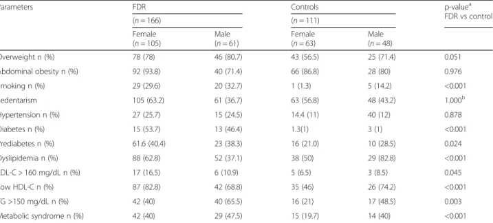
Table 2Clinical and laboratory characteristics of first-degree relatives (FDR) of patients with premature acute myocardial infarction and individuals with no family history of coronary artery disease (controls)
Parameters FDR Controls p-valuea
FDR vs control
(n= 166) (n= 111)
Female (n= 105)
Male (n= 61)
Female (n= 63)
Male (n= 48)
Overweight n (%) 78 (78) 46 (80.7) 43 (56.5) 25 (71.4) 0.051
Abdominal obesity n (%) 92 (93.8) 40 (71.4) 66 (86.8) 28 (80) 0.976
Smoking n (%) 29 (29.6) 20 (32.7) 1 (1.3) 5 (14.2) <0.001
Sedentarism 105 (63.2) 61 (36.7) 63 (56.8) 48 (43.2) 1.000b
Hypertension n (%) 27 (25.7) 15 (24.5) 14.4 (11) 40 (12) 0.878
Diabetes n (%) 15 (53.7) 13 (46.4) 1.3(1) 3 (1) <0.001
Prediabetes n (%) 61.6 (40.4) 23 (38.3) 16 (21.0) 10 (28.5) 0.024
Dyslipidemia n (%) 88 (62.8) 52 (37.1) 38 (50) 29 (82.8) <0.001
LDL-C > 160 mg/dL n (%) 17 (16.5) 6 (10.9) 5 (6.5) 3 (8.5) 0.045
Low HDL-C n (%) 87 (82.8) 42 (68.8) 35 (46) 26 (74.2) <0.001
TG >150 mg/dL n (%) 42 (40) 40 (65.5) 16 (21) 17 (48.5) 0.003
Metabolic syndrome n (%) 42 (40) 29 (47.5) 15 (19.7) 14 (40) <0.001
aChi-square test (FDR versus controls considering the whole population (independent of sex)bFisher exact test
Table 3Comparison of laboratory parameters between
first-degree relatives (FDR) of premature myocardial infarction individuals and controls
Variables FDR
(n= 166)
Controls (n= 111)
p-valuea
Fasting blood glucose (mg/dL) 103 ± 33 94 ± 13 0.521 Total cholesterol (mg/dL) 196 ± 44 190 ± 39 0.219 Non-HDL-C (mg/dL) 157 ± 43 141 ± 41 0.004 LDL-C (mg/dL) 122 ± 37 113 ± 35 0.031 HDL-C (mg/dL) 39 ± 10 48 ± 14 <0.001 Triglycerides (mg/dL) 179 ± 71 140 ± 74 0.002 TSH (mUI/L) 2.4 ± 1.6 1.9 ± 1 0.002
The National Health and Nutrition Examination Sur-vey III (NHANES III) suggested the reference value for TSH in the general population is between 0.4 and 4.12 mU/L [19]. However, there is evidence that TSH levels between 2.5 and 4.0 mU/L are related to metabolic alterations, and the discussion about normal TSH upper limit is increasing. Also, the National Academy of Clin-ical Biochemistry ratifies that over 95% of healthy eu-thyroid individuals present TSH concentrations between 0.4 and 2.5 mU/L. Thus, these considerations have raised the discussion that the upper limit of normal TSH values should be reduced to 2.5 mU/L [20].
Several studies reported elevated levels of cholesterol in individuals with subclinical hypothyroidism that were reverted with the replacement of levothyroxine [21, 22]. The HUNT Study (Nord-Trøndelag Health Study), which evaluated the association between TSH levels within the normal reference interval and serum lipid concentrations, showed a positive and linear association of total choles-terol, LDL-C, non-HDL-C and TG with TSH, and a nega-tive relation with HDL-C levels [23]. Whether the encountered elevated TSH levels contributed to the high atherosclerosis risk factor burden encountered in FDR of PMI patients in this study needs further evaluation.
One important finding of our study was that despite sta-tins were used by 90% of index cases, LDL-C was not ad-equately controlled and there was a marked presence of residual atherogenic dyslipidemia with predominance of low HDL-C concentrations. This pattern was similar in FDR where 85% of individuals presented dyslipidemia and 73% had low HDL-C. These findings corroborate previous studies that also reported this phenotype in individuals with early coronary heart disease onset and in their fam-ilies [3, 24]. Indeed, this pattern has been previously de-scribed to the so called Familial Combined Hyperlipidemia (FCH) phenotype [25]. FCH has been used as a term to describe a group of individuals within the same family with a phenotype of mixed dyslipidemia, or isolated
hypercholesterolemia, presenting or not low HDL-C and with an early onset of cardiovascular disease. The dyslipid-emia pattern is variable among members of the same family and is influenced by diet, exercise and weight status. In addition, many of the so called “FCH patients”also devel-oped dysglycemia, type 2 diabetes and hypertension. How-ever, the lack of a common and homogeneous genetic background in affected families has casted doubt on the very existence of that condition [26]. Indeed, a consensus docu-ment of the European Atherosclerosis Society on hypertri-glyceridemic states did not consider the existence of FCH as a cause of hypertriglyceridemia or mixed dyslipidemia [27]. However, this does not exclude a possible genetic compo-nent for the common phenotype of index cases and FDR.
This study has several limitations: its cross-sectional de-sign; difficulties for FDR’s adherence to attend medical ap-pointments and exams, and the consequent loss of 55% of relative patients; the evaluation of thyroid function in FDR did not include the analysis of free T4 and anti-thyroid antibody profile. However, its value is related to study of individuals with AMI at a very early age and clearly shows a similar atherosclerosis risk factor pattern in index cases and FDR that was clearly more severe than in controls.
Conclusions
FDR of patients with PMI not presenting the familial hypercholesterolemia phenotype had an unfavorable metabolic profile characterized by the elevated presence of type 2 diabetes, atherogenic dyslipidemia, metabolic syndrome and its components. The metabolic abnormal-ities were like the ones of index cases and the predicted cardiovascular risk was significantly higher than in con-trols. In addition, these individuals were severely under-treated regarding dyslipidemia control. This study clearly shows clustering of risk factors in relatives of individuals with PMI. FDR presenting those abnormalities must be submitted to intensive cardiovascular disease risk factor modification programs.
Abbreviations
AMI:Acute myocardial infarction; BMI: Body mass index; CAD: Coronary artery disease; FCH: Familial combined hyperlipidemia; FDR: First-degree relatives; HbA1c: Glycated hemoglobin; HDL-C: High-density lipoprotein-cholesterol; LDL-C: Low-density lipoprotein-lipoprotein-cholesterol; PMI: Premature myocardial infarction; SD: Standard deviation; TG: Triglycerides; TSH: Thyroid stimulant hormone
Acknowledgements
We thank the study participants and the Hospital Carlos Alberto Studart Gomes cardiometabolic unit, Fortaleza, Ceara, Brazil.
Funding
The data collection was funded under CAPES and FUNCAP research grant.
Availability of data and materials
Please contact author for data requests.
Authors’contributions
MHCG, RDS and RMMJ conceived the study, participated in its design and coordination, and manuscript preparation. MHCG, TCSS and LSB participated in the design of the study and collected the data. CMPP, PGBS, ACP, MHCG, LAAB and RDS participated in the design of the study and analyzed the data. All authors read and approved the final manuscript.
Ethics approval and consent to participate
Ethical approval was obtained from the Ethics Committee of Hospital do Coração Dr. Carlos Alberto Studart Gomes, protocol #824/11, in Fortaleza (CE), Brazil. All subjects gave informed consent.
Consent for publication
Not applicable.
Competing interests
RDS declares that received honoraria for consulting and or speaker activities and research from: Amgen, Astra Zeneca, Biolab, Kowa, Merck, Pfizer, Sanofi/ Regeneron and Procaps. The other authors declare that they have no competing interests.
Publisher’s Note
Springer Nature remains neutral with regard to jurisdictional claims in published maps and institutional affiliations.
Author details
1Heart Institute (InCor), University of Sao Paulo Medical School Hospital, São
Paulo, Brazil.2Federal University of Ceará, Fortaleza, Brazil.3Christus Medical
School, Fortaleza, Brazil.4Dr. Carlos Aberto Studart Gomes Hospital, Fortaleza,
Brazil.5Professor Costa Mendes, 1608, Zip-code: 60416-200. Rodolfo Teófilo,
Fortaleza, Ceará, Brazil.
Received: 14 September 2017 Accepted: 2 November 2017
References
1. Kang MK, Chang HJ, Kim YJ, Park AR, Park S, Jang Y, et al. Prevalence and determinants of coronary artery disease in first-degree relatives of premature coronary artery disease. Coron Artery Dis. 2012;23(3):167–73.
2. Wang TJ, Nam BH, D'Agostino RB, Wolf PA, Lloyd-Jones DM, MacRae CA, et al. Carotid intima-media thickness is associated with premature parental coronary heart disease: the Framingham heart study. Circulation. 2003; 108(5):572–6.
3. Jamil G, Jamil M, Alkhazraji H, Haque A, Chedid F, Balasubramanian M, et al. Risk factor assessment of young patients with acute myocardial infarction. Am J Cardiovasc Dis. 2013;3(3):170–4. eCollection 2013
4. Perk J, De Backer G, Gohlke H, et al. European Association for Cardiovascular Prevention & Rehabilitation (EACPR); ESC Committee for Practice Guidelines (CPG). European guidelines on cardiovascular disease prevention in clinical practice (version 2012). The fifth joint task force of the European Society of Cardiology and Other Societies on cardiovascular disease prevention in clinical practice (constituted by representatives of nine societies and by
invited experts). Eur Heart J. 2012;33(13):1635-1701. Erratum in: Eur Heart J. 2012;33(17):2126.
5. Santos RD, Watts GF. Familial hypercholesterolaemia: PCSK9 inhibitors are coming. Lancet. 2015;385(9965):307–10.
6. Nanchen D, Gencer B, Auer R, Räber L, Stefanini GG, Klingenberg R, et al. Prevalence and management of familial hypercholesterolaemia in patients with acute coronary syndromes. Eur Heart J. 2015;36(36): 2438–45.
7. Thygesen K, Alpert JS, Jaffe AS, Simoons ML, Chaitman BR, White HD, et al. Joint ESC/ACCF/AHA/WHF task force for universal definition of myocardial infarction; third universal definition of myocardial infarction. J Am Coll Cardiol. 2012;60(16):1581–98.
8. Dobson AJ, Alexander HM, Heller RF, Lloyd DM. How soon after quitting smoking does risk of heart attack decline? J Clin Epidemiol. 1991;44(11): 1247–53.
9. de Cardiologia SB, Xavier HT, Izar MC, Faria Neto JR, Assad MH, Rocha VZ, et al. V Brazilian guidelines on Dyslipidemias and prevention of atherosclerosis. Arq Bras Cardiol. 2013;101(4 Suppl 1):1–20. Portuguese
10. V Diretrizes Brasileiras de Hipertensão Arterial. Arq Bras Cardiol. 2007;89: e24–79.
11. American Diabetes Association. Executive summary: standards of medical care in diabetes–2014. Diabetes Care. 2014;37(Suppl 1):S5–13.
12. Associação Brasileira para o Estudo da Obesidade e Síndrome Metabólica (ABESO). Diretrizes Brasileiras de Obesidade 2009–2010 [Internet]. ABESO;
2010 [cited 2015 jul 7]. Available from: http://www.abeso.org.br/pdf/ diretrizes_brasileiras_obesidade_2009_2010_1.p df.
13. Donnelly JE, Blair SN, Jakicic JM, Manore MM, Rankin JW, Smith BK, American College of Sports Medicine. Appropriate physical activity intervention strategies for weight loss and prevention of weight regain for adults. Med Sci Sports Exerc. 2009;41(2):459–71.
14. International Diabetes Federation (IDF). Diabetes Atlas [Internet]. 6th ed. 2. Belgium: IDF; 2013 [cited 2015 aug 11]. Available from: http://www.idf.org/ diabetesatlas
15. Framingham Heart Study. Hard Coronary Disease (10-year risk) [Internet]. [cited 2015 aug 11]. Available from: https://www.framinghamheartstudy.org/ risk-functions/coronary-heartdisease/hard-10-year-risk.php
16. Roos A, Bakker SJ, Links TP, Gans RO, Wolffenbuttel BH. Thyroid function is associated with components of the metabolic syndrome in euthyroid subjects. J Clin Endocrinol Metab. 2007;92(2):491–6.
17. Pantos C, Mourouzis I, Galanopoulos G, Gavra M, Perimenis P, Spanou D, et al. Thyroid hormone receptor alpha1 downregulation in postischemic heart failure progression: the potential role of tissue hypothyroidism. Horm Metab Res. 2010;42(10):718–24.
18. Pingitore A, Chen Y, Gerdes AM, Iervasi G. Acute myocardial infarction and thyroid function: new pathophysiological and therapeutic perspectives. Ann Med. 2012;44(8):745–57.
19. Spencer CA, Hollowell JG, Kazarosyan M, Braverman LE. National Health and nutrition examination survey III thyroid-stimulating hormone (TSH)-thyroperoxidase antibody relationships demonstrate that TSH upper reference limits may be skewed by occult thyroid dysfunction. J Clin Endocrinol Metab. 2007;92(11):4236–40.
20. Demers LM, Spencer CA. Laboratory medicine practice guidelines: laboratory support for the diagnosis and monitoring of thyroid disease. Clin Endocrinol. 2003;58(2):138–40.
21. Caraccio N, Ferrannini E, Monzani F. Lipoprotein profile in subclinical hypothyroidism: response to levothyroxine replacement, a randomized placebo-controlled study. J Clin Endocrinol Metab. 2002;87(4):1533–8.
22. Iqbal A, Jorde R, Figenschau Y. Serum lipid levels in relation to serum thyroid-stimulating hormone and the effect of thyroxine treatment on serum lipid levels in subjects with subclinical hypothyroidism: the Tromso study. J Intern Med. 2006;260(1):53–61.
23. Asvold BO, Vatten LJ, Nilsen TI, Bjøro T. The association between TSH within the reference range and serum lipid concentrations in a population based study. Eur J Endocrinol. 2007;156(2):181–6.
24. Wang J, Liu ZQ, He PY, Yang YC, Zhang L, Muhuyati. Analysis of the risk factors and characteristics of coronary artery disease of Han, Uygur and Kazak patients with acute myocardial infarction in Xinjiang district. Int J Clin Exp Med. 2015;8(2):2831–8. eCollection 2015
26. Lewis GF, Xiao C, Hegele RA. Hypertriglyceridemia in the genomic era: a new paradigm. Endocr Rev. 2015;36(1):131–47.
27. Hegele RA, Ginsberg HN, Chapman MJ, Nordestgaard BG, Kuivenhoven JA, Averna M, Borén J, Bruckert E, Catapano AL, Descamps OS, Hovingh GK, Humphries SE, Kovanen PT, Masana L, Pajukanta P, Parhofer KG, Raal FJ, Ray KK, Santos RD, Stalenhoef AF, Stroes E, Taskinen MR, Tybjærg-Hansen A, Watts GF, Wiklund O, European Atherosclerosis Society Consensus Panel. The polygenic nature of hypertriglyceridaemia: implications for definition, diagnosis, and management. Lancet Diabetes Endocrinol. 2014; 2(8):655–66.
• We accept pre-submission inquiries
• Our selector tool helps you to find the most relevant journal • We provide round the clock customer support
• Convenient online submission
• Thorough peer review
• Inclusion in PubMed and all major indexing services
• Maximum visibility for your research
Submit your manuscript at www.biomedcentral.com/submit