IDENTIFICATION OF GRP75 AS A NOVEL PRES1 BINDING PROTEIN USING A PROTEOMICS STRATEGY
Lunbiao Cui$*; Yiyue Ge$; Yuhua Qi; Zhiyang Shi; Yongjun Jiao; Xian Qi; Xiangjun Zhai; Hua Wang
Institute of Microbiology, Jiangsu Provincial Center for Diseases Prevention and Control, Nanjing 210009, China
Submitted: April 13, 2009; Returned to authors for corrections: May 19, 2009; Approved: May 27, 2009.
ABSTRACT
The PreS1 region of the L protein is important in cell attachment and consequently in hepatitis B virus
(HBV) infectivity. To identify novel PreS1 interacting protein, we performed Glutathione-S-transferase
(GST) pull-down, two-dimensional gel electrophoresis (2-DE) and mass spectrometry assays.
Glucose-regulated proteins (GRP) 78 and 75 were found to bind PreS1. The interactions between PreS1 and GRP75
were confirmed by a co-immunoprecipitation experiment. GRP78 and GRP75 may play important roles in
mediating HBV virion entering into hepatocyte and regulating proper folding of the L protein due to their
critical functions in protein folding and trafficking. The finding of novel PreS1 binding protein enriches
our knowledge about molecular mechanism of HBV infection.
Key words: HBV, PreS1, Proteomics, GRP75
INTRODUCTION
The hepatitis B virus (HBV) is a worldwide cause of
hepatic disease with variations in prevalence from region to
region (13). China is one of the highest HBV prevalence areas.
There are approximately 130 million HBV carriers in China.
Current treatment for HBV infection has limited efficacy. The
attachment to hepatocytes by HBV during infection has long
been proposed to be a potential target for antiviral intervention.
However, little is known about the molecular events mediating
HBV attachment to hepatocytes. HBV PreS1 region,
specifically, the aa 21-47 segment, is believed to play an
essential role in mediating HBV attachment to the putative
receptor on hepatocytes (9,14). In the past two decades, Human
immunoglobulin A (IgA) receptor, a 31-kDa protein,
interleukin-6, a 44-kDa protein (HBV-BP), Homology to
SCCA1, a human squamous cell carcinoma antigen 1 (human
serpin), p80 (GRP78), a 35-kDa protein homolous to
Glycerinaldehyde-3-phoshate-dehydrgenase (GAPD), a serum
glycoprotein of 50-kDa, and an asialoglycoproteinreceptor
(ASGPR) have been proposed as PreS1 binding proteins (4),
but none of these molecules has been identified as receptor in
HBV infection.
With the rapid development in screening technology and
bioinformatics, novel receptor or coreceptor candidates have
been discovered recently. Deng found that lipoprotein lipase
(LPL), a key enzyme in lipoprotein metabolism, might interact
with PreS and HBV particles by using phage display library
(3). Li et al. screened NACA as a novel PreS1 associated
protein by using yeast two-hybrid system (10). Recently,
proteomic techniques have been used as a new tool for
studying protein-protein interaction. In this study, to identify
*Corresponding Author. Mailing address: Institute of Microbiology, Jiangsu Provincial Center for Diseases Prevention and Control, Nanjing 210009, China.; Fax: 86-25-83759371.; E-mail: [email protected]
novel PreS1 interacting protein, we performed
Glutathione-S-transferase (GST) pull-down, high-resolution two-dimensional
gel electrophoresis (2-DE) and mass spectrometry assays.
GRP75 was identified as a novel PreS1 binding protein and its
possible roles in virus infection are discussed. GRP78, a
protein previously described to interact with preS1, was also
identified in these assays.
MATERIALS AND METHODS
GST fusion construct of PreS1
The PreS1 fragment was amplified by PCR using HBV
DNA as template extracted from serum of a chronically
infected patient with HBsAg, anti-HBc, and HBeAg positive
serology. PCR were performed using the following primers, 5
-AGCGGATCCATGGGAGGTTGGTCTTCCA-3 (forward)
and 5 -ATATCTCGAGTTAGGCCTGAGGATGACTGT-3
(reverse). The PCR products were subsequently cloned as
BamH I-Xho I fragments into pGEX-4T2 (Pharmacia) and
recombinant vectors were introduced into the E. coli BL21
(DE3) (Invitrogen). The GST fusion proteins were induced as
described previously and were purified from the bacterial cell
lysates by affinity binding to glutathione sepharose beads
(Pharmacia) as detailed elsewhere (11).
GST pull-down assay
HepG2 cells were lysed in binding buffer (50
mMTris-HCl, pH 8.0, 150 mM NaCl, 5 mM EDTA, 0.5% NP-40, 1
g/mL leupeptin, 1 g/mL aprotinin and 0.1 mM PMSF.), and
triplicate independent soluble protein fraction was incubated
with GST and GST-PreS1 fusion proteins overnight at 4°C.
Then, co-precipitations were performed by addition of 80 µl
glutathione–sepharose 4B resin. After washing five times with
600 µl of binding buffer, binding proteins were eluted with 200
µl of 10 mM reduced glutathione in PBS. Eluted samples were
precipitated with 4 volumes of cold acetone, centrifuged and
the pellets were washed with 200 µl of 20% (v/v) cold
methanol. After centrifugation for 30 min at 12,000×g, pellets
were air-dried and dissolved in rehydrating solution (8 M urea,
2% CHAPS, 18mM dithiothreitol (DTT), 0.5% IPG buffer pH
3-10) for further 2-DE analysis.
Two-dimensional gel electrophoresis (2-DE)
2-DE was performed as described previously (2). Briefly,
samples were subjected to isoelectric focusing (IEF) using IPG
DryStrips with immobilized pH gradient, pH range 3-10, 17
cm, linear (Bio-Rad). IEF was performed in an IPGphor
(Amersham Biosciences) according to the following protocol:
30 V 6 h, 60 V 6 h, 500 V 1 h, 1000 V 1 h, 8000 V 3-4 h to 30
000 Vh. After IEF, strips were equilibrated in 50 mM
Tris-HCl, pH 8.8, 6 M urea, 2.0% SDS, 30% glycerol with 1% DTT
for 10 min, and then for 10 min in the same buffer without
DTT but with 4% iodoacetamide. Equilibrated strips were
placed on top of 10% (w/v) polyacrylamide gel to separate the
proteins by molecular mass.
Protein identification
Protein spots were excised from the gels, destained, and
subjected to in-gel digestion with trypsin (modified, sequence
grade porcine; Promega), as described earlier (2). Tryptic
peptides were dissolved in 2 µl of 0.5% TFA containing the
matrix (a-cyano-4-hydroxycinnamic acid) and analyzed by
matrix-assisted laser desorption/ionization-time of flight-mass
spectrometry (MALDI-TOF-MS) (Bruker Daltonics,
Germany). Data were screened against the NCBInr databases
using the MASCOT search program
(www.matrixscience.com).
Immunoprecipitation
PreS1 gene was cloned into the BamH I and Hind
restriction sites of pXJ40 (pXJ40-PreS1). Encoding domain of
GRP75 gene was generated by using the forward primer
(5’-CTTAAGCTTGCCATGATAAGTGCCAGCCGAGCTG-3’)
and reverse primer
(5’-CGCCTCGAGTATTACTGTTTTTCCTCCTTTTGATC-3’)
and cloned into Hind and Xho I restriction sites of pXJ40
(pXJ40-GRP75). pXJ40-PreS1 and pXJ40-GRP75 were
(Invitrogen). 48 h after transfection, cells were washed with
phosphate-buffered saline and lysed in 0.5 ml lysis buffer (20
mM Tris-HCl pH 8.0, 100 mM NaCl, 1 mM EDTA, 0.5 %
NP-40, 100 mol/L PMSF, 1 g/mL leupeptin, 1 g/mL aprotinin).
After brief sonication, the lysate was centrifugated at 15000
rpm for 15 min at 4°C. The supernatant was incubated with
mouse monoclonal anti-PreS1 antibody (Feipon Biotech Inc,
China) overnight at 4°C, then incubated with a slurry of
protein-A-sepharose with rotation for 2 h at 4°C. The beads
were pelleted and washed five times with cell lysis buffer.
Finally, proteins were solubilized in sodium dodecyl sulphate–
polyacrylamide gel electrophoresis (SDS-PAGE) loading
buffer by boiling.
Western blot
Samples were resolved in 8% or 17.5% polyacrylamide
gel, and proteins were transferred to a nictrocellulose
membrane. Immunoblotting was carried out using rabbit
anti-GRP75 antibody (Santa Cruz) or mouse anti-PreS1 antibody as
indicated above and anti-rabbit or anti-mouse IgG-horseradish
peroxidase (HRP) conjugates. After rinsing with PBS-T
(phosphate-buffered saline [PBS] containing 0.1% Tween 20),
the blots were visualized by the enhanced chemiluminescence
procedure as recommended by the supplier (Pierce).
RESULTS
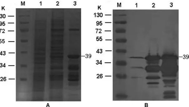
Preparation of GST-fusion construct of PreS1
We sought to identify novel PreS1-binding proteins using
a GST pull-down assay. To this end, an expression plasmid,
pGEX4T2-PreS1 was generated carrying a full-length preS1
sequence. The GST-PreS1 fusion protein was expressed in E.
coli BL21. SDS-PAGE analysis revealed that GST-PreS1 can
be expressed and be co-purified with other proteins (Fig. 1A,
lane 3). Similar to the previous study (11), Western blot
analysis with anti-preS1 monoclonal antibody indicated a
major band of a 39 K protein corresponding to the intact
GST-PreS1, several bands of lower molecular weight proteins that
most likely represent degradation products of the GST-preS1
fusion protein (Fig. 1B, lane 3).
Figure 1. Expression and purification of the preS1. (A) 10% SDS–PAGE. B. Immunoblotting with anti-preS1 monoclonal antibody. Uninduced recombinant cell lysates, induced recombinant cell lysates, and GST-PreS1 protein purified using
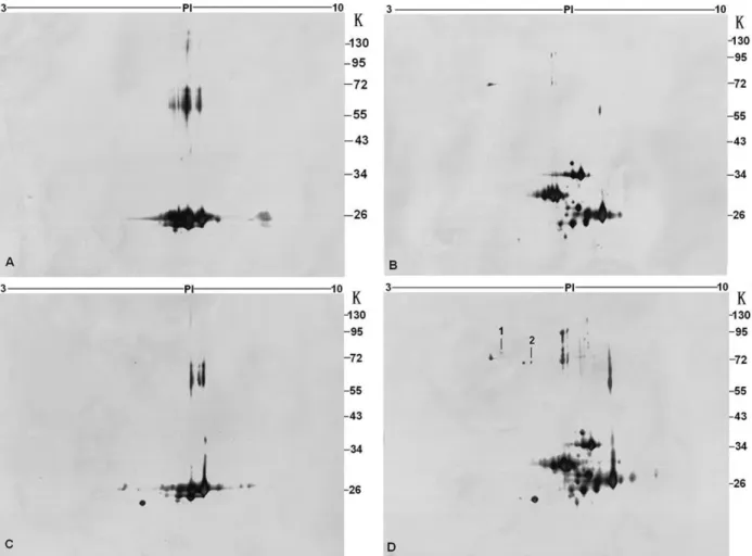
2-DE analysis of proteins binding with PreS1
To identify proteins associated with PreS1, the high
resolving power of 2-DE was applied to the analysis. The GST
and GST-PreS1 samples incubated with and without HepG2
cell lysate were separated using IPG strip in first dimension
and 10% polyacrylamide gels in the second dimension (Fig.2).
GST and GST-PreS1 samples incubated with binding buffer
(Fig. 2A, B), GST sample incubated with HepG2 cell lysate
(Fig. 2C) were set as control. Those protein spots appeared in
the pull-down sample gel while not in the control gels were
excised and further analyzed by MALDI-TOF-MS. Database
searching showed that two proteins of HepG2 could bind with
GST-PreS1, including GRP78 and GRP75 (Point 1, 2, Fig.
2D). Peptide masses and further data concerning protein
identity are listed in Table 1.
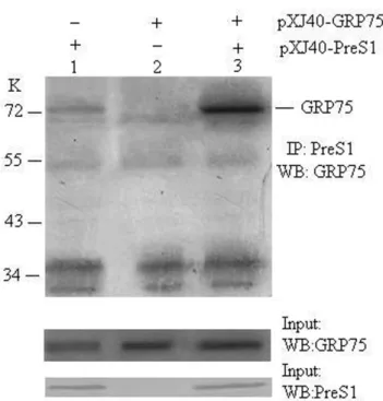
GRP75 associates with PreS1 in vivo
It has been previously demonstrated that GRP78 bound
specifically to the PreS1 in vitro and in vivo (1). In this study,
we therefore opted to confirm the interaction between GRP75
and PreS1 in vivo. The coding sequence of PreS1 and GRP75
was cloned into pXJ40. COS-7 cells were co-transfected
pXJ40-GRP75, pXJ40-PreS1, or empty vector. As shown in
figure 3, anti-PreS1 antibody coimmunoprecipitated PreS1 and
GRP75. It is thus possible that GRP75 is PreS1-associated
protein.
Figure 2. 2-DE analysis of GST-PreS1 binding protein. GST pull down using GST (A), GST-PreS1 (B) with binding buffer, GST (C) and GST-PreS1 (D) with HepG2 cell lysate. IEF on Immobiline DryStrips (pH range 3-10) followed by SDS-PAGE and silver
staining. Protein spots 1 and 2 (D) were identified by MALDI-TOF-MS, one of which was GRP78 (point 1), another was GRP75
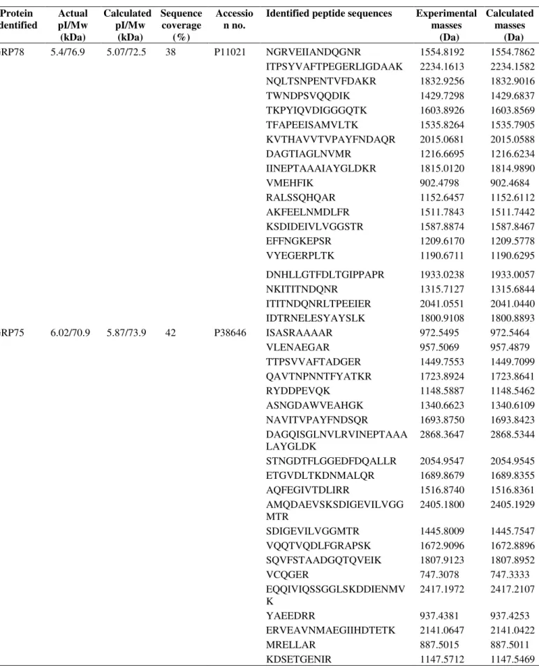
Table 1. Protein identities and peptide masses
Protein identified
Actual pI/Mw (kDa)
Calculated pI/Mw
(kDa)
Sequence coverage
(%)
Accessio n no.
Identified peptide sequences Experimental masses
(Da)
Calculated masses
(Da)
GRP78 5.4/76.9 5.07/72.5 38 P11021 NGRVEIIANDQGNR 1554.8192 1554.7862
ITPSYVAFTPEGERLIGDAAK 2234.1613 2234.1582
NQLTSNPENTVFDAKR 1832.9256 1832.9016
TWNDPSVQQDIK 1429.7298 1429.6837
TKPYIQVDIGGGQTK 1603.8926 1603.8569
TFAPEEISAMVLTK 1535.8264 1535.7905
KVTHAVVTVPAYFNDAQR 2015.0681 2015.0588
DAGTIAGLNVMR 1216.6695 1216.6234
IINEPTAAAIAYGLDKR 1815.0120 1814.9890
VMEHFIK 902.4798 902.4684
RALSSQHQAR 1152.6457 1152.6112
AKFEELNMDLFR 1511.7843 1511.7442
KSDIDEIVLVGGSTR 1587.8874 1587.8467
EFFNGKEPSR 1209.6170 1209.5778
VYEGERPLTK 1190.6711 1190.6295
DNHLLGTFDLTGIPPAPR 1933.0238 1933.0057
NKITITNDQNR 1315.7127 1315.6844
ITITNDQNRLTPEEIER 2041.0551 2041.0440
IDTRNELESYAYSLK 1800.9108 1800.8893
GRP75 6.02/70.9 5.87/73.9 42 P38646 ISASRAAAAR 972.5495 972.5464
VLENAEGAR 957.5069 957.4879
TTPSVVAFTADGER 1449.7553 1449.7099
QAVTNPNNTFYATKR 1723.8924 1723.8641
RYDDPEVQK 1148.5887 1148.5462
ASNGDAWVEAHGK 1340.6623 1340.6109
NAVITVPAYFNDSQR 1693.8750 1693.8423
DAGQISGLNVLRVINEPTAAA LAYGLDK
2868.3647 2868.5344
STNGDTFLGGEDFDQALLR 2054.9547 2054.9545
ETGVDLTKDNMALQR 1689.8679 1689.8355
AQFEGIVTDLIRR 1516.8740 1516.8361
AMQDAEVSKSDIGEVILVGG MTR
2405.1800 2405.1929
SDIGEVILVGGMTR 1445.8009 1445.7547
VQQTVQDLFGRAPSK 1672.9096 1672.8896
SQVFSTAADGQTQVEIK 1807.9123 1807.8952
VCQGER 747.3078 747.3333
EQQIVIQSSGGLSKDDIENMV K
2417.1972 2417.2107
YAEEDRR 937.4381 937.4253
ERVEAVNMAEGIIHDTETK 2141.0647 2141.0422
MRELLAR 887.5015 887.5011
Figure 3. GRP75 binds to PreS1 in vivo. The COS7 cell lysates, co-transfected with PreS1 (lane 1) and
pXJ40-GRP75 (lane 2) either alone or both (lane 3), were
immunoprecipitated with anti-PreS1 antibody. Lysates from
the transfected cell and the immunoprecipitates were subjected
to Western blot analysis using the anti-GRP75 antibody
DISCUSSION
GST pull down combining with proteomic techniques
provided a powerful tool for studying protein-protein
interaction in vitro. By using these methods, some novel
protein-protein interaction partners have been clarified, such as
PRS1 associated with p300 (6), HBx protein of HBV binding
to mitochondrial HSP60 and HSP70 (18), PKC and CtBP
interacted with BMPR-II (5).
Both GRP78 and GRP75 belong to HSP70 family
(HSP-70s) molecular chaperones which play critical roles in protein
folding and trafficking (8). The major difference between
GRP78 and GRP75 is the cellular localization. GRP78 almost
localized in endoplasmic reticulum (ER) (8). While GRP75
mainly resided in mitochondria but also resided in ER and both
of these protein are reported expressing in cell surface (15,17).
HSP-70s are highly conserved and demonstrate a 60–78% base
identity among eukaryotic cells. Biochemical studies have
demonstrated that all HSP70s have N-terminal 44-kDa and
18-kDa domain, C-terminal 10-18-kDa fragment. N-terminal 44-18-kDa
domain is ATPase domain. 18-kDa domain is peptide-binding
domain. C-terminal 10-kDa fragment carries highly conserved
EEVD terminal sequence (8). Therefore, it is likely that both
GRP78 and GRP75 bound to PreS1 through N-terminal 18-kDa
domain. GRP78 has been proved specifically bound to the
PreS1 (1,4). Using the receptor binding assay they determined
that amino acid residues 12 to 20 and 82 to 90 are essential for
the binding of pre-S1 to GRP78. Which site of PreS1 mediates
its attachment to GRP75 should be established.
Functional meaning of PreS1 interacting with both
proteins may be explained in two ways. First, GRP78 and
GRP75 mediate HBV virion binding to hepatocyte and
internalization. The other is regulating proper folding of the L
protein. The former possibility comes from the fact GRP78
bound specifically to the pre-S1 of native HBV particles. The
association of GRP75 with the IL-1 receptor type was detected
and proposed to play an important role in receptor
internalization (16). GRP75 was also isolated as a FGF-1
binding protein by FGF-1 affinity chromatography and was
shown to aid in its intracellular trafficking (12). In view of the
in vivo biological relevance of GRP78 and GRP75, combined
with the cell surface location, we postulate that one of role of
GRP78 and GRP75 may be act as an adjunctive carrier in
bridging HBV virion entry into the cell during viral infection.
L protein are synthesized at ER. It has also been suggested that
one of function of HSP-70s is to prevent transport of
incompletely assembled, misfolded, or aggregated proteins
from the ER (1,7,8). By analogy, we may therefore infer
GRP78 and GRP75 may interact with the incompletely
assembled L protein particles and retains them in the lumen of
ER to prevent their secretion. Whether GRP78 and GRP75
involved in HBV morphogenesis by regulating proper folding
of the L protein warrants further study.
In summary, we identified GRP75 as a novel PreS1
described to interact with preS1, using GST pull down
combining with proteomic techniques. We postulate that
GRP78 and GRP75 may play important roles in mediating
HBV virion entering into hepatocyte and regulating proper
folding of the L protein due to their critical functions in protein
folding and trafficking.
ACKNOWLEDGEMENTS
This study was supported by the Natural Science
Foundation of Jiangsu Province (No. BK2007608) and Jiangsu
Provincial Grant (No.BE2008683).
REFERENCES
1. Cho, D.Y.; Yang, G..H.; Ryu, C.J.; Hong, H.J. (2003). Molecular chaperone GRP78/BiP interacts with the large surface protein of hepatitis B virus in vitro and in vivo. J. Virol., 77, 2784-2788.
2. Cui, L.; Wang, Y.; Shi, Y.; Zhang, Z.; Xia, Y.; Sun, H.; Wang, S.; Chen, J.; Zhang, W.; Lu, Q.; Song, L.; Wei, Q.; Zhang, R.; Wang, X. (2007). Overexpression of annexin a1 induced by terephthalic acid-calculi in rat bladder cancer. Proteomics., 7, 4192-4202.
3. Deng, Q.; Zhuang, M.; Kong, Y.Y.; Xie, Y.H.; Wang, Y. (2005). Screening for PreS specific binding ligands with a phage displayed peptides library. World. J. Gastroenterol., 11, 4018-4023.
4. Glebe, D.; Urban, S.; (2007). Viral and cellular determinants involved in hepadnaviral entry. World. J. Gastroenterol., 13, 22-38.
5. Hassel, S.; Eichner, A.; Yakymovych, M.; Hellman, U.; Knaus, P.; Souchelnytskyi, S. (2004). Proteins associated with type II bone morphogenetic protein receptor (BMPR-II) and identified by two-dimensional gel electrophoresis and mass spectrometry. Proteomics., 4, 1346-1358.
6. Kaida, A.; Ariumi, Y.; Baba, K.; Matsubae, M.; Takao, T.; Shimotohno, K. (2005). Identification of a novel p300-specific-associating protein,
PRS1 (phosphoribosylpyrophosphate synthetase subunit 1). Biochem. J., 391, 239-247.
7. Kaul, S.C.; Deocaris, C.C.; Wadhwa, R. (2007). Three faces of mortalin: a housekeeper, guardian and killer. Exp. Gerontol., 42, 263-274. 8. Kiang, J.G.; Tsokos, G.C. (1998). Heat shock protein 70 kDa: molecular
biology, biochemistry, and physiology. Pharmacol. Ther., 80,183-120. 9. Le Seyec, J.; Chouteau, P.; Cannie, I.; Guguen-Guillouzo, C.; Gripon, P.
(1999). Infection process of the hepatitis B virus depends on the presence of a defined sequence in the pre-S1 domain. J. Virol., 73, 2052-2057. 10. Li, D.; Wang, X.Z.; Chen, Z.X.; Huang, Y.H. (2003). Screening the
hepatitis B virus PreS1 associated protein by the yeast two-hybrid system. Chin. J. Hepatol (Chin)., 11, 334-337.
11. Maeng, C.Y.; Oh, M.S.; Park, I.H.; Hong, H.J. (2001). Purification and structural analysis of the hepatitis B virus PreS1 expressed from escherichia coli. Biochem. Biophys. Res. Commun., 282, 787-792. 12. Mizukoshi, E.; Suzuki, M.; Loupatov, A.; Uruno, T.; Hayashi, H.;
Misono, T.; Kaul, S.C.; Wadhwa, R.; Imamura, T. (1999). Fibroblast growth factor-1 interacts with the glucose-regulated protein GRP75/mortalin. Biochem. J., 343, 461-466.
13. Paiva1, E.M.M.; Tiplle, A.F.V.; Silva, E.P.; Cardoso, D.D.P. (2008). Serological markers and risk factors related to hepatitis B virus in dentists in the Central West region of Brazil. Braz. J. Microbiol., 39, 251-256.
14. Pontisso, P.; Alberti, A. (1991). The role of preS1 in the interaction of hepatitis B virus with human hepatocytes. Hepatology., 14, 405-406. 15. Ran, Q.; Wadhwa, R.; Kawai, R.; Kaul, S.C.; Sifers, R.N.; Bick, R.J.;
Smith, J.R.; Pereira-Smith, O.M. (2000). Extramitochondrial localization of mortalin ⁄ mthsp70 ⁄ PBP74 ⁄ GRP75. Biochem. Biophys. Res.
Commun., 275, 174-179.
16. Sacht, G..; Brigelius-Flohé, R.; Kiess, M.; Sztajer, H.; Flohé, L. (1999). ATP-sensitive association of mortalin with the IL-1 receptor type I. Biofactors., 9, 49-60.
17. Singh, B.; Soltys, B.J.; Wu, Z.C.; Patel, H.V.; Freeman, K.B.; Gupta, R.S. (1997). Cloning and some novel characteristics of mitochondrial Hsp70 from Chinese hamster cells. Exp. Cell.Res., 234, 205-216. 18. Zhang, S.M.; Sun, D.C.; Lou, S.; Bo, X.C.; Lu, Z.; Qian, X.H.; Wang,