online | memorias.ioc.fiocruz.br
Efficacy of benznidazol treatment for asymptomatic chagasic patients
from state of Rio Grande do Sul evaluated during a three
years follow-up
CD Fernandes¹, FM Tiecher¹, MM Balbinot¹, DB Liarte³, D Scholl², M Steindel², A Romanha³/+
1Instituto de Pesquisa Biológica, Laboratório Central, Fundação Estadual de Produção e Pesquisa em Saúde, Porto Alegre, RS, Brasil 2Departamento de Microbiologia e Parasitologia, CCB, Universidade Federal de Santa Catarina, Florianópolis, SC, Brasil
3Instituto de Pesquisa René Rachou-Fiocruz, Belo Horizonte, MG, Brasil
The efficacy of benznidazol on the treatment of chagasic patients from the state of Rio Grande do Sul was evalu-ated during a three-year follow-up. A cohort of 80 asymptomatic chronic chagasic patients or blood bank donors (49 male and 31 female) was studied. Their ages varied from 17-42 years, with a mean and a median of 30 and 35 years, respectively. The 80 patients presented positive serology, hemoculture and polymerase chain reaction (PCR). They were treated with 5 mg/Kg benznidazol twice a day for 60 days. Serological, parasitological and PCR methods were used to evaluate response. Serology was performed using commercial ELISA and indirect immunofluorescence (IFI) tests, parasitemia was monitored by hemoculture in LIT medium and PCR with primers S35/S36 was used to amplify a Trypanosoma cruzi 330 bp kDNA repetitive sequence. PCR positivity of 240 seropositive individuals was com-pared using DNA preparations from whole blood/guanidine EDTA (GE), buffy-coat/GE and frozen buffy-coat. Fifty non-chagasic individuals were used as negative controls. PCR positivity was 86.7% for the frozen buffy-coat, 71.7% for the GE/buffy-coat and 69.2% for the GE/whole blood. The hemocultures became negative just after treatment and remained negative during the three years of follow-up. In the third year after treatment, 9/80 (11.3%) patients presented negative PCR and, from those, four also presented negative serological tests. Furthermore, a reduction in three serological titers was observed in 27/80 (33.8%) of the patients treated. Taken together, the results show that four of the 80 (5.0%) chronic chagasic patients from the state of Rio Grande do Sul were cured after treatment with benznidazol.
Key words: Chagas disease - diagnosis - polymerase chain reaction - benznidazol treatment - cure
Trypanosoma cruzi, the aetiological agent of Chagas disease, is a hemoflagellate protozoan parasite that af-fects about 15-16 million individuals and it is estimated that 75-90 million people are exposed to infection in Latin America (Coura 2007). In Brazil, the vector con-trol program, established in 1975 and continuing with the South Cone Initiative, eliminated the main vector,
Triatoma infestans, from the country (Massad 2007). The seroprevalence of T. cruzi infection in Brazil among children aged 7-14 years fell 99.8% from 1980-1999 and the absence of seropositivity among young children (aged 0-4 years) is evidence of the interruption of vecto-rial transmission (Massad 2007). In Brazil, the highest prevalence of T. cruzi infection was found in the states of Rio Grande do Sul, Minas Gerais, São Paulo, Paraná, Goiás and Bahia (Silveira & Rezende 1994, Dias 1997).
Human Chagas disease presents two distinct phases: the acute phase, which appears just after infection, and the chronic phase, which may last several years. After a long asymptomatic phase, around 30% of infected in-dividuals develop chronic disease with severe damage
Financial support: IPB/LACEN, CNPq, Fiocruz + Corresponding author: romanha@cpqrr.fiocruz.br Received 4 June 2008
Accepted 11 December 2008
to the heart and digestive system (Prata 2001). During the acute phase, T. cruzi trypomastigotes are usually de-tected by microscopic examination of fresh or stained blood-smears, as well as by xenodiagnosis and hemocul-ture (Luquetti & Rassi 2000). In contrast, during the chronic phase, diagnosis is based on the detection of cir-culating antibodies (Luquetti & Rassi 2000). However, due to the long-lasting maintenance of circulating anti-bodies, it is difficult to use serology as a marker for cure of the disease even after the successful treatment of T. cruzi infection (Luquetti & Rassi 2000).
PCR comparative studies of whole blood were per-formed using traditional methods, such as xenodiagno-sis and serology, for detecting T. cruzi; these showed that amplification of T. cruzi kDNA may substitute for xeno-diagnosis in assessing parasitemia in chronic chagasic patients and may also be used as a complement to sero-logical tests in blood banks (Avila et al. 1993, Marcon et al. 2002). Other studies showed that polymerase chain reaction (PCR) is a sensitive method that may detect the parasite DNA in up to 95% of blood samples from chronic chagasic patients (Britto et al. 1995a, Silber et al. 1997). Furthermore, PCR was shown to be a very useful tool for confirmation of diagnosis in patients with doubt-ful serology (Marcon et al. 2002).
considering that PCR sensitivity depends mainly on the target and source of the DNA, we compared different forms of biological sample preservation as sources of
T. cruzi kDNA.
PATIENTS, MATERIALS AND METHODS
Clinical samples and procedures - A cohort of 80 asymptomatic chronic chagasic patients or blood bank donors (49 males and 31 females) from the state of Rio Grande do Sul presenting positive serology and hemo-culture was studied. Their ages varied from 17-42 years old, with a mean and median age of 30-35 years, respec-tively. Patients were treated with 5 mg/Kg benznidazol twice a day for 60 days. Our cohort was obtained from an initial group of 240 seropositive chagasic individuals. A negative control group, consisting of 50 individuals with negative serology, hemoculture and PCR for T. cru-zi infection was included in the study. Chagasic patients were followed annually for a three-year period by serol-ogy, hemoculture and PCR.
Blood collection was performed with a Vacutainer®
system (Becton Dickinson, Franklin Lakes) based on the following volumes and objectives: i) 10 mL of blood for serology, ii) 30 mL of blood with heparin for hemoculture and iii) 15 mL of blood in EDTA Na2 for PCR. Five mil-liliters of whole blood was mixed (v/v) with 6 M guanidine hydrochloride/0.2M EDTA buffer pH 8.0 (GE buffer) im-mediately after collection and was stored at 4oC until use.
For the remaining 10 mL of whole blood, the buffy-coat was separated by centrifugation in a histopaque gradient®
(Sigma, St. Louis), collected and divided into two tubes; one was stored at -70oC and the other was added to 250 μL
of GE buffer and stored at 4oC until use.
Serological assays - The enzyme-linked immuno-sorbent assay (ELISA) was performed using the com-mercial Chagatest-Wienner kit (ELISA recombinant v. 3.0). Briefly, sera were diluted 1/20 in phosphate-buff-ered saline-tween 20 (0.05%) containing 1% bovine se-rum albumin. Two hundred microliters of diluted sera was added to microwells containing T. cruzi antigens. The plates were incubated at 37ºC for 30 min. After five washes in 0.1M phosphate buffer to remove unbound immunoglobulin, the samples were incubated at 37ºC for 30 min with a 1:40,000 dilution of peroxidase-labelled anti-human IgG conjugate. After another wash step, the plates were revealed with 60 mM hydrogen peroxide in 50mM citrate buffer, pH 3.2 and 0.01mM tetramethyl-benzidine in 0.1N chloride acid. After 30 min incuba-tion at rt, the reacincuba-tion was stopped by the addiincuba-tion of 2N sulphuric acid. The plates were read at a wavelength of 450-620 nm in a microplate reader (Anthos Labetec Instruments, Austria). The mean absorbance of the nega-tive controls plus 0.3 OD was used for cut off determina-tion according to the manufacturer’s instrucdetermina-tions.
Indirect immunofluorescence (IFI) assays were per-formed using the (IFI)-Chagas Bio Manguinhos Kit, FIOCRUZ, RJ, according to the manufacturer’s instruc-tions. Serum dilutions (1/40-1/1280) in phosphate buffer saline, pH 7.2 (PBS), were incubated for 30 min at 37oC
with total antigen pre-adsorbed onto the slide surface.
Unbound immunoglobulins were removed by washing the slides twice with PBS. Following incubation with fluorescein-labeled anti-human IgG conjugate for 30 min at 37oC, unbound conjugate was removed by two
washes with PBS. Slides were mounted with buffered glycerine, pH 9.5, and observed under a fluorescence microscope (Labophot Nikon, Japan). Positive chagasic and negative sera were used as controls. The cut off value for IFI was 1/40.
Hemoculture - Hemoculture was performed as de-scribed by Galvão et al. (1989). Briefly, 30 mL of hepa-rinized venous blood was collected from each individual in a vacutainer system (Vacuum II) and centrifuged at 600 g for 30 min at 4oC. Plasma was collected and stored
at -20oC. The pellet was resuspended in 15 mL of LIT
(liver infusion tryptose) medium and centrifuged as described above. After removal of the supernatant, the pellet was resuspended in 30 mL of LIT and distributed into six tubes (18 x 150 mm), which were maintained at 28oC and examined monthly until the fourth month.
Only patients with positive hemocul tures were treated. They received 5 mg/kg benznidazol twice a day for 60 days, followed by yearly serology, hemoculture and PCR analyses over the three years.
DNA extraction and PCR - The buffy coatwas added
to 200 μL of extraction buffer (50 mM-Tris HCl, pH 8.0/50
mM EDTA/100 mM NaCl/0.5% SDS) and incubated with
20 μg/mL proteinase K (Sigma) for 2 h at 45oC. Following
a phenol/chloroform extraction and ethanol/3M sodium acetate precipitation, the DNA was washed twice with
70% ethanol, resuspended in 50 μL of TE buffer
(Tris-HCl 10 mM and EDTA 1 mM), pH 8.0, and incubated
with RNAse A (10 μg/mL) at 42oC for 1 h. Blood lysates
were boiled for 15 min for kDNA decatenation (Britto et al. 1993) and a 0.5 mL sample was subjected to phenol/ chloroform extraction as already described.
Primers S-35 (5´ -AAA TAA TGT ACG GGT GAG ATG CAT GA-3´) and S-36 (5´ -GGG TTC GAT TGG GGT TGG TGT-3´) (Sturm et al. 1989) were used to am-plify a 330 bp fragment corresponding to the T. cruzi
minicircle. Amplification reactions were performed in
a final volume of 10 μL containing 1 U Taq DNA poly -merase (Biotools), 200 µM of each dNTP, 1.5 mM MgCl2, and 10mM Tris-EDTA, pH 9.5, 5 pmoles of each primer
and 2 μL of DNA preparation diluted 1/10 in TE buffer.
DNA amplification was carried out in an Eppendorf Mas-tercycler using the following temperature profile: an ini-tial step at 95oC/for 5 min, followed by 34 cycles at 95oC/1
min, 60oC/1 min and 72oC/1 min, and a final extension
step at 72oC/5 min. Positive controls consisted of 10 pg
of T. cruzi DNA (Y strain) and 2 µL of an artificial mix-ture of human + T. cruzi DNA (diluted 1:10). Negative controls consisted of 2 µL of non-chagasic human DNA (diluted 1:10). The amplified products were subjected to 1.5% agarose gel electrophoresis, visualized under UV light after ethidium bromide staining and digital-ly recorded in a Digidoc-it UVP® system (Cambridge,
ampli-fy a 280 pb DNA fragment corresponding to the human gene sequence # gi164518913. The thermal conditions and reagent concentrations were the same as above ex-cept that the number of cycles was 30 and DNA prepara-tion diluprepara-tion was 1:100.
Ethics - Informed written consent was obtained from all individuals enrolled in this study. This work was ap-proved by the Ethical Committee IPB/LACEN/FEPPS (protocol n. 05/2003) and fulfills resolution number 196/1996 from the Brazilian National Health Council for research involving human beings.
RESULTS
From the initial cohort of 240 seropositive asymp-tomatic chagasic patients, 57.5% were male and 42.5% were female with ages ranging from 17-53 years old (mean age 38 years). Anti-T. cruzi IgG antibodies were confirmed by both ELISA and IFI tests. Antibody titra-tion by IFI varied from 1/40-1/640. A serological titer of 1/40 was found in 27.5%, 1/80 in 20.0%, 1/160 in 23.3%, 1/320 in 12.1% and 1/640 in 17.1% of patients, respec-tively. From those, a group of 80 individuals, 49 men and 31 women with positive serology, hemoculture and PCR, aged 17-42 years with a mean of 30 years and a median of 35 years, received treatment with benznidazol. They were followed annually by serology for a period of three years (Table I). At the third year after treatment, a de-crease of three, two and one, titer in the IFI test was observed in 27 (33.8%), 33 (41.2%) and 12 (15.0%) pa-tients, respectively. Four (5.0%) patients remained with the same title and four (5%) became negative (data not shown). The last four patients (one in the 2nd year and three in the 3rd) presented negative serology by both IFI and ELISA tests, showing evidence of being cured. The hemocultures became negative after treatment and re-mained negative throughout the three years of follow-up. As little as 0.1 fg of pure T. cruzi DNA could be detect-ed by PCR. In artificial mixtures, T. cruzi k-DNA was specifically detected even in the presence of an excess
of 106-fold human DNA. Pre-treatment PCR positivity
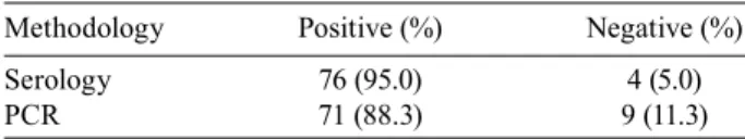
for T. cruzi k-DNA detection in blood by using different methods for preserving biological samples is shown in Table II. The results show that preservation of buffy coat at -70°C provides the highest PCR positivity (86.7%). All 50 individuals used as negative controls were PCR nega-tive. A comparison between serology and PCR results from the 80 patients treated with benznidazol after three years of follow-up is shown in Table III. In the third year after treatment, nine patients with positive PCR before treatment presented negative PCR and, from those, four presented negative serology as well. All the patients who were PCR-negative for T. cruzi DNA presented the spe-cific band (280 bp) for the human hypoxanthine phos-phorybosil transferase (HGPRT) gene, excluding the possibility of PCR reaction inhibition.
TABLE I
Indirect immunofluorescence titers for the follow-up of 80 chagasic patients from the state of Rio Grande do Sul
following treatment with benznidazol
After treatment
Before
treatment 1styear 2ndyear 3rd year
Serology titer n n n n
Negative 0 0 1 4
1/40 6 6 10 39
1/80 6 15 38 35
1/160 30 33 24 2
1/320 14 24 7 0
1/640 24 2 0 0
Total 80 80 80 80
TABLE II
Pre-treatment PCR positivity for T. cruzi k-DNA detection in blood by using different methods for preserving
biological samples
PCR positivity (%) Source of T. cruzi DNA (n = 240)
Buffy-coat -70ºC 208/240 (86.7)a
Buffy-coat GE buffer 172/240 (71.7) Whole blood GE buffer 156/240 (69.2)
a: p < 0.05, Chi square test; GE: guanidine/EDTA.
DISCUSSION
Different tools have been used to diagnose chronic
T. cruzi infection. Most of the serological commercial kits are highly sensitive for the detection of anti-T. cruzi
antibodies, but in Latin American countries where other parasites such as Leishmania spp. and Trypanosoma rangeli are found, false-positive results have been re-ported (Saldaña & Sousa 1996, Caballero et al. 2007). In the state of Rio Grande do Sul, asymptomatic infection by Leishmania Viannia parasites in humans has been re-cently reported (Fagundes et al. 2007).
In the present study, 240 chronic chagasic patients with an indeterminate form of Chagas disease were evaluated by serology, hemoculture and PCR methods. According to the WHO proposal, at least two distinct tests should be performed for a final serodiagnosis conclusion. In our study, 100% concordance between
TABLE III
Comparison between serology and PCR results from the 80 patients treated with benznidazol after a three-years follow-up
Methodology Positive (%) Negative (%)
Serology 76 (95.0) 4 (5.0)
IFI and ELISA assays was found. However, distinct antibody titers varying from 1/40-1/640 were found among these patients. It is well established that the T. cruzi taxon presents two major lineages (TcI and TcII) that may influence parasite antigenic composition and, consequently, patient antibody responses (Di Noia et al. 2002, Buscaglia & Di Noia 2003). This fact must be con-sidered when low or borderline antibody titers are found since most of the commercial kits use the T. cruzi TcII lineage as the antigen. Gutierrez et al. (2004) compared four different serological tests and reported discordant results for 15 out of 94 sera from Colombian chagasic patients. According to the authors, an in-house ELISA using an autochthonous T. cruzi TcI as the antigen shows the highest sensitivity, probably due to the fact that the TcI genotype is the most prevalent in that region. The TcII lineage is found most frequently in both acute and chronic chagasic patients in the south cone countries (Miles et al. 1980, Yeo et al. 2005, Steindel et al. 2008). In Brazil, distinct zymodemes are associated with acute and chronic Chagas disease (Luquetti et al. 1986).
The state of Rio Grande do Sul showed one of the highest prevalence rates (8.84%) of T. cruzi infection in humans (Camargo et al. 1984). Despite this high in-fection prevalence, it is noteworthy that manifestations of cardiac or digestive disease are rare in the state of Rio Grande do Sul (Vinhaes & Dias 2000). It has been speculated that this apparent difference may be due to the characteristics of the T. cruzi populations circulating there. In the southern cone regions where the TcII geno-type is most frequently found in humans, chagasic mega syndromes are common. In comparison, they are absent in the North of the Amazon (Miles et al. 1981). It seems that the TcII genotype is more virulent than TcI (Di Noia et al. 2002). Studies of 29 T. cruzi strains isolated from chagasic patients from the state of Rio Grande do Sul showed that these strains exhibit low virulence patterns in mice (Fernandes et al. 1997).
According to the Brazilian Ministry of Health, treat-ment of T. cruzi infection is recommended during the acute phase, congenital infection, early chronic phase (children under 15 years old and elderly patients with ev-idence of early chronic infection) and reactivation of the infection in individuals infected with HIV. At the Centro de Cardiologia do Rio Grande do Sul, specific treatment for T. cruzi infection is offered to all patients having a positive hemoculture. In the present study, 80 patients with positive hemoculture received treatment with benz-nidazol (5 mg/kg of body weight for 60 days taken twice a day) and were followed up for a three-year period by means of serological, parasitological and PCR methods.
Differences in the sensitivity of hemoculture have been reported. Luz et al. (1993) found 90% positiv-ity among untreated chronic chagasic patients from the state of Minas Gerais. Fernandes et al. (1999) reported 76% positivity among children from the state of Rio Grande do Sul. Lower rates of sensitivity for chronic patients have been shown by other authors (Chiari et al. 1989, 55.8%, Gomes et al. 1999, 36.5%, Lages-Silva et al. 2006, 53.0%). Such differences may represent dis-tinct levels of parasitemia that may depend on the phase
of the disease, the parasite strain and the host immune response. In the present study, 80/240 (33.3%) positiv-ity by hemoculture was found in the chronic chagasic patients with positive serology.
Longitudinal studies in endemic and non-endemic areas have shown that anti-T. cruzi antibodies persisted in infected individuals for many years (Coura et al. 1996, Gomes et al. 1999, Francolino et al. 2003). On the other hand, serology became negative after successful para-sitological treatment of both acute and chronic infec-tion, indicating cure from the infection (Luquetti 1999). A negative parasitological test alone does not represent treatment success; however, its positivity after chemo-therapy represents therapeutic failure. In a study of 27 chronic chagasic patients from the state of Goiás sub-mitted to chemotherapy with benznidazol, 11.1% of the patients presented positive hemoculture, demonstrating treatment failure (de Castro et al. 2006). In the present study, none of the 80 treated patients showed positive hemocultures in the three-year follow-up. Seventy-six out of the 80 (95.0%) patients showed positive serology with decreasing titers during the follow-up and four pa-tients (5.0%) revealed negative serology by both methods - evidence of being cured. One of those patients negated the serology in the second year after treatment and the other three negated the serology in the third year.
Due to the long persistence of anti-T.cruzi antibodies after chemotherapy and the low sensitivity of parasito-logical methods, PCR has been suggested to be a very useful tool for treated patients’ follow-up (Britto et al. 1995b). In our study, we analyzed the performance of PCR for the detection of T. cruzi k-DNA in the blood of two panels of patients using three distinct sample preservation methods. PCR performed with the frozen buffy coat as the T. cruzi DNA source showed a higher positivity than the GE-preserved buffy coat or whole blood. The higher PCR positivity on buffy coat prepara-tions is due to similar density between leucocytes and T. cruzi. Therefore, the bloodstream parasites concentrate on that zone after differential centrifugation. It is well known that hemoglobin is a strong PCR inhibitor. In our experiments using primers for the detection of the hu-man HGPRT gene, we observed the expected band in all PCR-negative patients, revealing that the PCR reaction was not inhibited.
REFERENCES
Avila HA, Pereira JB, Thiemann O, de Paiva E, De Grave W, Morel CM, Simpson L 1993. Detection of Trypanosoma cruzi in blood specimens of chronic chagasic patients by polymerase chain reac-tion amplificareac-tion of kinetoplast minicircle DNA: comparison with serology and xenodiagnosis. J Clin Microbiol31: 2421-2426.
Britto C, Cardoso A, Silveira C, Macedo V, Fernandes O 1995a. Poly-merase chain reaction (PCR) as a laboratory tool for the evalua-tion of the parasitological cure in Chagas disease after specific treatment. Medicina (B Aires) 59: 176-178.
Britto C, Cardoso MA, Vanni CM, Hasslocher-Moreno A, Xavier SS, Oelemann W, Santoro A, Pirmez C, Morel CM, Wincker P 1995b. Polymerase chain reaction detection of Trypanosoma cruzi in hu-man blood samples as a tool for diagnosis and treatment evalua-tion. Parasitology110: 241-247.
Britto C, Cardoso MA, Winker P, Morel CM 1993. A simple protocol for the physical cleavage of Trypanosoma cruzi kinetoplast DNA present in blood samples and its used in polimerase chain reac-tion (PCR) based diagnosis of Chagas disease. Mem Inst Oswaldo Cruz 88: 1711-1712.
Britto C, Silveira C, Cardoso MA, Marques P, Luquetti A, Macêdo V, Fernandes O 2001. Parasite persistence in treated chagasic pa-tients revealed by xenodiagnosis and polymerase chain reaction.
Mem Inst Oswaldo Cruz96: 823-826.
Buscaglia CA, Di Noia J 2003. Trypanosoma cruzi clonal diversity and the epidemiology of Chagas’ disease. Microb Infec 5: 419-427.
Caballero ZC, Sousa OE, Marques WP, Saez-Alquezar A, Umezawa ES 2007. Evaluation of serological tests to identify Trypanosoma cruzi infection in humans and determine cross-reactivity with
Trypanosoma rangeli and Leishmania spp. Clin Vaccine Immunol 14: 1045-1049.
Camargo ME, Silva GR, Castilho EA, Silveira AC 1984. Inquérito sorológico da prevalência da infecção chagásica no Brasil, 1975-1980. Rev Inst Med Trop São Paulo 26: 192-204.
Chiari E, Dias JC, Lana M, Chiari CA 1989. Hemocultures for the parasitological diagnosis of human chronic Chagas’ disease. Rev Soc Bras Med Trop22: 19-23.
Coura JR 2007. Chagas disease: what is known and what is needed - A background article. Mem Inst Oswaldo Cruz102 (Suppl 1): 113-122.
Coura JR, Fernandes O, Arboleda M, Barett TV, Carrara N, Degrave W, Campbell DA 1996. Human infection by Trypanosoma rangeli
in the Brazilian Amazon. Trans R Soc Trop Med Hyg 90: 278-279.
de Castro AM, Luquetti AO, Rassi A, Chiari E, Galvão LM 2006. Detection of parasitemia profiles by blood culture after treat-ment of human chronic Trypanosoma cruzi infection. Parasitol Res99: 379-383.
Di Noia JM, Busgaglia CA, Marchi CR, Almeida IC, Frasch AC 2002. A Trypanosoma cruzi small surface molecule provides the first immunological evidence that Chagas’ disease is due to single parasite lineage. J Exp Med 195: 401-413.
Dias JCP 1997. Controle da doença de Chagas. In JPC Dias, JR Cou-ra (eds.). Clínica e terapêutica da doença de Chagas. Editora Fiocruz, Rio de Janeiro, p. 453-467.
Fagundes A, Marzochi MC, Fernandes O, Perez MA, Schubach AO, Schubach TM, Amendoeira MR, Mouta-Confort E, Marzochi KB 2007. First encounter of subclinical human Leishmania ( Vi-annia) infection in state of Rio Grande do Sul, Brazil. Mem Inst Oswaldo Cruz102: 1003-1005.
Fernandes CD, Murta SMF, Cerávolo IP, Krug LP, Vidigal PG,
Stein-del M, Nardi N, Romanha AJ 1997. Characterization of Trypano-soma cruzi strains isolated from chronic patients, triatomines and opossums naturally infected from the state of Rio Grande do Sul, Brazil. Mem Inst Oswaldo Cruz 92: 343-351.
Fernandes CD, Tiecher FM, Fernandes DD, Pinheiro NM, Steindel M 1999. High rates of positive hemocultures in children and teenag-ers infected by Trypanosoma cruzi in the state of Rio Grande do Sul, Brazil. Mem Inst Oswaldo Cruz 94: 7-8.
Francolino SS, Antunes AF, Talice R, Rosa R, Selanikio J, de Re-zende JM, Romanha AJ, Dias JC 2003. New evidence of spon-taneous cure in human Chagas’ disease. Rev Soc Bras Med Trop 36: 103-107.
Galvão LMC, Cançado JR, Rezende DF, Krettli AU 1989. Hemocul-ture from chronic chagasic pasient using EDTA or heparin as an-ticoagulants. Braz J Med Biol Res 22: 841-843.
Gomes ML, Galvão LM, Macedo AM, Pena SD, Chiari E 1999. Cha-gas’ disease diagnosis: comparative analysis of parasitologic, mo-lecular and serologic methods. Am J Trop Med Hyg60: 205-210.
Gutierrez R, Angulo VM, Tarazona Z, Britto C, Fernandes O 2004. Comparison of four serological tests for the diagnosis of Chagas disease in a Colombian endemic area. Parasitology 129: 439-444.
Lages-Silva E, Ramírez LE, Pedrosa AL, Crema E, da Cunha Galvão LM, Junho Pena SD, Macedo AM, Chiari E 2006. Variability of kinetoplast DNA gene signatures of Trypanosoma cruzi II strains from patients with different clinical forms of Chagas’ disease in Brazil. J Clin Microbiol44: 2167-2171.
Luquetti AO 1999. Evolution of knowledge on the etiological diagno-sis of chagasic infection. Mem Inst Oswaldo Cruz94: 283-284.
Luquetti AO, Miles MA, Rassi A, de Rezende JM, de Souza AA, Po-voa MM, Rodrigues I 1986. Trypanosoma cruzi: zymodemes as-sociated with acute and chronic Chagas disease in central Brazil.
Trans R Soc Trop Med Hyg 80: 462-470.
Luquetti AO, Rassi A 2000. Diagnóstico laboratorial da infecção pelo
Tripanossoma cruzi. In Z Brener, ZA Andrade, M Barral-Netto (eds). Trypanosoma cruzi e doença de Chagas, Guanabara Koo-gan, Rio de Janeiro, p. 344-378.
Luz ZMP, Coutinho MG, Cançado JR 1993. Alta positividade de hemoculturas repetidas em pacientes chagásicos. Rev Soc Bras Med Trop 26: 66-67.
Marcon GEB, Andrade PD, Albuquerque DM, Wanderley JS, Almei-da EA, Guariento ME, Costa SCB 2002. Use of a nested poly-merase chain reaction (N-PCR) to detect Trypanosoma cruzi in blood samples from chronic chagasic patients and patients with doubtful serologies. Diagn Microbiol Infect Dis 43: 39-43.
Massad E 2007. The elimination of Chagas’ disease from Brazil. Epi-demiol Infect4: 1-12.
Miles MA, Cedillos RA, Póvoa MM, de Souza AA, Prata A, Ma-cedo V 1981. Do radically dissimilar Trypanosoma cruzi strains (zymodemes) cause Venezuelan and Brazilian forms of Chagas’ disease? Lancet1: 1338-1340.
Miles MA, Lanhan SM, De Souza AA, Povoa DG 1980. Further en-zymic characters of Trypanosoma cruzi and their evaluation for strain identification. Trans R Soc Trop Med Hyg 74: 221-242.
Prata A 2001. Clinical and epidemiological aspects of Chagas disease.
Lancet Infect Dis1: 92-100.
Saldaña A, Sousa OE 1996. Trypanosoma rangeli and Trypanosoma cruzi: cross-reaction among their immunogenic components.
Mem Inst Oswaldo Cruz91: 81-82.
humans and vectors using a set of primers (BP1/BP2) targeted to a nuclear DNA sequence. Exp Parasitol 85:225-232.
Silveira AC, Rezende DF 1994. Epidemiologia e controle da trans-missão vetorial da doença de Chagas. Rev Soc Bras Med Trop 27: 11-22.
Steindel M, Kramer Pacheco L, Scholl D, Soares M, de Moraes MH, Eger I, Kosmann C, Sincero TC, Stoco PH, Murta SM, de Car-valho-Pinto CJ, Grisard EC 2008. Characterization of Trypano-soma cruzi isolated from humans, vectors and animal reservoirs following an outbreak of acute human Chagas disease in Santa Catarina state, Brazil. Diag Microbiol Infect Dis60: 25-32.
Sturm NR, Degrave W, Morel CM, Simpson L 1989. Sensitive detec-tion and schizodeme classificadetec-tion of Trypanosoma cruzi cells by amplification of kinetoplast minicircles DNA sequences: use in di-agnosis of Chagas disease. Mol Biochem Parasitol33: 205-214.
Vinhaes MC, Dias JC 2000. Chagas disease in Brazil. Cad Saude Publica 16 (Suppl. 2): 7-12.