CAMPYLOBACTER JEJUNI IN FOOD AND FAECAL SAMPLES
Harkanwaldeep Singh1; R.S. Rathore1; Satparkash Singh2; Pawanjit Singh Cheema2*
1
Division of Veterinary Public Health, Indian Veterinary Research Institute, Izatnagar, Bareilly-243122, India; 2Division of
Bacteriology and Mycology, Indian Veterinary Research Institute, Izatnagar, Bareilly-243122, India.
Submitted: August 29, 2009; Approved: June 21, 2010.
ABSTRACT
In the present study, the efficacy of polymerase chain reaction (PCR) based on mapA gene of C. jejuni was
tested for detection of Campylobacter jejuni in naturally infected as well as spiked faecal and food samples
of human and animal origin. Simultaneously, all the samples were subjected to the cultural isolation of
organism and biochemical characterization. The positive samples resulted in the amplification of a DNA
fragment of size ~589 bp in PCR assay whereas the absence of such amplicon in DNA extracted from E.
coli, Listeria, Salmonella and Staphylococcus confirmed the specificity of the primers. Of randomly
collected 143 faecal samples comprising human diarrheic stools (43), cattle diarrheic faeces (48) and
poultry faecal swabs (52) only 4, 3 and 8, respectively, could be detected by isolation whereas 6, 3 and 10,
respectively, were found positive by PCR. However, among food samples viz. beef (30), milk (35), cheese
(30), only one beef sample was detected both by culture as well as PCR. Additionally, PCR was found to
be more sensitive for C. jejuni detection in spiked faecal and food samples (96.1% each) as relative to
culture isolation which could detect the organism in 86.7% and 80% samples, respectively. The results
depicted the superior efficacy of PCR for rapid screening of samples owing to its high sensitivity,
specificity and automation potential.
Key words: Campylobacter jejuni, isolation, PCR, spiking
INTRODUCTION
Different species within the genus Campylobacter have
emerged over the last three decades as important clinical
pathogens of human and veterinary concern. The majority of
acute bacterial intestinal infections in human beings in the
western countries are caused by these organisms, particularly
due to thermotolerant campylobacters (11). Among these, C.
jejuni and C. coli are the most common pathogens responsible
for the majority of human enteritis cases (2, 15). C. jejuni
subsp. jejuni has also been reported to cause abortion and
mastitis in bovines. Besides, zoonotic campylobacters have
been found associated with potentially life threatening
complications like Guillain-Barre syndrome, reactive arthritis,
hemolytic uraemic syndrome and meningitis etc. (7, 16, 17).
The prime cause of campylobacter infections is considered to
be contaminated food as the organism is a part of normal flora
in various animal species such as poultry, pigs, and cattle.
In the recent past, number of enteritis cases in humans due
to campylobacters has exceeded to those caused by Salmonella
and Shigella, especially in developed world. However, in
developing countries, the true incidence of campylobacteriosis
is often underestimated because of lack of adequate laboratory
infrastructure. The conventional methods of detection of
campylobacters are based on cultural isolation followed by
various genus and species specific biochemical tests which are
cumbersome and time consuming. The hippurate hydrolysis, a
routinely performed test for identification of C. jejuni has its
own limitations as false negative as well as false positive
results with this test have been reported due to emergence of
some hippurate negative C. jejuni strains and some hippurate
positive non-C. jejuni strains, respectively (5, 12).
Additionally, Campylobacter spp. can survive as viable but non
culturable (VBNC) forms which may not grow on selective
media. Subsequently, the refrigerated storage under reduced
oxygenated conditions that occur in modified atmospheric
packaging or vacuum packaging of food products may allow
resuscitation of injured or VBNC Campylobacter spp, hereby
rendering a potential threat to human health.
Polymerase chain reaction (PCR) assays have been widely
employed for identification of the pathogens owing to their
sensitivity and cost effectiveness (8). A number of PCR assays
have been described for the detection of campylobacters from
food and faecal samples(1, 4, 6, 13, 14). The present study was
carried to access the prevalence of C. jejuni in various food and
faecal samples and to compare the efficacy of cultural and
biochemical tests with PCR for detection of the organism.
MATERIALS AND METHODS
Sample collection
A total of 238 faecal and food samples of human and
animal origin belonging to Uttar Pradesh state of India were
included in the study. Of these, faecal samples comprised
human diarrhoeic stools (43), cattle diarrhoeic faeces (48) and
poultry faecal swabs (52) where as the food samples included
beef (30), milk (35) and cheese (30). The samples were
collected over ice maintaining all the sterility measures and
brought to the laboratory in enrichment medium.
Cultural and biochemical examination
For cultural isolation of the organism, modified selective
media (3) was employed. Briefly, the human and cattle
diarrhoeic stool samples and faecal swabs of poultry were
inoculated into modified enrichment broth and incubated at
37oC for 48 hr under microaerophilic conditions (5% O2, 10%
CO2 and 85% N2) using CampyPak (BD, Oxoid) gas
generating packs. The grown cultures from broth were streaked
onto respective agar plates, incubated as above and were
regularly observed for 5-7 days for any bacterial growth.
Characteristic Campylobacter colonies were tested for genus
specific phenotypic and biochemical characters i.e., Gram’s
staining, motility, oxidase, catalase and nitrate reduction tests
followed by species specific characters i.e., hippurate
hydrolysis, growth in 1% glycine, H2S production on triple
sugar iron agar, growth at 25oC and 42oC and sensitivity to
nalidixic acid.
Preparation of samples for PCR
Faecal samples, five gram each from cattle, poultry and
human were mixed with 50 ml of enrichment broth so as to
make a homogeneous suspension. The mixture was incubated
under microaerophillic conditions at 37oC for 3 h and then for
18 h at 42oC. Subsequently, it was centrifuged passively to
remove the debris and 1 ml of supernatant obtained was further
centrifuged at 10000xg for 10 min. The resulting pellet was
resuspended in 100 µl of TE buffer (10mM Tris-HCl, 1mM
EDTA, pH 8.0), boiled for 10 min followed by immediate
chilling on ice. After its centrifugation at 10000xg for 10 min,
a five µl of the supernatant was directly used as template in 25
µl PCR reaction. For preparation of template DNA from beef
and cheese, five gram minced sample from either of these was
diluted ten times (w/v) in enrichment broth and later on
processed in same way as that of stools. As regards milk
samples, 10 ml milk was added to 90 ml enrichment broth and
centrifuged at 10000xg for 10 min and the resulting pellet was
resuspended in 100 µl of TE buffer (pH 8.0). Subsequent
processing was carried out in similar manner as that of faecal
samples.
PCR assay
The primers based on mapA gene of C. jejuni (5) were got
custom synthesized. The sequences of forward and reverse
oligonucleotide primers were as follows:
Forward 5’-CTATTTTATTTTTGAGTGCTTGTG-3’
Reverse 5’-GCTTTATTTGCCATTTGTTTTATTA-3’
The cyclic conditions for PCR were same as those
described by Denis et al. (5) which were as follows: initial
denaturation at 94oC for 2 min followed by 30 cycles of
denaturation at 94oC for 40 sec, annealing at 54oC for 40 sec
and extension at 72oC for 1 min and a final extension at 72oC
for 5 min. The reaction mixture comprised of 1x PCR buffer
[50 mM Tris-HCl, 10 mM KCl, 5 mM (NH4)2SO4, pH8.3], 1.0
mM MgCl2 (MBI fermentas, USA), 0.2 mM dNTP mix (MBI
fermentas, USA), 16 p mol of each of the primers (Integrated
DNA Technologies, Inc, IA, USA), 1 U of Taq DNA
polymerase enzyme (MBI fermentas, USA), 5 l of template
DNA in 25 l of reaction mixture. The PCR products were
analyzed by 1.5% agarose gel (Amersham Pharmacia Biotech
AB, Uppsala, Sweden) electrophoresis and photographed using
a gel documentation system (Alpha Imager, Germany).
Specificity and sensitivity of PCR
To test the specificity of primers, the PCR assay was also
applied on E. coli, Salmonella, Listeria and Staphylococcus
organisms. PCR reaction mixture and cyclic conditions were
kept same as described above.
For testing the sensitivity of PCR, freshly grown pure
culture of Campylobacter was taken and the concentration of
cells in liquid culture was estimated to be 109 cells/ml. From
this culture, 10 fold serial dilutions were made from 109 to 104
cells/ml. Each of these dilutions was further diluted 1:10 in TE
buffer (pH 8.0), boiled for 10 min followed by chilling over ice
and centrifugation at 10000xg for 10 min. A 5 µl of the
supernatant was used as template in the PCR, resulting in a
final concentration ranging from 5x10⁵ to 51 cells per PCR.
Artificial inoculation/Spiking studies
The experimental inoculation studies were carried out to
assess the efficacy of the standardized PCR method for the
detection of C. jejuni in spiked faecal and food samples. Faecal
samples from cattle, poultry and human were inoculated with
overnight grown culture of C. jejuni so as to make a final
concentration of 106 bacterial cells/ml of stools and the
resulting mixture was centrifuged passively to remove the
debris. The resulting supernatant was diluted ten times in TE
buffer (pH 8.0) and boiled for 10 min followed by immediate
chilling on ice. Afterwards, it was centrifuged at 10000xg for
10 min and a five µl of the supernatant was used as template in
PCR. For spiking of milk, overnight grown culture of C. jejuni
was added to the pasteurized whole milk in order to achieve a
final concentration of 106 bacterial cells/ml of milk. One ml of
this spiked milk was diluted ten times in TE buffer (pH 8.0)
and later on processed in a way same as that of stools for
preparation of template DNA. As regards beef and cheese
samples, one gram of minced beef or cheese was mixed with
10 ml of enrichment broth and C. jejuni cells were added to it
making a final concentration of 106 cells/ml of suspension.
Subsequently, the debris was removed by passive
centrifugation and the supernatant was processed for the
preparation of template DNA as described elsewhere.
Simultaneously, the spiked samples were also streaked onto the
modified selective solid media and plates were incubated at
42oC for 3-5 days under microaerophilic conditions for cultural
isolation of C. jejuni.
RESULTS AND DISCUSSION
Of 143 faecal samples, 15 were found positive for C.
jejuni by cultural and biochemical examination, out of which 4
regards food samples, only one (beef) was found positive
whereas all the milk and cheese samples were found negative.
Biochemically, the organisms were positive for oxidase,
catalase, nitrate reduction tests as well as species specific tests
like hippurate hydrolysis, growth in 1% glycine and were
sensitive to nalidixic acid.
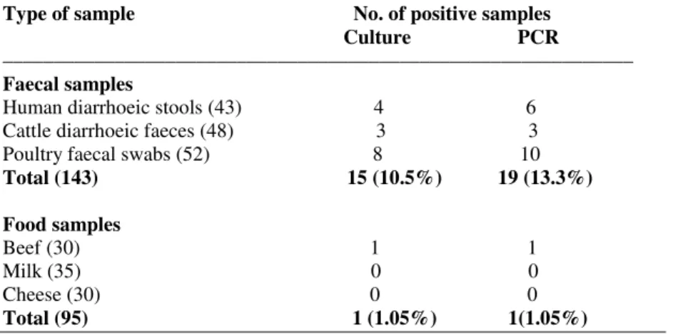
Table 1. Results of cultural isolation and PCR for detection of C.jejuni in randomly collected faecal and food samples
Type of sample No. of positive samples Culture PCR
–––––––––––––––––––––––––––––––––––––––––––––––––––––––––––––– Faecal samples
Human diarrhoeic stools (43) 4 6 Cattle diarrhoeic faeces (48) 3 3 Poultry faecal swabs (52) 8 10 Total (143) 15 (10.5%) 19 (13.3%)
Food samples
Beef (30) 1 1 Milk (35) 0 0 Cheese (30) 0 0 Total (95) 1 (1.05%) 1(1.05%)
PCR detected all those faecal samples found positive by
cultural examination as a fragment of size ~589 bp (Fig. 1) was
amplified from these samples. Additionally, four more samples
were found positive which were culturally negative; 2 each
from human diarrhoeic stools and poultry faecal swabs,
respectively. Hence, PCR was found more efficient for
detecting C. jejuni from faecal samples (10.5% by culture
versus 13.3% by PCR). Earlier workers have also demonstrated
the superior efficacy of PCR in detection of the campylobacters
from faecal samples declared negative by selective cultural and
biochemical tests (9, 10).
Regarding food samples, no difference was observed in
PCR and culture isolation methods for detection of organism as
only one beef sample was detected by PCR that was found
positive by selective culture method also. Low incidence of C.
jejuni in raw beef (3.2%) and raw bulk tank milk samples
(1.6%) has been reported earlier also (19).
As isolation and identification of the campylobacters
based on selective culture and biochemical differentiation upto
species level is tedious, time consuming and has been proved
time and again not very reliable. Hence, on the basis of our
study, we can say that PCR based methods are more rapid and
reliable, particularly while processing a large number of
samples.
No amplification was observed in PCR using DNA
extracted from E. coli, Listeria, Salmonella and Staphylococcus
organisms (Fig. 2). The absence of desired amplicon from
these organisms confirmed the specificity of the primers.
Regarding sensitivity of PCR on DNA extracted from pure
culture of C. jejuni by heat lysis method, upto a minimum of 50
cells per PCR reaction (corresponding to 105 cells/ml of
culture) were detected. However, the intensity of amplicons
gradually improved with increase in concentration of cells (Fig. Figure 1. PCR amplification
of mapA gene from
representative samples for
detection of Campylobacter
jejuni. Lane M: DNA ladder.
Lanes 1 to 3: human
diarrhoeic stool, cattle
diarrhoeic faeces and poultry
faecal swab samples,
3). High sensitivity of PCR assay for detection of C. jejuni
from pure culture was in agreement with observation of
Perssson and Olsen (13) who detected 10-100 cells per PCR
reaction.
Figure 2. Specificity study of PCR amplification of mapA gene. Lane
M: DNA ladder. Lane 1: Campylobacter jejuni. Lane 2: E. coli. Lane 3: Salmonella. Lane 4: Listeria. Lane 5: Staphylococcus.
Figure 3. Sensitivity assay of PCR by 10 fold dilution of bacterial
DNA derived from Campylobacter jejuni culture. Lane M: DNA ladder. Lanes 1 to 6: 104, 105, 106, 107, 108, 109 cells/ml respectively.
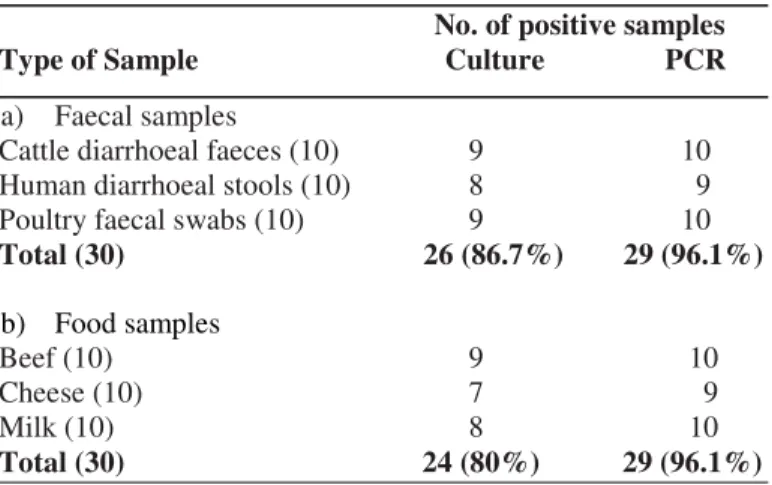
As regards artificial inoculation studies, culture and
biochemical identification could detect 26 (86.7%) of 30 faecal
samples and 24 (80%) of 30 food samples whereas 29 (96.1%)
each of faecal and food samples were found positive in PCR
assay (Table 2). Persson and Olsen (13) and Waage et al. (18)
have also found PCR to be quite effective in detection of C.
jejuni from spiked samples. Our study on spiked samples
further underscores the better efficacy of PCR over cultural
identification of the organism and bolsters the application of
technique in randomly collected faecal and food samples.
Table 2. Comparison of culture and PCR for detection of C.
jejuni in spiked faecal and food samples
No. of positive samples Type of Sample Culture PCR
a) Faecal samples
Cattle diarrhoeal faeces (10) 9 10 Human diarrhoeal stools (10) 8 9 Poultry faecal swabs (10) 9 10 Total (30) 26 (86.7%) 29 (96.1%)
b) Food samples
Beef (10) 9 10
Cheese (10) 7 9
Milk (10) 8 10
Total (30) 24 (80%) 29 (96.1%)
REFERENCES
1. Al Amri, A.; Senok, A.C.; Ismaeel, A.Y.; Al-Mahmeed, Ali E.; Botta, Giuseppe A. (2007). Multiplex PCR for direct identification of Campylobacter spp. in human and chicken stools. J. Med. Microbiol. 56, 1350-1355.
2. Allos, B.M.; Blaser, M.J. (1995). C. jejuni and expanding spectrum of related infections. Clin. Infect. Dis. 20, 1092-1099.
3. Barua, R.; Rathore, R.S. (2006). Development of modified selective media for the isolation of Campylobacter jejuni from poultry. J. Food Sci. Technol. 43, 305-307.
4. Debruyne, L.; Samyn, E.; De Brandt, E.; Vandenberg, O.; Heyndrickx, M.; Vandamme, P. (2008). Comparative performance of different PCR assays for the identification of Campylobacter jejuni and Campylobacter coli. Res. Microbiol. 159, 88-93.
5. Denis, M.; Soumet, C.; Rival, K.; Ermel, G.; Salavat, G.; Colin, P. (1999). Development of a mPCR assay for simultaneous identification of Campylobacter jejuni and C. coli. Lett. Appl. Microbiol. 29, 406-410. 6. Jackson, C.J.; Fox, A.J.; Jones, D.M. (1996). A novel polymerase chain
reaction assay for the detection and speciation of thermophilic Campylobacter spp. J. Appl. Bacteriol. 81, 467-473.
8. Kricka, L.J. (1998). Prospects for chemiluminescent and bioluminescent immunoassay and nucleic acid assays in food testing and the pharmaceutical industry. J. Biolumin. Chemilumin. 13, 189-193. 9. Kulkarni, S.P.; Lever, S.; Logan, J.M.; Lawson, A.J.; Stanley, J.; Shafi,
M. (2002). Detection of Campylobacter species: a comparison of culture and polymerase chain reaction based methods. J. Clin. Pathol. 55, 749-753.
10. Lawson, A.J.; Shafi, M.S.; Pathak, K.; Stanley, J. (1998). Detection of Campylobacter in gastroenteritis: Comparison of direct PCR assay of faecal samples with selective culture. Epidemiol. Infect. 121, 547-553. 11. Maher, M.; Finnegan, C.; Collins, E.; Ward, B.; Carroll, C.; Cormican,
M. (2003). Evaluation of culture methods and a DNA probe-based PCR assay for detection of Compylobacter spp. in clinical specimens of faeces. J. Clin.Microbiol. 41, 2980-2986.
12. Morris, G.K.; El Sherbeeny, M.R.; Patton, C.M.; Kodaka, H.; Lombard, G.L.; Edmonds, P.; Hollis, D.G.; Brenner, D.J. (1985). Comparison of four hippurate hydrolysis methods for identification of thermophilic Campylobacter spp. J. Clin. Microbiol. 22, 714-718.
13. Persson, Søren; Olsen, Katharina E.P. (2005). Multiplex PCR for identification of Campylobacter coli and Campylobacter jejuni from pure
cultures and directly on stool samples. J. Med. Microbiol. 54, 1043-1047. 14. Sails, Andrew D.; Fox, A.J.; Bolton, F.J.; Wareing, D.R.A.; Greenway, D.L.A. (2003). A Real-Time PCR Assay for the detection of Campylobacter jejuni in foods after enrichment culture. Appl. Environ. Microbiol. 69, 1383-1390.
15. Skirrow, M.B. (1994). Disease due to Campylobacter, Helicobacter and related bacteria. J. Comp. Pathol., 111, 113-149.
16. Stanfford, T.D.; Tenkate, R.J. (2001). Risk factors for Campylobacter infection in infants and young children: A matched case control study. Epidemiol.Infect. 127, 399-404.
17. Stern, N.D.; Line, J.E. (2000). Campylobacter. In: The microbiological safety and quality of food, pp. 1040-1056.
18. Waage, A.S.; Vardund, T.; Lund, V.; Kapperud, G. (1999). Detection of small numbers of Campylobacter jejuni and Campylobacter coli cells in environmental water, sewage, and food samples by a seminested PCR assay. Appl. Environ. Microbiol. 65, 1636-1643.