www.cbpv.com.br/rbpv
Ovary histology and quantification of hemolymph proteins of
Rhipicephalus
(
Boophilus
)
microplus
treated with
Melia azedarach
Histologia do ovário e quantificação de proteínas na hemolinfa de
Rhipicephalus
(
Boophilus
)
microplus
tratado com
Melia
azedarach
Lorena Alessandra Dias de Sousa1; Thiago Lopes Rocha2; Simone Maria Teixeira Sabóia-Morais2; Lígia Miranda Ferreira Borges1*
1Centro de Parasitologia Veterinária, Instituto de Patologia Tropical e Saúde Pública, Escola de Veterinária e Zootecnia,
Universidade Federal de Goiás – UFG, Goiânia, GO, Brasil
2Laboratório de Comportamento Celular, Departamento de Morfologia, Instituto de Ciências Biológicas – ICB,
Universidade Federal de Goiás – UFG, Goiânia, GO, Brasil
Received January 11, 2013 Accepted July 4, 2013
Abstract
This study aimed to analyze ovary histology and quantify total protein in the hemolymph of Rhipicephalus (Boophilus) microplus females treated with hexane extracts from green fruits of Melia azedarach. Eight engorged females were immersed in the extract at 0.25% concentration, and eight in water containing 5% acetone (control). The females were dissected 72 hours after treatment, and the ovaries were weighed and subjected to standard histological techniques. The total protein concentration was measured in the hemolymph of 200 females, of which 100 were treated as described above and 100 served as a control. In the treated group, ovary weight reduction and predominance of immature oocytes were observed. In addition, there were decreases in the diameters of the cytoplasm and germ vesicle of the oocytes in the treated group, compared with the controls. The protein concentration in the hemolymph was higher in the treated group than in the controls. The morphological changes observed in the treated ovaries included: presence of vacuolization; alteration of oocyte morphology, which changed from rounded to elongated; deformation of the chorion; and disorganization of the yolk granules. These results demonstrate the action of M. azedarach fruit extracts on R. (B.) microplus oogenesis.
Keywords:Rhipicephalus (Boophilus) microplus, oocytes, ovary, histology, hemolymph proteins, Melia azedarach.
Resumo
Este estudo foi desenvolvido, visando analisar a histologia do ovário e quantificar as proteínas totais na hemolinfa de fêmeas de Rhipicephalus (Boophilus) microplus tratadas com extrato hexânico de frutos verdes de Melia azedarach. Oito fêmeas ingurgitadas foram tratadas por imersão com o extrato na concentração de 0,25%, e oito com água contendo 5% de acetona (controle). As fêmeas foram dissecadas 72 horas após o tratamento e os ovários foram pesados e submetidos a técnicas histológicas padrões. A concentração total de proteína foi mensurada na hemolinfa de 200 fêmeas, sendo 100 tratadas como descrito anteriormente e 100 como controle.Foi observada redução do peso dos ovários, predomínio de ovócitos imaturos e houve diminuição nos diâmetros do citoplasma e da vesícula germinal dos ovócitos do grupo tratado em comparação ao controle. A concentração de proteína na hemolinfa foi mais alta no grupo tratado que no controle. As alterações morfológicas observadas nos ovários tratados foram a presença de vacuolizações, alteração da morfologia dos ovócitos que mudaram de redondos para alongados, deformação do córion e desorganização dos grânulos de vitelo. Estes resultados demonstram a ação do extrato de M. azedarach na ovogênese de R. (B.) microplus.
Palavras-chave: Rhipicephalus (Boophilus) microplus, ovócitos, ovário, histologia, proteínas na hemolinfa, Melia azedarach.
*Corresponding author: Lígia Miranda Ferreira Borges
Centro de Parasitologia Veterinária, Instituto de Patologia Tropical e Saúde Pública, Escola de Veterinária e Zootecnia, Universidade Federal de Goiás – UFG, CP 131, CEP 74001-970, Goiânia, GO, Brasil
e-mail: [email protected]
Introduction
Rhipicephalus (Boophilus) microplus is a major parasitic problem for cattle producers, because of the economic losses caused by blood spoliation, predisposition towards myiasis, damage to leather, retardation of calf development, decreased meat production, excessive expenditure on control measures and transmission of pathogens that cause diseases such as anaplasmosis and babesiosis (JONSSON, 2006).
The main method for controlling R. (B.) microplus is based on use of synthetic acaricides. However, frequent use of these compounds can lead to presence of toxic residues in food, biological imbalance, environmental contamination, poisoning of human beings and animals, and selection of resistant ticks (SONENSHINE, 1991).
The main motivation for searching for plant molecules as a possible tick control method arose from the perceived need to minimize the adverse effects of prolonged and intensive use of chemical acaricides (OLIVO et al., 2009). In this context, some plants such as Melia azedarach and Azadirachta indica (Meliaceae)
stand out through contributing bioactive extracts for R. (B.)
microplus control. Particularly with M. azedarach, Borges et al. (1994, 2003) and Sousa et al. (2008, 2011) observed high efficacy against tick reproduction, from fruit extracts. It is known that this plant has an effect on the neuroendocrine system of insects, which has been described by authors such as Schmidt et al. (1998) and Mordue and Nisbet (2000). However, there is no information on its interference with the physiology of oogenesis in ticks.
The ovary of R. (B.) microplus is classified as panoistic because it does not contain follicular and nourishing cells. It consists of a single tubular structure with oocytes of various sizes and at different developmental stages, attached to the epithelial wall through a pedicel. The latter structure is responsible for synthesizing and providing substances for oocyte development (SAITO et al., 2005). In ticks, vitellogenin synthesis occurs mainly outside of the ovaries: it is released into the hemolymph for subsequent ovary uptake (DIEHL; DOTSON, 1986).
Oogenesis in R. (B.) microplus is divided into six stages from I to VI that are defined in terms of cytoplasm appearance and presence of the germ vesicle, yolk granules and chorion (SAITO et al., 2005).
Given the above, this study aimed to analyze the histology of the ovary and quantify the total protein in the hemolymph of R. (B.) microplus females treated with hexane extracts from green fruits of M. azedarach.
Materials and Methods
Tick collection
Engorged females of R. (B.) microplus were obtained from naturally infested Holstein cattle that had been left without acaricide treatment for at least 30 days. After collection, the ticks were washed with water, dried and selected based on their external morphological condition. They were weighed individually in order to standardize the individuals examined at between 200 and 235 mg.
Preparation of extracts of Melia azedarach and
immersion test
Unripe fruits of M. azedarach were collected in Goiânia (16° 34’ 24” S and 49° 17’ 32” W), because these have been found to be bioactive against the reproductive efficiency of R. (B.) microplus (BORGES et al., 2003; SOUSA et al., 2008). Dry and powdered ripe fruits were extracted by means of percolation in a Soxhlet apparatus using hexane as the solvent. The solvent was then evaporated using a rotary evaporator (Rotavapor®).
Eight females were weighed and immersed for 5 min in 10 mL of solution containing the extract at 0.25% concentration diluted in water plus 5% acetone. A control group with the same number of females was immersed in distilled water plus 5% acetone. They were then placed in a Petri dish and conditioned for 72 hours under controlled conditions (T = 27 °C; RH > 80%).
Histology and morphometry of the ovary
Before extraction of the ovaries, the females were anesthetized in a freezer at –5° C for 5 min. Dissection and extraction of the ovaries was performed under an stereomicroscope (Leica®), using a saline solution (NaCl 7.5 g/L, Na2HPO4 2.38 g/L and KH2PO4 2.72 g/L). The ovaries were weighed for subsequent calculation of the gonadosomatic index (= ovarian weight / weight of females) and were deposited individually into Eppendorf tubes with buffered paraformaldehyde solution for 24 h (pH 7.4; 4% formaldehyde). After this period, the solution was discarded and 70% alcohol was added and the material was stored at 4 °C.
Subsequently, the ovaries were dehydrated in increasing ethanol concentrations (70% to 100%); embedded in Historesin® (Leica, USA) in accordance with the manufacturer’s specifications; and subjected to microtomy to obtain sections with 2.0 µm thickness, using an ultra-microtome (Leica Ultracut UCT, USA). The sections were stained with toluidine blue (TB) 1% (w/v) at pH 8.4 (VETEC, Brazil). Each ovary was cut at a distance of 0.5 cm from the proximal portion of the oviduct.
The histopathological analyses were performed on micrographs of the ovaries, which were obtained under a photonic microscope (Leica DMLB, USA) coupled to the image capture system (Samsung SHC 410 NAD digital color camera), using objective lenses of 10, 20 and 40X with a 100 µm scale. Oocytes I to V of each selected field were first ranked in accordance with the phases described by Saito et al. (2005) and then counted. Observations on the presence of toxic effects due to actions by the M. azedarach extract on the oocytes were also based on characterization of the stages of oogenesis carried out by the latter author.
ovary. From these, one section per slide was selected and three images of each stage of oogenesis were obtained.
Quantification of protein in tick hemolymph
One hundred engorged females were treated with the extract and one hundred with distilled water plus acetone (control group), and were conditioned for 72 h, as described earlier. The dorsal surface of each female was perforated using a glass microtube (outer diameter = 1.0 mm, internal diameter = 0.78 mm and tip diameter = 10-20 µm), prepared in a puller (PC-10, Narishige, Japan). The tick body was subjected to gentle pressure to allow the hemolymph to extravasate. The hemolymph was then aspirated to the same microtube using a 1 mL disposable syringe connected to the tube. Hemolymph samples contaminated with blood due to leakage of intestinal contents were discarded.
The pooled hemolymph samples were placed into tubes containing 30 µL of a protease inhibitor cocktail (Sigma-Aldrich Inhibit ®) and 82 µL of saline buffer (1.5 M NaCl and 50 mM EDTA) plus phenylthiourea and were kept on ice throughout sample collection. After collection, the tubes were centrifuged at 5040 × g for 10 minutes at 4 °C. To quantify total protein in hemolymph,
the modified Lowry method was followed (MARKWELL et al., 1978), using bovine serum albumin (Merck®) as a standard.
Statistical analysis
Statistical analyses were performed using the Statistica 7.0 software (STATSOFT, 2005), by means of the one-way ANOVA test followed by Student t test comparison. Results were considered significant when p < 0.05.
Results
The ovaries of the treated females were of lower weight than those in the control group, which resulted in a significantly lower gonadosomatic index score (0.076 ± 0.015) than in the
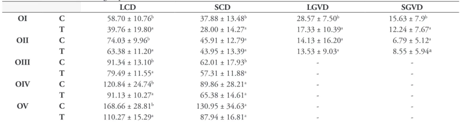
control group (0.148 ± 0.024). The highest total oocyte count was observed in the control group. From the individual scores, immature oocytes predominated and there was less vitellogenesis in the treated group, compared with the controls (Table 1). There was a decrease in cytoplasm diameter in all the oocytes evaluated in the treated group. The diameters of the germ vesicle were also lower in oocyte I in the treated group but there was no change in oocyte II (Table 2).
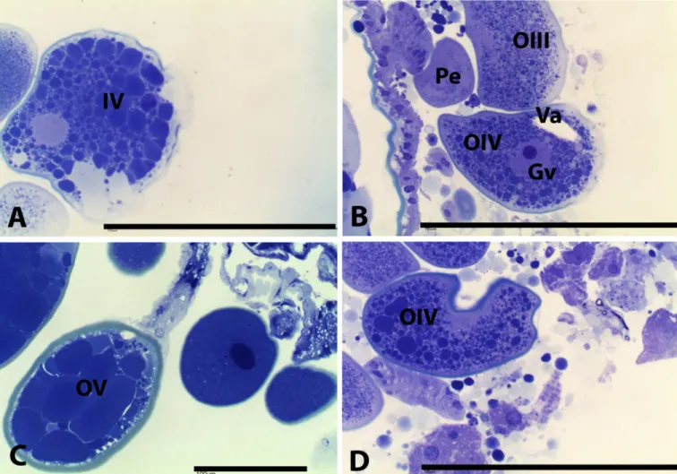
The morphological characteristics for oocytes from the control group (Figure 1) were as follows:
• Oocyte I – rounded with homogeneous cytoplasm and no yolk granules, and the germ vesicle has prominent nucleolus. At this stage, the germ vesicle occupies a large area of the cytoplasm;
• Oocyte II – higher cellular volume compared with oocyte I. At this stage, the oocyte does not have evident granules in the cytoplasm and the germ vesicle and nucleolus are still prominent;
• Oocyte III – cytoplasm is rich in coarse and evenly distributed granulation and the germ vesicle has shifted to the periphery. At this stage, it is possible to identify the chorion surrounding the oocyte, which has a sky-blue color;
Table 1. Mean ± standard deviation of oocyte numbers in each stage (OI to OV) and total counts in the ovaries of Rhipicephalus
(Boophilus) microplus females treated with a hexane extract of unripe fruits of Melia azedarach at 0.25% concentration. The control group was treated with water + acetone.
Oocytes Control Treated
OI 13.54 ± 4.26a 18.8 ± 3.58b
OII 12.67 ± 3.37ª 22.83 ± 3.63b
OIII 15.75 ± 3.15ª 20.79 ± 3.52b
OIV 31.25 ± 7.9b 15.00 ± 2.94ª
OV 26.71 ± 5.82b 12.33 ± 2.82ª
Total 99.92 ± 10.81b 89.54 ± 7.94a
Means followed by different lowercase letter in the lines differ significantly according to Student t test (p < 0.05).
Table 2. Mean (µm) ± standard deviation of largest (LCD) and smallest (SCD) cytoplasm diameters, and largest (LGVD) and smallest (SGVD) germ vesicle diameters of Rhipicephalus (Boophilus) microplus oocytes treated with a hexane extract of unripe fruits of Melia azedarach at 0.25% concentration (T) and control group (C).
LCD SCD LGVD SGVD
OI C 58.70 ± 10.76b 37.88 ± 13.48b 28.57 ± 7.50b 15.63 ± 7.9b
T 39.76 ± 19.80a 28.00 ± 14.27a 17.33 ± 10.39a 12.24 ± 7.67a
OII C 74.03 ± 9.96b 45.91 ± 12.79a 14.13 ± 16.20a 6.79 ± 5.12a
T 63.38 ± 11.20a 43.95 ± 13.39a 13.53 ± 9.03a 8.55 ± 5.94ª
OIII C 91.34 ± 13.10b 62.01 ± 17.93b -
-T 79.49 ± 11.55a 57.31 ± 11.88a -
-OIV C 120.84 ± 24.74b 89.86 ± 28.21a -
-T 91.13 ± 10.27a 65.38 ± 14.61a -
-OV C 168.66 ± 28.81b 130.95 ± 34.63a -
-T 110.27 ± 15.29a 87.94 ± 16.81a -
• Oocyte IV – cytoplasm with large and small yolk granules. In this phase, the germ vesicle and nucleolus are peripheral and the chorion is evident;
• Oocyte V – cytoplasm is completely filled with large yolk
granules and the chorion is thicker;
• Oocyte VI – cytoplasm is disorganized and the chorion has folds, making it irregular.
from rounded to elongated, with deformation of the chorion and disorganization of yolk granules (Figure 2).
The engorged females treated with M. azedarach extract had
higher protein concentrations in the hemolymph (130.57 µg/µL) than shown by females in the control group (112.7 µg/µL).
Discussion
The results from this study demonstrate the effect of crude
hexane extracts from the fruits of M. azedarach on R. (B.)
microplus oogenesis. These results corroborate investigations
that have already been carried out using M. azedarach extracts,
which described interference from the plant compounds on the
reproductive parameters of females of R. (B.) microplus, with
inhibition of egg conversion and larva hatching (BORGES et al., 2003; SOUSA et al., 2008).
The main changes observed here were decreased numbers of mature oocytes, increased numbers of immature oocytes, decreased cytoplasm and germ vesicle diameters and cellular deformities. The observed predominance of immature oocytes
at the expense of vitellogenic oocytes revealed that blockage of
the development process of oogenesis in R. (B.) microplus had
occurred. Morphological alterations such as vacuoles and cellular disorganization indicated cell intoxication caused by the extract of M. azedarach. Vacuolization is an attempt by the cell to isolate the toxic substances or even the cytoplasm that was damaged by the action of the compound, so that the cell can still perform its metabolic processes. In addition to toxic substances and debris, many vacuoles can also contain entire organelles that are unable to perform their metabolic functions (ARNOSTI et al., 2011).
The toxic effect observed in oocytes and nearby cell structures have also been observed in other studies using plants. Denardi et al. (2011) and Arnosti et al. (2011) evaluated the activity of neem (A. indica) and ricinoleic acid on R. sanguineus, and observed that these had inhibitory effects on oocytes, thereby preventing them from reaching advanced stages of development. In addition, there was morphological damage such as vacuolization, cellular disorganization and deformation. Likewise, these findings are similar to those found by Friesen and Kaufman (2003, 2004) in an evaluation on the inhibitory effect of cypermethrin against 20-hydroxyecdysone. This
acaricide caused suppression of vitellogenesis and egg development in Amblyomma hebraeum.
The effects attributed to M. azedarach are largely ascribed to the action of limonoids, especially azadirachtin, salannin, meliantrol and nimbin (YAMASAKI et al., 1988). These compounds can inhibit the molting process through interfering with the neuroendocrine system, thereby altering the hemolymph ecdysteroid levels
(SCHMIDT et al., 1998). In Drosophila melanogaster, Aedes
aegypti and Manduca sexta, azadirachtin, salannin, nimbin and 6-desacetyl nimbin were found to inhibit the activity of ecdysone in a dose-dependent manner (MITCHELL et al., 1997).
In the present study, higher protein concentration was observed in the hemolymph of engorged females treated with hexane extract of M. azedarach than in the controls. Vitellogenin is the most abundant protein in tick eggs and represents approximately 11% of the total protein in hemolymph (SONENSHINE, 1991). Its concentration in hemolymph increases with engorgement, and is regulated by its uptake in oocytes (SEIXAS et al., 2010). Since the treated females predominantly presented immature oocytes and had fewer vitellogenic oocytes, it can be assumed that the accumulation of protein in the hemolymph occurred because vitellogenin was not taken up by oocytes.
Reproduction and embryogenesis in ticks are regulated by ecdysteroid hormones (CABRERA et al., 2009). 20-hydroxyecdysone is the major ecdysteroid hormone, accompanied by its precursor, ecdysone (DELBECQUE et al., 1978; DEES et al., 1984).
However, the specific process that triggers vitellogenin uptake
has not been established. The most favored hypothesis is that a “vitellogenin capture factor” may exist (FRIESEN; KAUFMAN, 2004; SEIXAS et al., 2010). Thus, it can be suggested that this accumulation occurred through a direct toxic effect from the plant on oocytes, thereby making them unable to capture this protein, probably through interference with this capture factor.
Acknowledgements
We are grateful to Fundação de Apoio à Pesquisa do Estado de Goiás (FAPEG) for the financial support.
References
Arnosti A, Brienza PD, Furquim KCS, Chierice GO, Bechara GH, Calligaris IB et al. Effects of ricinoleic acid esters from castor oil of
Ricinus communis on the vitellogenesis of Rhipicephalus sanguineus
(Latreille, 1806) (Acari: Ixodidae). Exp Parasitol 2011; 127(2): 575-580. PMid:21070770. http://dx.doi.org/10.1016/j.exppara.2010.10.006
Borges LMF, Silva AC, Neves BP. Teste “in vitro” de eficácia de cinamomo (Melia azedarach, L.) sobre fêmeas ingurgitadas do Boophilus microplus, Can. (Acari: Ixodidae). Rev Pat Trop 1994; 23(2): 175-179.
Borges LMF, Ferri PH, Silva WJ, Silva WC, Silva JG. In vitro efficacy of extracts of Meliaazedarach against the tick Boophilus microplus. Med Vet Entomol 2003; 17(2): 228-231. PMid:12823842. http://dx.doi. org/10.1046/j.1365-2915.2003.00426.x
Cabrera AR, Donohue KV, Roe RM. Regulation of female reproduction in mites: a unifying model for the Acari. J Insect Physiol 2009; 55(12): 1079-1090. PMid:19698719. http://dx.doi.org/10.1016/j.jinsphys.2009.08.007
Delbecque JP, Diehl PA, O’Connor JD. Presence of ecdysone and ecdysterone in the tick Amblyomma hebraeum Koch.
Experientia 1978; 34(10): 1379-1381. http://dx.doi.org/10.1007/ BF01981487
Dees WH, Sonenshine DE, Breidling E. Ecdysteroids in the American dog tick, Dermacentor variabilis (Acari: Ixodidae), during different periods of tick development. J Med Entomol 1984; 21(5): 514-523. PMid:6502610.
Denardi SE, Bechara GH, Oliveira PR, Camargo Mathias MI. Inhibitory action of neem aqueos extract (Azadirachta indica A. Juss) on the vitellogenesis of Rhipicephalus sanguineus (Latreille, 1806) (Acari: Ixodidae) ticks. Microsc Res Tech 2011; 74(10): 889-899. PMid:21936022. http://dx.doi.org/10.1002/jemt.20973
Diehl PA, Dotson EM. Chemistry, function, and metabolism of tick ecdysteroids. In: Sauer JR, Hair JH. Morphology, Physiology and Behavioral Biology of Ticks. Chichester: Ellis Horwood; 1986. p. 165-193. Friesen KJ, Kaufman WR. Cypermethrin inhibits egg development in the ixodid tick, Amblyomma hebraeum. Pest Bioch Physiol 2003; 76(1): 25-35. http://dx.doi.org/10.1016/S0048-3575(03)00032-4
Friesen K, Kaufman WR. Effects of 20-hydroxyecdysone and other hormones on egg development, and identification of a vitellin-binding protein in the ovary of the tick, Amblyomma hebraeum. J Insect Physiol 2004; 50(6): 519-529. PMid:15183281. http://dx.doi. org/10.1016/j.jinsphys.2004.03.008
Jonsson NN. The productivity effects of cattle tick (Boophilus microplus) infestation on cattle, with particular reference to Bos indicus cattle and their crosses. Vet Parasitol 2006; 137(1-2): 1-10. PMid:16472920. http:// dx.doi.org/10.1016/j.vetpar.2006.01.010
Markwell MAK, Haas SM, Bieber LL, Tolbert NE. A modification of the Lowry procedure to simplify protein determination in membrane and lipoprotein samples. Anal Biochem 1978; 87(1): 206-210. http:// dx.doi.org/10.1016/0003-2697(78)90586-9
Mitchell MJ, Smith SL, Johnson S, Morgan ED. Effects of the neem tree compounds azadirachtin, salannin, nimbin, and 6-desacetylnimbin on ecdysone 20-monooxygenase activity. Arch Insect Biochem Physiol 1997; 35(1-2): 199-209. http://dx.doi.org/10.1002/(SICI)1520-6327(1997)35:1/2<199::AID-ARCH18>3.0.CO;2-6
Mordue AJ, Nisbet AJ. Azadirachtin from the Neem tree Azadirachta indica: its actions against insects. An Soc Ent Brasil 2000; 29(4): 615-632. http://dx.doi.org/10.1590/S0301-80592000000400001
Olivo CJ, Heimerdinger A, Ziech MF, Agnolin CA, Meinerz GR, Both FR, et al. Extrato aquoso de fumo em corda no controle do carrapato de bovinos. Cienc Rural 2009; 39(4): 1131-1135. http://dx.doi. org/10.1590/S0103-84782009000400026
Saito KC, Bechara GH, Nunes ET, Oliveira PR, Denardi SE, Camargo-Mathias MI. Morphological, histological, and ultrastructural studies of the ovary of the cattle-tick Boophilus microplus (Canestrini, 1887) (Acari: Ixodidae). Vet Parasitol 2005; 129(3-4): 299-311. PMid:15845286. http://dx.doi.org/10.1016/j.vetpar.2004.09.020
StatSoft. Statistica 7.0 Software. Tucksa: USA; 2005.
Sousa LAD, Soares SF, Pires HB Jr, Ferri PH, Borges LMF. Avaliação
da eficácia de extratos oleosos de frutos verdes e maduros de cinamomo (Melia azedarach) sobre Rhipicephalus (Boophilus) microplus (Acari: Ixodidae). Rev Bras Parasitol Vet 2008; 17(1): 36-40. PMid:18554439. Sousa LAD, Pires HB Jr, Soares SF, Ferri PH, Ribas P, Lima EM, et al. Potential synergistic effect of Melia azedarach fruit extract and Beauveria bassiana in the control of Rhipicephalus (Boophilus) microplus (Acari: Ixodidae) in cattle infestations. Vet Parasitol 2011; 175(3-4): 320-324. PMid:21055878. http://dx.doi.org/10.1016/j.vetpar.2010.10.012
Schmidt GH, Rembold H, Ahmed AAI, Breuer M. Effect of Melia azedarach fruit extract on juvenile hormone titer and protein content in the hemolymph of two species of Noctuid Lepidopteran Larvae [Insecta: Lepidoptera: Noctuidae]. Phytoparasitica 1998; 26(4): 283-291. http:// dx.doi.org/10.1007/BF02981442