Vol.47, n. 4 : pp. 643-648, August 2004
ISSN 1516-8913 Printed in Brazil
BRAZILIAN ARCHIVES OF
BIOLOGY AND TECHNOLOGY
A N I N T E R N A T I O N A L J O U R N A L
Validation of an Ovarian Biopsy Method for Monitoring
Oocyte Development in the Fat Snook,
Centropomus
parallelus
Poey, 1860 in Captivity
Eduardo Medeiros Ferraz
1∗, Luis Alvarez-Lajonchère
2, Vinicius Ronzani Cerqueira
3and
Sidinei Candido
31 Instituto de Pesca; APTA; SAA/SP; Av. Francisco Matarazzo, 455; 05001-900; [email protected]; São Paulo -
SP - Brazil. 2 Departamento de Aqüicultura; UFSC; Pesquisador - Grupo Piscimar; Calle 41 nº. 886 e/ 24 y Ave.
N. Vedado; Plaza; La Habana - Cuba. 3 Departamento de Aqüicultura;CCA; UFSC; C. P. 476; 88040.970;
Florianópolis - SC - Brazil
ABSTRACT
The validation of an ovarian biopsy method for in vivo assessment of oocyte maturation in Centropomus parallelus was studied. Diameters of intra-ovarian oocytes siphoned with cannula were analyzed fresh and preserved with 1% formalin in 0.7% NaCl solution. Oocytes in different stages were present along the ovaries, up to the tertiary yolk globule stage, which had a unimodal diameter frequency distribution. The oocyte diameter means were not significantly different at four sites along the ovaries (P > 0.05). Samples obtained with cannula were representative of the ovary central portion, in vivo and in vitro samples of the seven females examined were not significantly different (P > 0.05). An estimate of the coefficient of variation corrected for bias (P < 0.05) for 8 repeated in vivo samples was 1.9 ± 0.6. The results demonstrated that for the species, the biopsy method was satisfactory, providing representative samples of the ovaries.
Key words: Ovarian Biopsy, Fat Snook, Centropomus parallelus
∗
INTRODUCTION
Indo-Pacific and American species of the family Centropomidae, mainly the barramundi Lates calcarifer and the snook Centropomus spp. respectively, are valuable game and commercial fishes (Tucker, 1987; Barlow et al., 1993). The commercial culture of L. calcarifer is well established, with annual production based on reliable technologies for mass production of juveniles (National Institute of Coastal Aquaculture, 1986; Dhert et al., 1992). However,
although Centropomus spp., shows good potential for culture (Tucker, 1987), production of juveniles is still at an experimental level (Edwards and Henderson, 1987; Tucker, 1987; Amador del Angel and Cabrera Rodriguez, 1994).
probabilities of positive results, and to determine correct hormonal dosages. In previous spawning experiments with fat snook, intra-ovarian oocytes were taken with a catheter to estimate their diameter, based on studies done on other species (Shehadeh et al., 1973; Garcia, 1989-a). The aim of the present study was to validate this biopsy method for the assessment of intra-ovarian oocyte development of fat snook spawners.
MATERIALS AND METHODS
The study was done in March 1998, during the natural breeding season for fat snook C. parallelus in Florianópolis, Santa Catarina (Brazil). Females were collected from a captive broodstock held in floating cages within an undrainable tide-pond containing brackish water (Lagoa da Conceição). The biopsies were taken during the morning (09.00-10.00 hours) from females anesthetized with benzocaine (50 ppm), using a 0.8-mm diameter polyethylene cannula inserted through the oviduct to approximately the central portion of one of the ovaries. The intra-ovarian oocyte samples were drawn by suctioning while the cannula was slowly withdrawn and then either measured immediately or preserved in 10-mL vials with a solution of 1% formalin in 0.9% NaCl (Shehadeh et al., 1973). The oocytes were examined on a 60 mm-diameter Petri dish, under stereomicroscope and their diameter individually measured with an ocular micrometer to the smallest division, which measured 25 µm. Particular attention was given to those oocytes in the tertiary yolk globule stage with an opaque appearance. The sample size for the diameter measurements was estimated with the iterative procedure described by Sokal and Rolf (1981). The effect of fixation on the oocyte diameter was analyzed by measuring the diameter of 100 individual fresh oocytes, and further measuring them again 1, 2, 4, and 24h after they were placed in the preservative solution.
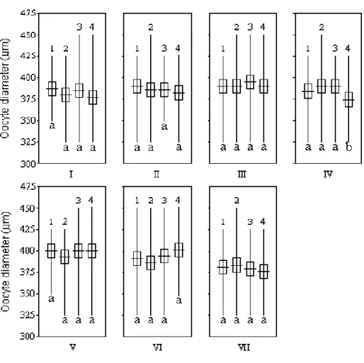
The external morphology of the ovaries of this species is peculiar, because the oviduct of a sexually mature female is located approximately at the posterior forth portion of the ovary length. Taking this into consideration, it was determined if there were differences in oocyte development across the length of the ovary. Seven females were
sacrificed and from one ovary of each, oocyte samples were taken from four sites: (1) near the cloacal opening, (2) on the anterior end, (3) on the central portion, and (4) on the posterior end.
Paired samples were taken from the central portion of the ovary of seven females, one in vivo with the cannula and one in vitro from the central portion of one ovary after each female was sacrificed, to determine if the in vivo oocyte samples were representative of the ovaries.
The precision of the oocyte mean diameter estimates was calculated by the coefficient of variation corrected for bias with the confidence limits (Sokal and Rolf, 1981) of 8 in vivo samples obtained with a cannula from one female. Statistical differences between mean diameters were calculated by one-way analysis of variance (ANOVA) followed by Duncan’s multiple range test, and Student’s t-test in the case of the paired in vivo and in vitro samples at P = 0.05, after the homogeneity of variances was demonstrated by Bartlett’s test (Sokal and Rolf, 1981).
RESULTS
Females examined, 375 - 410 mm in total length and 547 - 771 g in total weight, had oocytes in different prematuration stages in the four ovary sites studied. Those in the tertiary yolk-globule stage had a unimodal frequency distribution of their diameter. A sample size of 50 oocytes was estimated by the iterative procedure for the mean diameter to be 90% certain of detecting a 5% difference between two means of the four sites of the ovaries at the 1% level of significance with a previous coefficient of variation estimate of 6 %. There were no detected effects of the preservative solution on the oocyte diameter (P > 0.05) between fresh and those preserved over the 24-h interval of the test (Table 1).
statistically significant differences (P > 0.05). The estimate of the coefficient of variation corrected for
bias (V*) with its confidence limits for the 8 means of repeated in vivo samples from one female was:
V* ± t 0.05[7] SV* = 1.9 ± 0.6.
Table 1 - Oocyte mean diameters (n=100) ± standard error of the mean (SEM) from fresh and fixed (1% formalin in 0.9% NaCl solution) samples, and Student t-test comparisons with 0 h (P = 0.05) of biopsied snook
Centropomus parallelus.
Fixation period (h) Mean diameter ± SEM (µm) t0.05 Statistics
0 (Fresh) 379 ± 5 - -
1 381 ± 6 0.14 NS
2 385 ± 4 0.29 NS
4 384 ± 5 0.24 NS
24 382 ± 4 0.21 NS
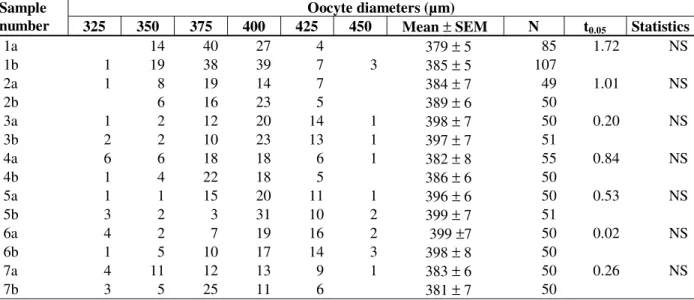
Table 2 - Oocyte diameter-frequency distributions of in vivo (a) and in vitro (b) paired samples, mean ± standard error of the mean (SEM) of oocytes, and Student t-test comparisons of paired means (P = 0.05) from each of seven snook Centropomusparallelus females.
Oocyte diameters (µm) Sample
number 325 350 375 400 425 450 Mean ± SEM N t0.05 Statistics
1a 14 40 27 4 379 ± 5 85 1.72 NS
1b 1 19 38 39 7 3 385 ± 5 107
2a 1 8 19 14 7 384 ± 7 49 1.01 NS
2b 6 16 23 5 389 ± 6 50
3a 1 2 12 20 14 1 398 ± 7 50 0.20 NS
3b 2 2 10 23 13 1 397 ± 7 51
4a 6 6 18 18 6 1 382 ± 8 55 0.84 NS
4b 1 4 22 18 5 386 ± 6 50
5a 1 1 15 20 11 1 396 ± 6 50 0.53 NS
5b 3 2 3 31 10 2 399 ± 7 51
6a 4 2 7 19 16 2 399 ±7 50 0.02 NS
6b 1 5 10 17 14 3 398 ± 8 50
7a 4 11 12 13 9 1 383 ± 6 50 0.26 NS
7b 3 5 25 11 6 381 ± 7 50
DISCUSSION
The preservative solution did not affect the oocyte diameters within 24 h. The same was observed in other species (Shehadeh et al., 1973; Alvarez-Lajonchère et al., 1983, 2001; Tamaru et al., 1988), although Garcia (1989-a) reported an increase in L. calcarifer oocyte diameters after one hour in a buffered 5% formalin solution. Ovarian biopsy methods have been validated on synchronous as well as asynchronous oocyte-development species (Shehadeh et al., 1973, Markmann and Doroshov, 1983; Alvarez-Lajonchère et al., 1983, 2001; Rodriguez and Garzo, 1986).
Figure 1 - Oocytes measurements (mean oocyte diameter ± standard error of 50 oocytes mean at P = 0.05, and data range) from four ovary sites (1 to 4) in seven snook
Centropomus parallelus females (I to VII). Different letters (a and b) correspond to significant difference at P < 0.05.
Hormonal treatments for induced spawning of C. parallelus were applied to females with mean oocyte diameter of at least 390 µm (Cerqueira, 1995), which were within the 95% confidence intervals of estimated means of 67% of the females in the present study. Tucker and Campbell (1988) reported a mean diameter for sectioned yolked oocytes of C. undecimalis in the range of the vitellogenic or postvitellogenic opaque oocytes found in the present study. Wallace et al. (1993) reported that to induce final maturation in C. undecimalis fresh oocytes should have diameters larger than 0.5 mm.
Based on the present data, to further improve the established biopsy method and effectively select fat
snook females for spawning induction treatments, complementary studies are recommended with an in vitro oocyte maturational competence test (Greeley et al., 1987; Patiño and Thomas, 1990; Wallace et al., 1993) to estimate the minimum oocyte diameter that would positively respond to an acute hormonal treatment.
The in vivo method for monitoring ovarian sexual
specially the same characteristics of the most advanced oocyte group (late vitellogenic and postvitellogenic).
ACKNOWLEDGEMENTS
This study was part of a project (Brazilian Mariculture Linkage Program) supported by Canadian International Development Agency (CIDA) and CNPq. The authors are grateful to CNPq for providing a scholarship to one of the authors (E.M.F), to the staff of Laboratório de Piscicultura Marinha (Jaqueline Araújo, Antônio Sayão and Israel Silva), and to Shelby Banner for reviewing the English manuscript.
RESUMO
A validação de um método de biópsia ovariana para determinação in vivo da maturação ovocitária em Centropomus parallelus foi descrita. Os diâmetros de ovócitos, obtidos de amostras intra-ovarianas sifonadas por cânula, foram analisados a fresco e preservados com formalina (1%) em solução de NaCl (0,7%). Ovócitos em diferentes estádios de maturação estavam presentes ao longo dos ovários, até o estádio de vitelogênese completa, apresentando uma distribuição de freqüência de diâmetros unimodal. O diâmetro médio dos ovócitos não diferiu significativamente entre as quatro regiões dos ovários (P > 0,05). Amostras obtidas com a cânula são representativas da porção central do ovário, uma vez que as amostras in vivo e in vitro das sete fêmeas examinadas não foram significativamente diferentes (P > 0,05). Uma estimativa do coeficiente de variação corrigido para “bias” (P < 0,05) para oito amostras repetidas in vivo foi de 1,9 ± 0,6. Os resultados demonstraram que para esta espécie, este método de biópsia é satisfatório, provendo amostras representativas dos ovários.
REFERENCES
Alvarez-Lajonchère, L.; Guerrero-Tortolero, D. and Perez-Urbiola, J. C. (2001), Validation of an ovarian biopsy method in a sea bass, Centropomus medius
Gunther. Aquaculture Research, 32, 379-384.
Alvarez-Lajonchère, L.; Báez Hidalgo, M. and Gotera, G. (1982), Estudio de la biología pesquera del robalo de ley Centropomus undecimalis (Bloch) (Pisces, Centropomidae) en Tunas de Zaza, Cuba.
Revista Investigaciones Marinas, Universidad de La Habana, 3, 159-200.
Alvarez-Lajonchère, L.; Berdayes Arritola, J.; Díaz Bellido, S. J. and Laiz Averhoff, O. (1983), Método de muestreo in vivo de ovocitos intraovaricos en las lisas Mugil liza y M. curema (Pisces, Mugilidae) y en el patao Eugerres brasilianus (Pisces, Gerreidae).
Revista Latinoamericana de Acuicultura, 18, 27-38. Amador del Angel, L. E. and Cabrera Rodríguez, P.
(1994), El robalo Centropomus undecimalis: una especie nativa con potencialidad en acuicultura.
Gaceta Universitaria, Universidad Autónoma del
Carmen, 11, 19-26.
Barlow, C. G.; Rodgers, L. J.; Palmer, P. J. and Longhurst, C. J. (1993), Feeding habits of hatchery reared barramundi Lates calcarifer (Bloch) fry.
Aquaculture, 109, 131-144.
Cerqueira, V. R. (1995), Testes de indução de desova do robalo, Centropomusparallelus, do litoral da ilha de Santa Catarina com Gonadotrofina Coriônica Humana (HCG). Proceedings of 7th Congresso Brasileiro de Engenharia de Pesca, AEP - PE, SUDENE, Recife. pp. 95-102.
Cerqueira, V. R.; Macchiavello, J. A. G. and Brügger, A. M. (1995), Produção de alevinos de robalo,
Centropomus parallelus Poey, 1860, através de
larvicultura intensiva em laboratório. Proceedings of 7th Simpósio Brasileiro de Aqüicultura, Peruíbe. ACIESP, São Paulo. pp. 191-197.
Dhert, P.; Lavens, P. and Sorgeloos, P. (1992), State of the art of Asian seabass Lates calcarifer
larviculture. Journal of the World Aquaculture Society 23, 317-329.
Edwards, R. E. and Henderson, B. D. (1987), An experimental hatchery project: studies of propagation, culture and biology of snook (Centropomus undecimalis). Proc. Gulf Carib. Fish. Inst., 38, 211-230.
Garcia, L. M. B. (1989a), Development of an ovarian biopsy technique in the sea bass, Lates calcarifer
(Bloch). Aquaculture, 77, 97-102.
Garcia, L. M. B. (1989b), Dose-dependent spawning response of mature female sea bass, Lates calcarifer
(Bloch), to pelleted luteinizing hormone-releasing hormone analogue (LHRHa). Aquaculture, 77, 85-96. Greeley Jr., M. S.; Calder, D. R. and Wallace, R. A.
Markmann, C. and Doroshov, S. I. (1983), Ovarian catheterization of the channel catfish, Ictalurus punctatus. Aquaculture, 35,163-169.
National Institute of Coastal Aquaculture (1986),
Technical manual for seed production of seabass. Kao Seng, Songkhla (Thailand), National Institute of Coastal Aquaculture. 49 pp.
Patiño, R. and Thomas, P. (1990), Effects of gonadotropin on ovarian intrafollicular processes during the development of oocyte maturational competence in a teleost, the Atlantic croaker: evidence for two distinct stages of gonadotropic control of final oocyte maturation. Biology of Reproduction, 43, 818-827.
Rodríguez, M. and Garzo, G. (1986), Biopsia ovárica por canulación, método eficiente para evaluar la madurez de las hembras de Cyprinus carpio. Revista Latinoamericana de Acuicultura, 38, 83-90.
Shehadeh, Z. H.; Kuo, C. M. and Milisen, K. K. (1973), Validation of an in vivo method for monitoring ovarian development in the grey mullet (Mugil cephalus L.). J. Fish Biology, 5, 489-496. Sokal, R. R. and Rohlf, F. J. (1981), Biometry. 2nd. ed.
New York : Freeman and Co. 859 pp.
Tamaru, C. S.; Lee, C. S.; Kelley, C. D.; Banno, J. E. P.; Ha, Y.; Aida, K. and Hanyn, I. (1988), Characterising the stage of maturity most receptive to an acute LHRH-analogue therapy for inducing milkfish (Chanos chanos) to spawn. Aquaculture, 74, 147-163.
Tucker Jr., J. W. (1987), Snook and tarpon snook culture and preliminary evaluation for commercial farming. Progr. Fish-Cult., 49, 49-57.
Tucker Jr., J. W. and Campbell, S. W. (1988), Spawning season of common snook along the East central Florida coast. Florida Scientist, 51, 1-6.
Wallace, R. A.; Boyle, S. M.; Grier, H. J.; Sellman, K. and Petrino, T. R. (1993), Preliminary observations on oocyte maturation and other aspects of reproductive biology in captive female snook, Centropomus undecimalis. Aquaculture, 116, 257-273.