Discloses Novel Targets for Immunodiagnosis of Tegumentary and
Visceral Clinical Forms of Leishmaniasis
Daniel Menezes-Souza,a
Tiago Antônio de Oliveira Mendes,a
Matheus de Souza Gomes,b
João Luís Reis-Cunha,a Ronaldo Alves Pinto Nagem,c
Cláudia Martins Carneiro,d
Eduardo Antônio Ferraz Coelho,e
Lúcia Maria da Cunha Galvão,f Ricardo Toshio Fujiwara,a
Daniella Castanheira Bartholomeua
Departamento de Parasitologia, Instituto de Ciências Biológicas, Universidade Federal de Minas Gerais, Belo Horizonte, Minas Gerais, Brazila
; Instituto de Genética e Bioquímica, Universidade Federal de Uberlândia, Patos de Minas, Minas Gerais, Brazilb
; Departamento de Bioquímica e Imunologia, Instituto de Ciências Biológicas, Universidade Federal de Minas Gerais, Belo Horizonte, Minas Gerais, Brazilc
; Departamento de Análises Clínicas, Escola de Farmácia, Universidade Federal de Ouro Preto, Ouro Preto, Minas Gerais, Brazild
; Programa de Pós-Graduação em Ciências da Saúde, Infectologia e Medicina Tropical, Faculdade de Medicina and Departamento de Patologia Clínica, COLTEC, Universidade Federal de Minas Gerais, Belo Horizonte, Minas Gerais, Brazile
; Programa de Pós-Graduação em Ciências da Saúde, Universidade Federal do Rio Grande do Norte, Natal, Rio Grande do Norte, Brazilf
Gold standard serological diagnostic methods focus on antigens that elicit a strong humoral immune response that is specific to
a certain pathogen. In this study, we used bioinformatics approaches to identify linear B-cell epitopes that are conserved among
Leishmania
species but are divergent from the host species
Homo sapiens
and
Canis familiaris
and from
Trypanosoma cruzi
, the
parasite that causes Chagas disease, to select potential targets for the immunodiagnosis of leishmaniasis. Using these criteria, we
selected heat shock protein 83.1 of
Leishmania braziliensis
for this study. We predicted three linear B-cell epitopes in its
se-quence. These peptides and the recombinant heat shock protein 83.1 (rHSP83.1) were tested in enzyme-linked immunosorbent
assays (ELISAs) against serum samples from patients with tegumentary leishmaniasis (TL) and visceral leishmaniasis (VL) and
from dogs infected with
Leishmania infantum
(canine VL [CVL]). Our data show that rHSP83.1 is a promising target in the
diag-nosis of TL. We also identified specific epitopes derived from HSP83.1 that can be used in the diagdiag-nosis of human TL (peptide 3),
both human and canine VL (peptides 1 and 3), and all TL, VL, and CVL clinical manifestations (peptide 3). Receiver operating
characteristic (ROC) curves confirmed the superior performance of rHSP83.1 and peptides 1 and 3 compared to that of the
solu-ble
L. braziliensis
antigen and the reference test kit for the diagnosis of CVL in Brazil (EIE-LVC kit; Bio-Manguinhos, Fiocruz).
Our study thus provides proof-of-principle evidence of the feasibility of using bioinformatics to identify novel targets for the
immunodiagnosis of parasitic diseases using proteins that are highly conserved throughout evolution.
L
eishmaniasis is a neglected vector-borne tropical disease that is
caused by parasites of the
Leishmania
genus. Currently, it has a
major impact on human health in tropical regions and affects
approximately 12 million people worldwide (
1
). Two million new
cases are reported annually, with an incidence of 1 to 1.5 million
cases of tegumentary leishmaniasis (TL) and 500,000 cases of
vis-ceral leishmaniasis (VL) (
1
). The clinical forms of leishmaniasis
range from self-healing cutaneous lesions to fatal visceral
infec-tions. In TL, a wide variety of skin manifestations, such as
cutane-ous leishmaniasis (CL) and mucocutanecutane-ous leishmaniasis (ML),
have been described (
2
). The CL form is characterized by one or
more painless ulcers with raised borders and a bed of granulation
tissue that appear near the area of the sand fly bite, while ML is
characterized by the progressive destruction of the
nasopharyn-geal mucosa (
3
,
4
). Although the determining factors involved in
the development of each clinical form have not been elucidated, it
is likely that host and parasite genetics are involved (
2
,
5
).
In Brazil, TL is distributed throughout the country, and among
the various
Leishmania
species that can cause the disease,
Leish-mania braziliensis
is responsible for the majority of the cases.
L.
braziliensis
infection results in higher clinical severity due to larger
ulcerated areas and a higher proportion of patients with mucosal
involvement in the upper airway (
6
,
7
). VL is caused by the
Leish-mania infantum
parasite and is a zoonotic disease that has shown
significant changes in transmission with progressive urbanization
and geographic expansion; it now affects regions in which it was
previously quite rare (
2
). Dogs are the main urban reservoirs and
represent the major source of infection for the vector due to the
high prevalence of canine infections and intense cutaneous
para-sitism that may contribute to urban spread of the disease (
8
,
9
).
The major prophylactic practice recommended by the World
Health Organization to control the human disease and canine
visceral leishmaniasis (CVL) (
8
) involves early accurate diagnosis,
systematic treatment of human cases, vector control with
insecti-cide, and the elimination of seropositive dogs (
10
). At present,
there is no gold standard serological test for diagnosing
leishman-iasis, and a combination of different techniques is frequently
nec-essary to obtain precise results. Therefore, the development of a
new serological technique with higher sensitivity and specificity
Received13 March 2014Returned for modification22 April 2014 Accepted1 May 2014
Published ahead of print7 May 2014
Editor:P. P. Wilkins
Address correspondence to Daniella Castanheira Bartholomeu, daniella@icb.ufmg.br.
Copyright © 2014, American Society for Microbiology. All Rights Reserved.
doi:10.1128/CVI.00151-14
on October 9, 2017 by UNIVERSIDADE FEDERAL DE OURO PRETO
http://cvi.asm.org/
than the available commercial tests and that is able to discriminate
postvaccination reactivity from active infections would represent
an important innovation in the serological diagnosis of
leishman-iasis (
11
). Additionally, due to the high conservation of proteins
among the various species of
Leishmania
, the selection of an
anti-gen that is able to simultaneously diagnose the various clinical
forms of the disease would represent an interesting strategy for the
technological development and large-scale production of tests for
diagnosis (
12
,
13
).
In this context, we performed genome-wide screening to
iden-tify linear B-cell epitopes in the predicted proteome of
L.
brazil-iensis
in an attempt to identify conserved targets within the
Leish-mania
genus for the serodiagnosis of the tegumentary and
visceral forms of leishmaniasis. The protein selected in this
study was heat shock protein 83.1 (HSP83.1), a highly
con-served molecule in prokaryotes and eukaryotes that plays
im-portant roles in protein folding, assembly of protein
com-plexes, and the translocation of proteins across cellular
compartments (
14
). We mapped three B-cell linear epitopes in
this protein whose sequences are divergent from its orthologs
in
Homo sapiens
and
Canis familiaris
and in
Trypanosoma cruzi
,
the etiologic agent of Chagas disease (CD), which is frequently
associated with cross-reactivity with leishmaniasis (
15
,
16
). The
reactivities of recombinant HSP83.1 (rHSP83.1) and three
de-rived B-cell epitopes with serum samples from TL and VL patients
and from
Leishmania
-infected dogs were evaluated in
enzyme-linked immunosorbent assays (ELISAs). rHSP83.1 demonstrated
excellent performance for diagnosing TL. Additionally, we
iden-tified specific epitopes derived from HSP83.1 that may be useful in
the diagnosis of human TL (peptide 3), both human and canine
VL (peptides 1 and 3), and all TL, VL, and CVL clinical
manifes-tations (peptide 3). Receiver operating characteristic (ROC)
curves confirmed the superior performance of rHSP83.1 and
pep-tides 1 and 3 compared to that of the soluble
L. braziliensis
antigen
and the reference test for diagnosing CVL in Brazil (EIE-LVC kit;
Bio-Manguinhos, Fiocruz) (
17
).
MATERIALS AND METHODS
Ethics statement and human and dog serum samples.All samples that were used were anonymous and were obtained from the serum bank of the Laboratory of Immunology and Genomics of Parasites, Federal University of Minas Gerais. Approval to use the samples was obtained from the Human Research Ethics Committee (protocol CAAE – 00842112.2.0000.5149) and the Committee on Ethics of Animal Ex-perimentation from the Federal University of Minas Gerais (protocol 44/2012).
The human serum panel consisted of 65 samples from tegumentary leishmaniasis patients infected withL. braziliensisthat presented cutane-ous (CL) (n⫽45) or mucosal (ML) (n⫽20) clinical manifestations obtained from the Centro de Referência em Leishmaniose (Januária, Mi-nas Gerais, Brazil) and 55 samples from visceral leishmaniasis patients infected withL. infantumobtained from the University Hospital (Montes Claros, Minas Gerais, Brazil). The infection was confirmed by micro-scopic analyses of biopsy specimens from cutaneous lesions (for TL) or bone marrow aspirate samples (for VL), followed by specific PCR assays for the kinetoplastid DNA (kDNA) ofLeishmaniaparasites (18). These individuals were known to be noninfected withT. cruzi. Information re-garding the clinical evaluation and PCR results were obtained from the medical records of the patients. To evaluate the cross-reactivity with Cha-gas disease, serum samples from patients with chronic ChaCha-gas disease (CD) (n⫽20) with infections confirmed by hemoculture or the Chagatest recombinant ELISA version 3.0 kit and the Chagatest hemagglutination
inhibition (HAI) were used in this study. Serum samples from healthy Brazilian individuals (controls [CT]) (n⫽50) from areas where TL and CD are not endemic (Belo Horizonte, Minas Gerais, Brazil) were used as negative controls in these assays. The resulting optical density (OD) values were compared with those obtained with the panel of TL and VL samples. Male and female beagle dogs from an area where VL is not endemic that hadLeishmania-negative tissue smears (bone marrow) were consid-ered to be noninfected and were used as the control group (CT,n⫽30). Serum samples fromLeishmania-infected dogs (CVL) (n⫽30) were ob-tained from the area where CVL is endemic in Minas Gerais in southeast-ern Brazil. The main inclusion criterion for the CVL serum samples used in this study was parasitological positivity forL. infantumconfirmed by microscopic analysis of bone marrow aspirate samples. Samples from dogs that were experimentally infected withT. cruzi(CD,n⫽15) but that were parasitologically negative forLeishmaniawere included in this study to evaluate possible cross-reactivity.
In silicoprediction of linear B-cell epitopes.Linear B-cell epitopes were predicted for the HSP83.1 protein ofL. braziliensis(TriTrypDB iden-tification [ID] [19] LbrM.33.0340) using the BepiPred 1.0 program with a cutoff of 1.3 (20). A BLASTp (21) search was performed against GeneDB (http://www.genedb.org/) to retrieve the HSP83.1 sequences ofT. cruzi, H. sapiens, andC. familiarisusingL. braziliensisHSP83.1 as a query. Mul-tiple alignments of HSP83.1 were performed using the Clustal X 2.0 pro-gram (22), with the default parameters (23).
SolubleL. braziliensisantigen.SolubleL. braziliensisantigen (SLbA) was prepared fromL. (V.)braziliensis(MHOM/BR/75/M2904) station-ary-phase promastigotes maintained in Schneider’s insect medium (Sig-ma-Aldrich) supplemented with 10% inactivated fetal bovine serum, 100 U/ml penicillin, and 100g/ml streptomycin (Gibco). The parasites were initially subjected to three cycles of freezing (with liquid nitrogen) and thawing (42°C), followed by ultrasonication (Ultrasonic processor model GEX-600) with 10 alternating cycles of 30 s on/off sonication in an ice water bath at 35 MHz. The lysate was then centrifuged at 6,000⫻gat 4°C for 15 min. The supernatant containing SLbA was collected, and the pro-tein content was quantified using the Pierce BCA propro-tein assay (Thermo Scientific).
Cloning, protein expression, and purification.The primers used to am-plify the HSP83.1 gene from theLeishmaniagenomic DNA were HSP83.1-Forward (5=-GCTAGCATGACGGAGACGTTCGCGTT) and HSP83.1-Reverse (5=-AAGCTTTCAGTCCACCTGCTCCATG). The sites for restriction enzymes (NheI and HindIII) that were added to the forward and reverse primers, respectively, to facilitate cloning, are shown in bold type. The DNA fragments obtained were excised from the gel, purified, digested with the restriction enzymes, and cloned into the pET28a-TEV vector (24). ElectrocompetentEscherichia coliBL21 Arctic Express (DE3) cells (Agilent Technologies, USA) were transformed with the recombi-nant plasmid pET28a-TEV-HSP83.1 by electroporation using a Micro-Pulser electroporation apparatus (Bio-Rad Laboratories, USA). Gene in-sertion was confirmed by colony PCR and sequencing using T7 primers (Macrogen, South Korea).
Protein expression and purification were performed as previously de-scribed (25). Briefly, expression was induced by adding isopropyl--D
-thiogalactopyranoside (IPTG) to a final concentration of 1.0 mM, and the bacterial culture was incubated for 24 h at 12°C with shaking at 200 rpm per min. The cells were then ruptured by sonication, the debris was re-moved by centrifugation, and the recombinant protein was purified using a HisTrap HP affinity column connected to an ÄKTAprime chromatog-raphy system (GE Healthcare, USA). The eluted fractions containing rHSP83.1 (705 amino acids, 80.6 kDa) were concentrated using the Ami-con Ultra 15 centrifugal filters 10,000 nominal molecular weight limit (NMWL) (Millipore, Germany) and further purified on a Superdex 200 gel filtration column (GE Healthcare Life Sciences, USA).
Soluble peptide synthesis.The soluble peptides were manually syn-thesized by 9-fluorenylmethoxy (Fmoc) chemistry in solid phase on a 30-mol scale (26). Briefly, Fmoc-amino acids were activated with a 1:2
on October 9, 2017 by UNIVERSIDADE FEDERAL DE OURO PRETO
http://cvi.asm.org/
solution of Oxyme toN,N=-diisopropylcarbodiimide (DIC). The active amino acids were then incorporated into Rink amide resin with a substi-tution degree of 0.61. Fmoc deprotection was then performed using 25% 4-methylpiperidine. These steps were repeated until the synthesis of each peptide was completed. The peptides were then deprotected and released from the resin by treatment with a solution of 95% trifluoroacetic acid, 2.5% water, and 2.5% triisopropylsilane and precipitated with cold diiso-propyl ether. The peptides were obtained withⱖ90% purity, as confirmed by matrix-assisted laser desorption ionization–tandem time of flight mass spectrometry (MALDI-TOF-TOF MS) Autoflex III equipment (Bruker Daltonics, USA). Instrument calibration was achieved using peptide cal-ibration standard II (Bruker Daltonics, USA) as a reference, and ␣-cyano-4-hydroxycinnamic acid was used as the matrix.
Serological assay and depletion ELISA.rHSP83.1 and SLbA were coated onto 96-well microplates (Nalge Nunc International, USA) over-night at 2 to 8°C at a concentration of 250 ng/well for rHSP83.1 and 50 ng/well for SLbA. The peptides were coated onto flat-bottom plates (CoStar, USA) overnight at 37°C at a concentration of 10g/well. The plates were blocked with 150l of phosphate-buffered saline (PBS) con-taining 5% bovine serum albumin (BSA) for 1 h at 37°C and then treated with 1:100 dilutions of human or canine serum samples for 1 h at 37°C. Peroxidase-conjugated antibodies specific for human or dog IgG (Sigma-Aldrich, USA) were diluted at 1:5,000 and added to the plates for 1 h at 37°C. The wells were washed, and the 3,3=,5,5=-tetramethylbenzidine (TMB) substrate (Sigma-Aldrich, USA) in citrate buffer containing hy-drogen peroxide was added to the plate. The plates were incubated for 30 min in the dark at 37°C. The reaction was halted by adding 4 N H2SO4,
and the absorbance at 450 nm was read with an automatic microplate reader (VersaMax; Molecular Devices, USA). Each serum sample was as-sayed in duplicate. The results of the ELISA using rHSP83.1 as an antigen were compared with those using SLbA. The results of the ELISA using rHSP83.1 as an antigen for the canine serum samples were compared with those with the EIE-LVC kit immunoassay (Bio-Manguinhos, Fiocruz, Brazil), which is the test currently recommended by the Brazilian Ministry of Health for the screening of seroreactive animals (17). The assays were carried out according to the manufacturer’s instructions.
Depletion ELISAs were performed as previously described (27,28). Briefly, flat-bottom plates (CoStar, USA) were coated overnight at 37°C with 10g/well of peptide 1 (EEDESKKKSCGDEGEPKVE), peptide 2 (VTEGGEDKKK), and peptide 3 (EVAEAPPAEAAPA) and then washed and blocked as described above. All serum samples from individuals in the TL, VL, and CVL groups that were parasitologically positive for Leishma-niaand reactive for the tested peptide were used in the assay. Pools of sera were added to the plates at a 1:100 dilution and incubated overnight at 2 to 8°C. The depleted and undepleted sera were transferred to plates coated overnight with rHSP83.1 (50 ng/well) after appropriate washing and blocking, and the ELISAs were performed as described above.
Statistical analysis. The lower limits of positivity (cutoff) for rHSP83.1, SLbA, and peptides 1, 2, and 3 were established for optimal sensitivity and specificity using the receiver operating characteristic (ROC) curve. The cutoff was chosen based on the point that provides the maximum of the sum of the sensitivity and specificity (29). The EIE-LVC cutoff was obtained according to the manufacturer (twice the average of the negative control provided by the kit). The performance of each test was evaluated according to the sensitivity (Se), specificity (Sp), positive predictive value (PPV), negative predictive value (NPV), area under the curve (AUC), and accuracy (AC). The degree of agreement between the ELISAs using rHSP83.1, SLbA, or the EIE-LVC kit with the parasitological test (biopsy specimen, aspirate sample, or PCR) was determined by the Kappa index (k) values with 95% confidence intervals and interpreted ac-cording to the following Fleiss scale: 0.00 to 0.20, poor; 0.21 to 0.40, fair; 0.41 to 0.60, moderate; 0.61 to 0.80, good; 0.81 to 0.99, very good; and 1.00, perfect (30). The one-sample Kolmogorov-Smirnov test was used to determine whether a variable was normally distributed. For the depletion assays, signif-icant differences were detected using a two-way analysis of variance
(ANOVA). The differences were considered statistically significant at aP value of⬍0.05. All of the statistical analyses were performed using GraphPad Prism (version 5.0) and the GraphPad QuickCalcs software.
RESULTS
Sequence divergence and prediction of B-cell linear epitopes in
L. braziliensis
HSP83.1 protein and its orthologs.
Gold standard
serological diagnostic methods focus on markers that are able to
elicit a strong antibody response specific to the pathogen.
Al-though HSP83.1 is conserved throughout evolution, B-cell
epitopes derived from
Leishmania
HSP83.1, with sequences that
are divergent from those of the parasite hosts and other closely
related parasites, may bind to specific antibodies and thus allow
for the specific identification of samples from patients with
leish-maniasis. To identify potential epitopes that can increase the
ac-curacy of the currently available serological tests, we scanned the
HSP83.1 sequence of
L. braziliensis
for B-cell linear epitopes that
are divergent in the host orthologs and that cooccur with
intrin-sically unstructured regions (
Fig. 1
). The presence of these
un-structured regions suggests that a given protein region is in an
unfolded structure and therefore possibly accessible for antibody
binding. As expected, the HSP83.1 ortholog of
L. braziliensis
,
LbrM.33.0340, displayed a high level of identity to the
ortholo-gous proteins from
L. infantum
,
T. cruzi
,
C. familiaris
, and
H.
sapiens
, with identities of
⬎
63.5%, as shown in
Fig. 1
and
Table 1
.
Despite this overall high level of similarity, we identified three
potential linear B-cell epitopes (peptide 1, EEDESKKKSCGDEG
EPKVE; peptide 2, VTEGGEDKKK; and peptide 3, EVAEAPPAE
AAPA) in the LbrM.33.0340 protein that are highly divergent in
T.
cruzi
as well as in dog and human orthologs, with identities
rang-ing from 21.4 to 60.0% (
Fig. 1
and
Table 1
). This result leads us to
speculate that despite the high overall sequence similarity between
HSP83 in
Leishmania
species and its orthologs in
T. cruzi
and in
humans and dogs, this protein and the three predicted B-cell
lin-ear epitopes might be potential targets in the immunodiagnosis of
leishmaniasis.
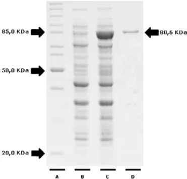
Expression and purification of rHSP83.1 protein.
To verify
whether the HSP83.1 protein is a candidate for the development of
new diagnostic assays, we first expressed this protein as a
His-tagged recombinant protein. The full-length coding region of the
LbrM.33.0340 gene was amplified by PCR and cloned into the
pET28a-TEV expression vector. rHSP83.1 was overexpressed in
E.
coli
BL21 Arctic Express (DE3) cells as a soluble protein and
pu-rified by Ni
2⫹affinity chromatography, followed by gel filtration
on a Superdex 200 column. The recombinant protein, with a
pre-dicted molecular mass of 80.6 kDa, was successfully expressed and
obtained at a high level of purity (
Fig. 2
).
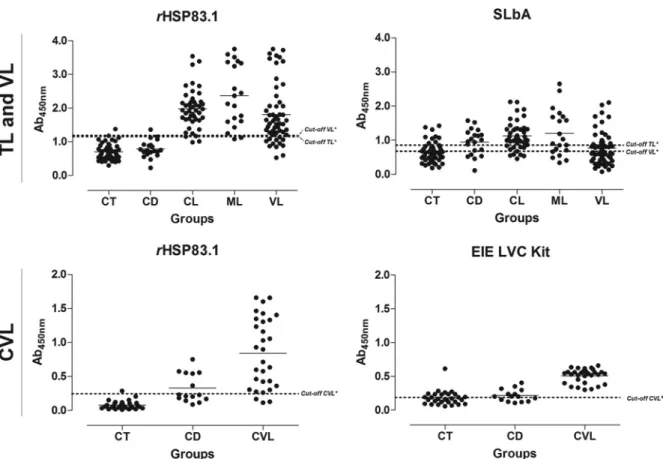
ELISA performance using rHSP83.1, peptides 1, 2, 3, and
SLbA in TL diagnosis.
To evaluate the specific antibody responses
against rHSP83.1 and the derived peptides 1, 2, and 3 in TL, ELISAs
with these antigens were compared to SLbA ELISAs (
Fig. 3
to
5
and
Tables 2
and
3
). When tested with serum samples from CL patients,
the rHSP83.1 and peptide 1, 2, and 3 antigens showed sensitivities of
95.55%, 71.11%, 64.44%, and 95.55%, respectively. The serum
sam-ples from ML patients presented lower sensitivity values to all
anti-gens tested (90.00%, 55.00%, 50.00%, and 75.00%, respectively). The
antigens presented total sensitivity (CL
⫹
ML) for rHSP83.1 and
sensitivities of 93.85% (95% confidence interval [CI], 84.99 to
98.30%), 63.08% (95% CI, 50.20 to 74.72), 63.08% (95% CI, 50.20 to
74.72), and 89.23% (95% CI, 79.06 to 95.56) for peptides 1, 2, and 3,
on October 9, 2017 by UNIVERSIDADE FEDERAL DE OURO PRETO
http://cvi.asm.org/
respectively (
Table 2
). ELISAs using SLbA extract presented
sensitiv-ities of 75.55% and 60.00% for detecting the CL and ML clinical
forms, respectively. The total sensitivity of SLbA in TL was 70.77%
(95% CI, 58.17 to 81.40%), which was lower than that for rHSP83.1
and peptide 3 (
Table 2
). To evaluate specificity (Sp), serum samples
from chagasic patients and negative-control individuals were also
tested. rHSP83.1 and peptides 1, 2, and 3 showed total specificity
values of 95.71% (95% CI, 87.98 to 99.11%), 94.29% (95% CI, 86.01
to 98.42), 90.00% (95% CI, 80.48 to 95.88%), and 91.43% (95% CI,
82.27 to 96.79%), respectively. The ELISA using SLbA presented a
much lower specificity value of 68.57% (95% CI, 56.37 to 79.15%)
(
Table 2
).
The maximum PPV was achieved with rHSP83.1 (95.31%),
followed by peptide 1 (91.11%), peptide 3 (90.63%), peptide 2
(85.42%), and SLbA (67.65%). High NPVs were also observed for
rHSP83.1 (94.37%), followed by peptide 3 (90.14%), peptide 1
(73.33%), peptide 2 (72.41%), and SLbA (71.64%) (
Table 2
).
Based on the ELISA results, ROC curves were calculated to
evaluate the capacity of the tested antigens to discriminate TL
patients from healthy volunteers (
Fig. 5
and
Table 3
). The area
under the curve (AUC) and accuracy (AC) were used to compare
the efficiencies of the different diagnostic antigens or tests (
29
)
(
Table 3
). rHSP83.1 presented the highest AUC value (0.989; 95%
FIG 1Sequence divergence and prediction of B-cell linear epitopes and intrinsically unstructured/disordered regions inL. braziliensisHSP83.1 and its orthologs. Alignment betweenL. braziliensisHSP83.1 (TriTrypDB ID LbrM.33.0340) and orthologous proteins present inL. infantum(TriTrypDB ID Linj.33.0360),T. cruzi(TriTrypDB ID TcCLB.509105.140),H. sapiens(RefSeq IDNP_005339.3), andC. familiaris(RefSeq IDXP_532154.4). The yellow boxes mark predicted B-cell epitopes, and the gray boxes mark predicted disordered regions. The continuous black underlined amino acid sequences represent three potential B-cell epitopes predicted by BepiPred in the LbrM.33.0340 protein, and red, green, and blue highlight amino acid conservations in theT. cruzi,C. familiaris, andH. sapienssequences in relation to theL. braziliensissequence.
TABLE 1Sequence identity of the B-cell linear epitopes predicted in LbrM.33.0340 HSP83 protein ofLeishmania braziliensisand its orthologs
Species
% identity with LbrM.33.0340
% identity with peptidea:
1 2 3
Leishmania infantum 93.9 52.3 70.0 71.4
Trypanosoma cruzi 85.2 31.6 50.0 50.0
Canis familiaris 64.1 36.8 60.0 35.7
Homo sapiens 63.5 36.8 40.0 21.4
aPeptide 1, EEDESKKKSCGDEGEPKVE; peptide 2, VTEGGEDKKK; peptide 3,
EEVAEAPPAEAAPA.
FIG 2Expression and purification of recombinant HSP83.1 protein. Protein samples were separated by 12.5% SDS-PAGE. Lane A, molecular weight stan-dard, lysate of culture before (lane B) and after (lane C) induction with IPTG and recombinant HSP83.1 protein (mass, 80.6 kDa) purified by gel filtration (lane D).
on October 9, 2017 by UNIVERSIDADE FEDERAL DE OURO PRETO
http://cvi.asm.org/
CI, 0.979 to 1.000), followed by peptide 3 (0.961; 95% CI, 0.933 to
0.989), peptide 1 (0.881; 95% CI, 0.826 to 0.936), peptide 2 (0.819;
95% CI, 0.748 to 0.889), and SLbA (0.753; 95% CI, 0.673 to 0.834).
The accuracy value for rHSP83.1 was also the highest (94.81%),
followed by peptide 3 (90.37%), peptide 1 (79.26%), peptide 2
(77.04%), and SLbA (69.63%) (
Table 2
).
ELISA performance using rHSP83.1, peptides 1, 2, and 3, and
SLbA in VL diagnosis.
We also tested the performance of ELISAs
using rHSP83.1 and the derived peptides 1, 2, and 3 in VL
diag-nosis and compared the results to those of the SLbA ELISA (
Fig. 3
to
5
and
Tables 2
and
3
). rHSP83.1 and peptides 1, 2, and 3
pre-sented total sensitivities of 78.18% (95% CI, 64.99 to 88.19%),
83.64% (95% CI, 71.20 to 92.23%), 85.45% (95% CI, 73.34 to
93.50%), and 87.27% (95% CI, 75.52 to 94.73%), respectively, for
detecting VL (
Table 2
). The total sensitivity of SLbA for detecting
VL was 52.73% (95% CI, 38.80 to 66.35%), which is much lower
than those for the other tested antigens. rHSP83.1 and peptides 1,
2, and 3 presented specificity values of 97.14% (95% CI, 90.06 to
99.65%), 95.71% (95% CI, 87.98 to 99.11), 92.86% (95% CI, 84.11
to 97.64%), and 94.29% (95% CI, 86.01 to 98.42%), respectively;
again, these values were much higher than that obtained for SLbA
(50.00%; 95% CI, 37.80 to 62.20%) (
Table 2
).
The maximum PPV was achieved with rHSP83.1 (95.56%),
followed by peptide 1 (93.88%), peptide 3 (92.31%), peptide 2
(90.38%), and SLbA (45.31%). The highest NPV was observed for
peptide 3 (90.41%), followed by peptide 2 (89.04%), peptide 1
(88.16%), rHSP83.1 (85.00%), and SLbA (57.38%). Peptide 3 also
presented the highest AUC value (0.943; 95% CI, 0.892 to 0.995),
followed by rHSP83.1 (0.937; 95% CI, 0.894 to 0.981), peptide 1
(0.934; 95% CI, 0.880 to 0.988), peptide 2 (0.924; 95% CI, 0.867 to
0.981), and SLbA (0.510; 95% CI, 0.405 to 0.614) (
Fig. 4
and
Table
3
). The accuracy value for peptide 3 was the highest (91.20%),
followed by peptide 1 (90.40%), peptide 2 (89.60%), rHSP83.1
(88.80%), and SLbA (51.20%) (
Table 2
).
ELISA performance using rHSP83.1, peptides 1, 2, and 3, and
the EIE-LVC kit in CVL diagnosis.
Next, we evaluated the
perfor-mance of ELISAs using rHSP83.1 and the derived peptides 1, 2,
and 3 in CVL diagnosis and compared the results to those
ob-tained with the EIE-LVC kit (
Fig. 3
to
5
and
Tables 2
and
3
).
Serological assays using rHSP83.1 and peptides 1, 2, and 3
pre-sented total sensitivities of 90.00% (95% CI, 73.47 to 97.89%),
96.67% (95% CI, 82.78 to 99.92%), 63.33% (95% CI, 43.86 to
80.07%), and 93.33% (95% CI, 77.93 to 99.18%), respectively, for
detecting CVL (
Table 2
). All of the tested antigens showed
sensi-tivity values for detecting CVL that were inferior to those obtained
with the EIE-LVC kit (100.00% sensitivity; 95% CI, 88.43 to
100.00%). In contrast, ELISAs using the antigens tested in this
study showed a better specificity value (84.44%; 95% CI, 70.54 to
93.51%) than that of the EIE-LVC kit (53.33%; 95% CI, 37.87 to
68.34%) (
Table 2
).
FIG 3Comparison of the reactivity of ELISAs against rHSP83.1 and SLbA and the EIE-LVC kit against serum samples from TL and VL patients and fromL. infantum-infected dogs. (Top) ELISAs were performed on samples from different groups of individuals (CT [control] group,n⫽50; CD [Chagas disease] patients,n⫽20; CL [cutaneous leishmaniasis],n⫽45; ML [mucosal leishmaniasis],n⫽20; VL [visceral leishmaniasis],n⫽55). (Bottom) ELISAs were performed on samples from different groups of dogs (CT [control group],n⫽30; CD [T. cruzi-infected dogs],n⫽15; CVL [canine visceral leishmaniasis],n⫽ 30). An asterisk indicates a cutoff value obtained by ROC curve; a pound sign indicates a cutoff value obtained according to the manufacturer. Dots represent individual absorbance values. The dotted line represents the cutoff value. The solid line corresponds to the mean values. Ab450nm, absorbance at 450 nm.
on October 9, 2017 by UNIVERSIDADE FEDERAL DE OURO PRETO
http://cvi.asm.org/
The maximum PPV was achieved with peptide 3 (96.65%),
fol-lowed by peptide 1 (90.63%), rHSP83.1 (79.41%), peptide 2
(67.86%), and the EIE-LVC kit (58.82%). The highest NPV was
ob-served for the EIE-LVC kit (100.00%), followed by peptide 1
(97.67%), peptide 3 (95.65%), rHSP83.1 (92.68%), and peptide 2
(76.60%) (
Table 2
). Peptide 3 presented the highest AUC value
(0.987; 95% CI, 0.967 to 1.007), followed by peptide 1 (0.950; 95% CI,
0.894 to 1.005), rHSP83.1 (0.924; 95% CI, 0.867 to 0.981), and
pep-FIG 4Comparison of the reactivities of the ELISAs against peptides 1, 2, and 3 against serum samples from TL and VL patients and fromL. infantum-infected dogs. (Top) ELISAs were performed on samples from the different groups of individuals (CT [control group],n⫽50; CD [Chagas disease] patients,n⫽20; CL [cutaneous leishmaniasis],n⫽45; ML [mucosal leishmaniasis],n⫽20; VL [visceral leishmaniasis],n⫽55). (Bottom) ELISAs were performed on samples from the different groups of dogs (CT [control] group,n⫽30; CD [T. cruzi-infected dogs],n⫽15; CVL [canine visceral leishmaniasis],n⫽30). An asterisk indicates a cutoff value obtained by ROC curve.
FIG 5Comparison of ROC curves obtained from rHSP83.1, peptides 1, 2, and 3, and SLbA. The ROC curves were used to determine the ELISA cutoff, sensitivity, specificity, and AUC. Ab450nm, absorbance at 450 nm.
on October 9, 2017 by UNIVERSIDADE FEDERAL DE OURO PRETO
http://cvi.asm.org/
tide 2 (0.755; 95% CI, 0.644 to 0.866). The AUC value was not
calcu-lated for the EIE-LVC kit because the cutoff value not was established
using an ROC curve but was instead calculated as twice the average of
the negative control provided by the kit, as recommended by the
manufacturer. The accuracy value for peptide 3 was the highest
(96.00%), followed by peptide 1 (94.67%), rHSP83.1 (86.67%),
pep-tide 2 (73.33%), and the EIE-LVC kit (72.00%) (
Table 2
).
Agreement between rHSP83.1, peptides 1, 2, and 3, SLbA,
and the EIE-LVC kit with parasitological assays.
The agreement
(Kappa index) values between the serological tests using
rHSP83.1, peptides 1, 2, and 3, SLbA, and the EIE-LVC kit with
parasitological assays are shown in
Table 3
. Regarding TL
diagno-sis, the best agreement score was observed for rHSP83.1 (0.896,
very good), followed by peptide 3 (0.807, very good), peptide 1
(0.580, moderate), peptide 2 (0.536, moderate), and SLbA (0.393,
fair). For the diagnosis of human and canine VL, the best
agree-ment scores were obtained for peptide 3 (0.820, very good for VL;
0.916, very good for CVL), followed by peptide 1 (0.803, very good
for VL, and 0.890, very good for CVL).
Linear B-cell epitopes identified in this study contribute
sig-nificantly to the overall antigenicity of rHSP83.1.
To determine
the contribution of the linear B-cell epitopes identified in this
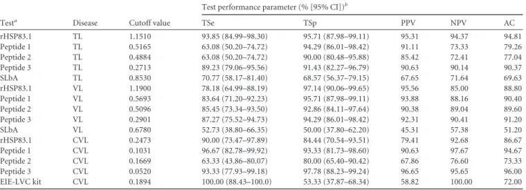
TABLE 2Measure of diagnostic performance for rHSP83.1, peptides 1, 2, and 3, SLbA, and the EIE-LVC kit
Testa Disease Cutoff value
Test performance parameter (% [95% CI])b
TSe TSp PPV NPV AC
rHSP83.1 TL 1.1510 93.85 (84.99–98.30) 95.71 (87.98–99.11) 95.31 94.37 94.81
Peptide 1 TL 0.5165 63.08 (50.20–74.72) 94.29 (86.01–98.42) 91.11 73.33 79.26
Peptide 2 TL 0.4884 63.08 (50.20–74.72) 90.00 (80.48–95.88) 85.42 72.41 77.04
Peptide 3 TL 0.2713 89.23 (79.06–95.56) 91.43 (82.27–96.79) 90.63 90.14 90.37
SLbA TL 0.8530 70.77 (58.17–81.40) 68.57 (56.37–79.15) 67.65 71.64 69.63
rHSP83.1 VL 1.1900 78.18 (64.99–88.19) 97.14 (90.06–99.65) 95.56 85.00 88.80
Peptide 1 VL 0.5693 83.64 (71.20–92.23) 95.71 (87.98–99.11) 93.88 88.16 90.40
Peptide 2 VL 0.5096 85.45 (73.34–93.50) 92.86 (84.11–97.64) 90.38 89.04 89.60
Peptide 3 VL 0.2901 87.27 (75.52–94.73) 94.29 (86.01–98.42) 92.31 90.41 91.20
SLbA VL 0.6780 52.73 (38.80–66.35) 50.00 (37.80–62.20) 45.31 57.38 51.20
rHSP83.1 CVL 0.2473 90.00 (73.47–97.89) 84.44 (70.54–93.51) 79.41 92.68 86.67
Peptide 1 CVL 0.1031 96.67 (82.78–99.92) 93.33 (81.73–98.60) 90.63 97.67 94.67
Peptide 2 CVL 0.1669 63.33 (43.86–80.07) 80.00 (65.40–90.42) 67.86 76.60 73.33
Peptide 3 CVL 0.0520 93.33 (77.93–99.18) 97.78 (88.23–99.24) 96.65 95.65 96.00
EIE-LVC kit CVL 0.1894 100.00 (88.43–100.0) 53.33 (37.87–68.34) 58.82 100.00 72.00
aCutoff values for all tests obtained by ROC curve, except that for the EIE-LVC kit, which was obtained according to the manufacturer. bParameters were calculated using all samples presented in this work for TL (CT⫹CD⫹CL⫹ML) (
n⫽135), VL (CT⫹CD⫹VL) (n⫽125), and CVL (CT⫹CD⫹LM⫹ CVL) (n⫽75). Tse, total sensitivity; CI, confidence interval; TSp, total specificity; PPV, positive predictive value; NPV, negative predictive value; AC, accuracy.
TABLE 3Diagnostic performance for rHSP83.1, peptides 1, 2, and 3, SLbA, and the EIE-LVC kit using ROC curves, data validation, and agreement using kappa index
Testa Disease AUCb(95% CIc)
No. with resultd:
e(95% CI) Agreementf
TP TN FP FN
rHSP83.1 TL 0.989 (0.979–1.000) 61 67 3 4 0.896 (0.821–0.971) Very good
Peptide 1 TL 0.881 (0.826–0.936) 41 66 4 24 0.580 (0.448–0.712) Moderate
Peptide 2 TL 0.819 (0.748–0.889) 41 63 7 24 0.536 (0.397–0.674) Moderate
Peptide 3 TL 0.961 (0.933–0.989) 58 64 6 7 0.807 (0.707–0.907) Very good
SLbA TL 0.753 (0.673–0.834) 48 46 22 19 0.393 (0.238–0.548) Fair
rHSP83.1 VL 0.937 (0.894–0.981) 43 68 2 12 0.768 (0.655–0.881) Good
Peptide 1 VL 0.934 (0.880–0.988) 46 67 3 9 0.803 (0.697–0.908) Very good
Peptide 2 VL 0.924 (0.867–0.981) 47 65 5 8 0.788 (0.679–0.897) Good
Peptide 3 VL 0.943 (0.892–0.995) 48 66 4 7 0.820 (0.719–0.922) Very good
SLbA VL 0.510 (0.405–0.614) 29 35 35 26 0.027 (⫺0.147–0.200) Poor
rHSP83.1 CVL 0.924 (0.867–0.981) 27 38 7 3 0.728 (0.572–0.884) Good
Peptide 1 CVL 0.950 (0.894–1.005) 29 42 3 1 0.890 (0.786–0.995) Very good
Peptide 2 CVL 0.755 (0.644–0.866) 19 36 9 11 0.438 (0.230–0.646) Moderate
Peptide 3 CVL 0.987 (0.967–1.007) 28 44 1 2 0.916 (0.823–1.000) Very good
EIE-LVC kit CVL NA 30 24 21 0 0.478 (0.316–0.639) Moderate
aCutoffs for all tests obtained by ROC curve, except that for the EIE-LVC kit, which was obtained according to the manufacturer. bAUC, area under the curve; NA, not applicable.
cCI, confidence interval.
dTP, true positive; TN, true negative; FP, false positive; FN, false negative.
eKappa index values were calculated using all samples presented in this work for TL (CT⫹CD⫹CL⫹ML) (n⫽135), VL (CT⫹CD⫹VL) (n⫽125), and CVL (CT⫹CD⫹
CVL) (n⫽75).
fAgreement was calculated using parasitological assays as a gold standard test.
on October 9, 2017 by UNIVERSIDADE FEDERAL DE OURO PRETO
http://cvi.asm.org/
study to the antigenicity of rHSP83.1, ELISA depletion assays were
performed using the synthetic peptides EEDESKKKSCGDEGEP
KVE, VTEGGEDKKK, and EVAEAPPAEAAPA, which
corre-spond to the predicted linear B-cell epitopes present in the protein
(
Fig. 6
). In this assay, the IgG reactivity against rHSP83.1 after the
depletion of antibodies against peptide 1 was reduced by 13% for
the CL (
P
⬍
0.01), 12% for the ML (
P
⬍
0.001), 8% for the VL
(
P
⬍
0.001), and 5% for the CVL (
P
⬍
0.05) group. For peptide 2,
we observed a reduction of 21% for the CL (
P
⬍
0.01), 17% for the
ML (
P
⬍
0.001), 8% for the VL (
P
⬍
0.001), and no reduction for
the CVL group. The depletion of antibodies against peptide 3
re-sulted in a reduction of 21% for the CL (
P
⬍
0.001), 18% for the
ML (
P
⬍
0.001), 16% for the VL (
P
⬍
0.001), and 15% for the CVL
(
P
⬍
0.001) group in the reactivity against rHSP83.1. No
signifi-cant differences in the reduction of reactivity after depletion were
observed in the human and canine CT and CD groups for any of
the peptides evaluated. Our results suggest that specific antibodies
against peptides 1 and 3 contribute significantly to the overall
antigenicity of rHSP83.1 in all
Leishmania
-infected groups;
how-ever, peptide 2 contributes to the antigenicity of this protein in the
TL and VL groups only.
DISCUSSION
Genome-wide epitope predictions for pathogenic microorganisms
have broadened the range of target epitopes and have provided clues
to enhance peptide immunogenicity. Among the methods of
selec-tion, epitope mapping is a very useful procedure that has vast
appli-cations in the areas of antibody production, immunodiagnostics,
epitope-based vaccine design, selective deimmunization of
therapeu-tic proteins, and autoimmunity. The identification of these targets
was facilitated by the publication of several parasite genomes that
together with the availability of algorithms for epitope prediction
allow for the use of large-scale approaches for the identification of
new antigens (
31–33
). As an attempt to identify new targets for
diagnosing multiple forms of leishmaniasis, we screened the
L.
braziliensis
proteome to predict linear B-cell epitopes that are
con-served in
Leishmania
spp. and divergent in
H. sapiens
,
C.
familia-ris
, and
T. cruzi
, the etiologic agent of Chagas disease, which often
presents cross-reactivity with leishmaniasis due to sharing
multi-ple common epitopes within the trypanosomatid species (
34
,
35
).
Heat shock proteins (HSPs) are highly conserved molecules in
prokaryotes and eukaryotes that play important roles in protein
folding, assembly of protein complexes, and the translocation of
proteins across cellular compartments (
14
). Despite the fact that
their sequences are highly conserved throughout evolution, it has
been proposed that the recognition of epitopes shared by HSPs
from different pathogens may provide the immune system with a
universal signal of infection (
36
). In this sense, HSP70 and HSP83
of
L. infantum
have been described as strong mitogens for B
lym-phocytes in a murine model (
37
). This phenomenon occurs even
in the absence of T lymphocytes and adherent cells, suggesting a
direct effect of
Leishmania
HSPs on B cells, and that
Leishmania
may serve to divert the immune response into the nonspecific
activation of immune cells (
37
).
In our study, we identified three linear B-cell epitopes in
HSP83.1 of
L. braziliensis
that are conserved within
Leishmania
species but are divergent from the orthologous proteins from
human and canine hosts and from
T. cruzi
, the etiological agent
of Chagas disease that has been responsible for several reports of
cross-reactivity in the serological diagnosis of leishmaniasis.
Pre-vious studies have described that heat shock proteins, such as
HSP83 and other chaperones, are among the most abundant
pro-teins detected in
Leishmania
antigenic extracts and have obtained
promising results that can be applied to the development of
diag-nostic kits (
38–44
). Here, the antigenicity of recombinant
FIG 6Immunodepletion assay showing specific IgG antibody recognition of the synthetic peptides with known reactivity to HSP83.1. Pools of serum (n⫽10) from the different groups were depleted with peptide 1, 2, or 3 (CT [control] group; CD [Chagas disease]; CL [cutaneous leishmaniasis]; ML [mucosal leishmaniasis]; VL [visceral leishmaniasis]; CVL [canine visceral leishmaniasis]). The mean antibody OD values are shown on theyaxis, and the error bars indicate the standard deviation. Significant differences are indicated on the graphs withPvalues (*,P⬍0.05; **,P⬍0.01; ***,P⬍0.001).
on October 9, 2017 by UNIVERSIDADE FEDERAL DE OURO PRETO
http://cvi.asm.org/
HSP83.1 (rHSP83.1) and three epitopes predicted in this protein
were tested with sera from the TL, VL, and CVL groups.
rHSP83.1 showed excellent performance in the diagnosis of
TL. Not all of the epitopes mapped in this study were
recog-nized by sera from all clinical forms of leishmaniasis.
Never-theless, our results allowed for the identification of epitopes
that presented high performance depending upon the species
of the parasite. Interestingly, it was possible to identify specific
epitopes derived from HSP83.1 that presented high
perfor-mance for the diagnosis of human TL (peptide 3), both human
and canine VL (peptides 1 and 3), and all TL, VL, and CVL
clinical manifestations (peptide 3).
In recent years, synthetic peptides have shown high
sensi-tivity and specificity when used as antigens in diagnostic tests
(
23
,
45
). These findings have also been associated with several
advantages over chemically purified or recombinant antigens,
because their production does not involve the manipulation of
living organisms, and the peptides can be obtained with a high
level of purity. Thus, it is appealing to use these peptides to
develop devices to diagnose one or multiple types of
leishman-iasis (
46
).
Comparing conventional and new techniques proposed to be
innovative is essential to justify the implementation of the novel
methods. In this sense, several authors have described ELISAs
us-ing soluble antigens from promastigotes for serological diagnosis
that showed high sensitivity for detecting
Leishmania
-infected
in-dividuals (
34
,
35
,
47
,
48
). Despite these findings, the use of these
antigens still represents a relevant obstacle because it is common
to observe a large number of false-positive reactions in individuals
infected with other trypanosomatids due to the sharing of
multi-ple common epitopes used in immunofluorescence assays or
ELISAs sensitized with crude antigens (
34
,
35
,
49
,
50
). As
ex-pected, using crude antigens, such as the soluble
L. braziliensis
antigen or the EIE-LVC kit, which uses antigen prepared from
Leishmania major
-like promastigotes, leads to a pronounced
number of false positives compared to with rHSP83.1. This is the
main limitation to their use in epidemiological surveys in areas
where leishmaniasis is endemic (
47
). Furthermore, different
spe-cies and strains of
Leishmania
can be used for the production of
crude antigen, thus limiting the standardization and
reproducibil-ity of test results (
51
,
52
). Therefore, there is a need to standardize
an antigen for use in different regions of the world (
47
).
The serologic enzyme-linked immunosorbent assay is an
im-portant tool in the diagnosis of leishmaniasis compared to
meth-ods currently used to directly reveal parasites in tissue smears that
are invasive, are risky, require considerable expertise, and are
in-appropriate for use in epidemiological surveillance (
53
,
54
). Given
the above, the rational development of a new serological
tech-nique featuring high sensitivity and specificity that is also able to
discriminate cross-reactivity with other human and canine
infec-tions, at the individual level, represents an important innovation
in the serological diagnosis of CVL (
11
).
The present study demonstrates that rHSP83.1 and the
epitopes mapped on its sequence might be among the target
mol-ecules that could be used in an immunodiagnostic test. This was
confirmed by the high sensitivity for diagnosing
Leishmania
infec-tions, the high specificity for discriminating between other human
and canine infections, and the high degree of agreement with
par-asitological techniques for the diagnosis of leishmaniasis. Further
prospective studies using large cohorts of negative and positive
individuals from areas where leishmaniasis is endemic are
neces-sary to better characterize this approach as a possible marker in the
diagnosis of TL, VL, and CVL and for monitoring posttherapeutic
cures of TL and VL.
ACKNOWLEDGMENTS
C.M.C., E.A.F.C., R.T.F., and D.C.B. thank the Conselho Nacional de Desenvolvimento Científico e Tecnológico, Brazil, for their fellowships. We thank Michele Silva de Matos for her technical support.
REFERENCES
1.Desjeux P.2001. Worldwide increasing risk factors for leishmania-sis. Med. Microbiol. Immunol.190:77–79.http://dx.doi.org/10.1007 /s004300100085.
2.Desjeux P.2004. Leishmaniasis: current situation and new perspectives. Comp. Immunol. Microbiol. Infect. Dis.27:305–318.http://dx.doi.org/10 .1016/j.cimid.2004.03.004.
3.Gontijo B, de Carvalho Mde L.2003. American cutaneous leishmaniasis. Rev. Soc. Bras. Med. Trop.36:71– 80. (In Portuguese.)http://dx.doi.org /10.1590/S0037-86822003000100011.
4.Martins AL, Barreto JA, Lauris JR, Martins AC.2014. American tegu-mentary leishmaniasis: correlations among immunological, histopatho-logical and clinical parameters. An. Bras. Dermatol.89:52–58.http://dx .doi.org/10.1590/abd1806-4841.20142226.
5.Costa JM, Marsden PD, Llanos-Cuentas EA, Netto EM, Carvalho EM, Barral A, Rosa AC, Cuba CC, Magalhães AV, Barreto AC.1986. Dis-seminated cutaneous leishmaniasis in a field clinic in Bahia, Brazil: a re-port of eight cases. J. Trop. Med. Hyg.89:319 –323.
6.Marsden PD.1985. Clinical presentations of Leishmania braziliensis bra-ziliensis. Parasitol. Today 1:129 –133. http://dx.doi.org/10.1016/0169 -4758(85)90057-2.
7.Grimaldi G, Jr, Tesh RB.1993. Leishmaniases of the New World: current concepts and implications for future research. Clin. Microbiol. Rev.
6:230 –250.
8.Tesh RB.1995. Control of zoonotic visceral leishmaniasis: is it time to change strategies? Am. J. Trop. Med. Hyg.52:287–292.
9.Molina R, Amela C, Nieto J, San-Andrés M, González F, Castillo JA, Lucientes J, Alvar J.1994. Infectivity of dogs naturally infected with Leishmania infantumto colonizedPhlebotomus perniciosus. Trans. R. Soc. Trop. Med. Hyg.88:491– 493.http://dx.doi.org/10.1016/0035-9203(94) 90446-4.
10. Chappuis F, Sundar S, Hailu A, Ghalib H, Rijal S, Peeling RW, Alvar J, Boelaert M.2007. Visceral leishmaniasis: what are the needs for diagnosis, treatment and control? Nat. Rev. Microbiol.5:S7–S16.http://dx.doi.org /10.1038/nrmicro1748.
11. de Andrade RA, Reis AB, Gontijo CM, Braga LB, Rocha RD, Araújo MS, Vianna LR, Martins-Filho OA. 2007. Clinical value of anti-Leishmania(Leishmania)chagasiIgG titers detected by flow cytometry to distinguish infected from vaccinated dogs. Vet. Immunol. Immuno-pathol.116:85–97.http://dx.doi.org/10.1016/j.vetimm.2007.01.002. 12. de Oliveira CI, Nascimento IP, Barral A, Soto M, Barral-Netto M.2009.
Challenges and perspectives in vaccination against leishmaniasis. Parasi-tol. Int.58:319 –324.http://dx.doi.org/10.1016/j.parint.2009.07.013. 13. Souza AP, Soto M, Costa JM, Boaventura VS, de Oliveira CI, Cristal JR,
Barral-Netto M, Barral A. 2013. Towards a more precise serological diagnosis of human tegumentary leishmaniasis usingLeishmania recom-binant proteins. PLoS One8:e66110.http://dx.doi.org/10.1371/journal .pone.0066110.
14. Henderson B.2010. Integrating the cell stress response: a new view of molecular chaperones as immunological and physiological homeostatic regulators. Cell Biochem. Funct.28:1–14.http://dx.doi.org/10.1002/cbf .1609.
15. Vexenat Ade C, Santana JM, Teixeira AR. 1996. Cross-reactivity of antibodies in human infections by the kinetoplastid protozoa Trypano-soma cruzi,Leishmania chagasiandLeishmania(Viannia)braziliensis. Rev. Inst. Med. Trop. Sao Paulo38:177–185.
16. Mancianti F, Pedonese F, Poli A.1996. Evaluation of dot enzyme-linked immunosorbent assay (dot-ELISA) for the serodiagnosis of canine leish-maniosis as compared with indirect immunofluorescence assay. Vet. Parasitol.65:1–9.http://dx.doi.org/10.1016/0304-4017(96)00946-6. 17. Alves WA, Bevilacqua PD.2004. Quality of diagnosis of canine visceral
on October 9, 2017 by UNIVERSIDADE FEDERAL DE OURO PRETO
http://cvi.asm.org/
leishmaniasis in epidemiological surveys: an epidemic in Belo Horizonte, Minas Gerais, Brazil, 1993–1997. Cad. Saude Publica.20:259 –265. (In Portuguese.)http://dx.doi.org/10.1590/S0102-311X2004000100043. 18. de Bruijn MH, Barker DC.1992. Diagnosis of New World leishmaniasis:
specific detection of species of theLeishmania braziliensiscomplex by am-plification of kinetoplast DNA. Acta Trop.52:45–58.http://dx.doi.org/10 .1016/0001-706X(92)90006-J.
19. Aslett M, Aurrecoechea C, Berriman M, Brestelli J, Brunk BP, Car-rington M, Depledge DP, Fischer S, Gajria B, Gao X, Gardner MJ, Gingle A, Grant G, Harb OS, Heiges M, Hertz-Fowler C, Houston R, Innamorato F, Iodice J, Kissinger JC, Kraemer E, Li W, Logan FJ, Miller JA, Mitra S, Myler PJ, Nayak V, Pennington C, Phan I, Pinney DF, Ramasamy G, Rogers MB, Roos DS, Ross C, Sivam D, Smith DF, Srinivasamoorthy G, Stoeckert CJ, Jr, Subramanian S, Thibodeau R, Tivey A, Treatman C, Velarde G, Wang H.2010. TriTrypDB: a func-tional genomic resource for theTrypanosomatidae. Nucleic Acids Res.
38:D457–D462.http://dx.doi.org/10.1093/nar/gkp851.
20. Larsen JE, Lund O, Nielsen M.2006. Improved method for predicting linear B-cell epitopes. Immunome Res. 2:2.http://dx.doi.org/10.1186 /1745-7580-2-2.
21. Altschul SF, Gish W, Miller W, Myers EW, Lipman DJ.1990. Basic local alignment search tool. J. Mol. Biol.215:403– 410.http://dx.doi.org/10 .1006/jmbi.1990.9999.
22. Larkin MA, Blackshields G, Brown NP, Chenna R, McGettigan PA, McWilliam H, Valentin F, Wallace IM, Wilm A, Lopez R, Thompson JD, Gibson TJ, Higgins DG.2007. Clustal W and Clustal X version 2.0. Bioinformatics23:2947–2948. http://dx.doi.org/10.1093/bioinformatics /btm404.
23. Mendes TA, Reis Cunha JL, de Almeida Lourdes R, Rodriguez Luiz GF, Lemos LD, dos Santos AR, da Câmara AC, Galvão LM, Bern C, Gilman RH, Fujiwara RT, Gazzinelli RT, Bartholomeu DC.2013. Identification of strain-specific B-cell epitopes inTrypanosoma cruziusing genome-scale epitope prediction and high-throughput immunoscreening with peptide arrays. PLoS Negl. Trop. Dis.7:e2524.http://dx.doi.org/10.1371/journal .pntd.0002524.
24. Carneiro FR, Silva TC, Alves AC, Haline-Vaz T, Gozzo FC, Zanchin NI.
2006. Spectroscopic characterization of the tumor antigen NY-REN-21 and identification of heterodimer formation with SCAND1. Biochem. Biophys. Res. Commun.343:260 –268.http://dx.doi.org/10.1016/j.bbrc .2006.02.140.
25. Coitinho JB, Costa DM, Guimarães SL, de Góes AM, Nagem RA.2012. Expression, purification and preliminary crystallographic studies of NahF, a salicylaldehyde dehydrogenase fromPseudomonas putidaG7 involved in naphthalene degradation. Acta Crystallogr. Sect. F Struct. Biol. Cryst. Commun.68:93–97.http://dx.doi.org/10.1107/S174430911105038X. 26. Wellings DA, Atherton E.1997. Standard Fmoc protocols. Methods
Enzy-mol.289:44–67.http://dx.doi.org/10.1016/S0076-6879(97)89043-X. 27. Santiago HC, Bennuru S, Boyd A, Eberhard M, Nutman TB. 2011.
Structural and immunologic cross-reactivity among filarial and mite tro-pomyosin: implications for the hygiene hypothesis. J. Allergy Clin. Immu-nol.127:479 – 486.http://dx.doi.org/10.1016/j.jaci.2010.11.007. 28. Bueno LL, Lobo FP, Morais CG, Mourão LC, Machado de Avila RA,
Soares IS, Fontes CJ, Lacerda MV, Chavez Olórtegui C, Bartholomeu DC, Fujiwara RT, Braga EM.2011. Identification of a highly antigenic linear B cell epitope withinPlasmodium vivaxapical membrane antigen 1 (AMA-1). PLoS One6:e21289.http://dx.doi.org/10.1371/journal.pone.0021289. 29. Linnet K, Bossuyt PM, Moons KG, Reitsma JB.2012. Quantifying the
accuracy of a diagnostic test or marker. Clin. Chem.58:1292–1301.http: //dx.doi.org/10.1373/clinchem.2012.182543.
30. Fleiss JL, Spitzer RL, Endicott J, Cohen J. 1972. Quantification of agreement in multiple psychiatric diagnosis. Arch. Gen. Psychiatry26:
168 –171.http://dx.doi.org/10.1001/archpsyc.1972.01750200072015. 31. Singh H, Ansari HR, Raghava GP.2013. Improved method for linear
B-cell epitope prediction using antigen’s primary sequence. PLoS One
8:e62216.http://dx.doi.org/10.1371/journal.pone.0062216.
32. Dudek NL, Perlmutter P, Aguilar MI, Croft NP, Purcell AW.2010. Epitope discovery and their use in peptide based vaccines. Curr. Pharm. Des.16:3149 –3157.http://dx.doi.org/10.2174/138161210793292447. 33. Bryson CJ, Jones TD, Baker MP.2010. Prediction of immunogenicity of
therapeutic proteins: validity of computational tools. BioDrugs24:1– 8.
http://dx.doi.org/10.2165/11318560-000000000-00000.
34. Badaró R, Reed SG, Barral A, Orge G, Jones TC.1986. Evaluation of the micro enzyme-linked immunosorbent assay (ELISA) for antibodies in
American visceral leishmaniasis: antigen selection for detection of infec-tion-specific responses. Am. J. Trop. Med. Hyg.35:72–78.
35. Barbosa-De-Deus R, dos Mares-Guia ML, Nunes AZ, Costa KM, Jun-queira RG, Mayrink W, Genaro O, Tavares CAP. 2002. Leishmania major-like antigen for specific and sensitive serodiagnosis of human and canine visceral leishmaniasis. Clin. Diagn. Lab. Immunol.9:1361–1366.
http://dx.doi.org/10.1128/CDLI.9.6.1361-1366.2002.
36. Kaufmann SH.1990. Heat shock proteins and the immune response. Immunol. Today11:129 –136.http://dx.doi.org/10.1016/0167-5699(90) 90050-J.
37. Rico AI, Gironès N, Fresno M, Alonso C, Requena JM.2002. The heat shock proteins, Hsp70 and Hsp83, ofLeishmania infantumare mitogens for mouse B cells. Cell Stress Chaperones7:339 –346.http://dx.doi.org/10 .1379/1466-1268(2002)007⬍0339:THSPHA⬎2.0.CO;2.
38. Coelho VT, Oliveira JS, Valadares DG, Chávez-Fumagalli MA, Duarte MC, Lage PS, Soto M, Santoro MM, Tavares CA, Fernandes AP, Coelho EA.2012. Identification of proteins in promastigote and amastigote-like Leishmaniausing an immunoproteomic approach. PLoS Negl. Trop. Dis.
6:e1430.http://dx.doi.org/10.1371/journal.pntd.0001430.
39. Costa MM, Andrade HM, Bartholomeu DC, Freitas LM, Pires SF, Chapeaurouge AD, Perales J, Ferreira AT, Giusta MS, Melo MN, Gazzinelli RT.2011. Analysis ofLeishmania chagasiby 2-D difference gel electrophoresis (2-D DIGE) and immunoproteomic: identification of novel candidate antigens for diagnostic tests and vaccine. J. Proteome Res.
10:2172–2184.http://dx.doi.org/10.1021/pr101286y.
40. Skeiky YA, Benson DR, Costa JL, Badaro R, Reed SG.1997. Association ofLeishmaniaheat shock protein 83 antigen and immunoglobulin G4 antibody titers in Brazilian patients with diffuse cutaneous leishmaniasis. Infect. Immun.65:5368 –5370.
41. Rey-Ladino JA, Joshi PB, Singh B, Gupta R, Reiner NE.1997. Leishma-nia major: molecular cloning, sequencing, and expression of the heat shock protein 60 gene reveals unique carboxy terminal peptide sequences. Exp. Parasitol.85:249 –263.http://dx.doi.org/10.1006/expr.1996.4137. 42. Quijada L, Requena JM, Soto M, Alonso C.1998. Analysis of the
anti-genic properties of theL. infantumHsp70: design of synthetic peptides for specific serodiagnosis of human leishmaniasis. Immunol. Lett.63:169 – 174.http://dx.doi.org/10.1016/S0165-2478(98)00071-6.
43. Celeste BJ, Angel SO, Castro LG, Gidlund M, Goto H.2004.Leishmania infantumheat shock protein 83 for the serodiagnosis of tegumentary leish-maniasis. Braz. J. Med. Biol. Res.37:1591–1593.http://dx.doi.org/10.1590 /S0100-879X2004001100001.
44. Kaur J, Kaur S.2013. ELISA and Western blotting for the detection of Hsp70 and Hsp83 antigens ofLeishmania donovani. J. Parasit. Dis.37:68 – 73.http://dx.doi.org/10.1007/s12639-012-0133-0.
45. Carmona SJ, Sartor PA, Leguizamón MS, Campetella OE, Agüero F.
2012. Diagnostic peptide discovery: prioritization of pathogen diagnostic markers using multiple features. PLoS One7:e50748.http://dx.doi.org/10 .1371/journal.pone.0050748.
46. Aguirre S, Silber AM, Brito ME, Ribone ME, Lagier CM, Marcipar IS.
2006. Design, construction, and evaluation of a specific chimeric antigen to diagnose chagasic infection. J. Clin. Microbiol.44:3768 –3774.http://dx .doi.org/10.1128/JCM.01043-06.
47. Rosário EY, Genaro O, Franca-Silva JC, da Costa RT, Mayrink W, Reis AB, Carneiro M.2005. Evaluation of enzyme-linked immunosorbent assay using crudeLeishmaniaand recombinant antigens as a diagnostic marker for canine visceral leishmaniasis. Mem. Inst. Oswaldo Cruz100:
197–203.http://dx.doi.org/10.1590/S0074-02762005000200015. 48. Mettler M, Grimm F, Capelli G, Camp H, Deplazes P.2005. Evaluation
of enzyme-linked immunosorbent assays, an immunofluorescent-antibody test, and two rapid tests (immunochromatographic-dipstick and gel tests) for serological diagnosis of symptomatic and asymptomatic Leishmaniainfections in dogs. J. Clin. Microbiol.43:5515–5519.http://dx .doi.org/10.1128/JCM.43.11.5515-5519.2005.
49. da Costa CA, Genaro O, de Lana M, Magalhães PA, Dias M, Michalick MS, Melo MN, da Costa RT, Magalhães-Rocha NM, Mayrink W.1991. Canine visceral leishmaniasis: evaluation of the serologic method used in epidemiologic studies. Rev. Soc. Bras. Med. Trop.24:21–25.http://dx.doi .org/10.1590/S0037-86821991000100004.
50. Roffi J, Dedet JP, Desjeux P, Garré MT.1980. Detection of circulating antibodies in cutaneous leishmaniasis by enzyme-linked immunosorbent assay (ELISA). Am. J. Trop. Med. Hyg.29:183–189.
51. Burns JM, Jr, Shreffler WG, Benson DR, Ghalib HW, Badaro R, Reed SG.1993. Molecular characterization of a kinesin-related antigen of
on October 9, 2017 by UNIVERSIDADE FEDERAL DE OURO PRETO
http://cvi.asm.org/
mania chagasithat detects specific antibody in African and American vis-ceral leishmaniasis. Proc. Natl. Acad. Sci. U. S. A.90:775–779.http://dx .doi.org/10.1073/pnas.90.2.775.
52. Martin SK, Thuita-Harun L, Adoyo-Adoyo M, Wasunna KM.1998. A diagnostic ELISA for visceral leishmaniasis, based on antigen from media conditioned byLeishmania donovanipromastigotes. Ann. Trop. Med. Parasitol.92:571–577.http://dx.doi.org/10.1080/00034989859267. 53. Pereira VR, Reis Lde C, Souza Mde A, de Oliveira AP, de Brito ME,
Lage PS, Andrade MC, Rocha RD, Martins-Filho OA.2012.
Evalu-ation of anti-lived and anti-fixedLeishmania(Viannia)braziliensis pro-mastigote IgG antibodies detected by flow cytometry for diagnosis and post-therapeutic cure assessment in localized cutaneous leishmaniasis. Diagn. Microbiol. Infect. Dis. 74:292–298. http://dx.doi.org/10.1016/j .diagmicrobio.2012.06.025.
54. Kumar S, Kumar D, Chakravarty J, Rai M, Sundar S.2012. Identifica-tion and characterizaIdentifica-tion of a novelLeishmania donovaniantigen for se-rodiagnosis of visceral leishmaniasis. Am. J. Trop. Med. Hyg.86:601– 605.
http://dx.doi.org/10.4269/ajtmh.2012.11-0261.