UNIVERSIDADE DE LISBOA
FACULDADE DE CIÊNCIAS
DEPARTAMENTO DE BIOLOGIA VEGETAL
A
1
and A
2A
Adenosine Receptors Expression in ALS
Transgenic Mice for the Human Gene SOD1
Gonçalo Luis Monteiro Ramos
Mestrado em Biologia Molecular e Genética
2012
UNIVERSIDADE DE LISBOA
FACULDADE DE CIÊNCIAS
DEPARTAMENTO DE BIOLOGIA VEGETAL
A
1
and A
2A
Adenosine Receptors Expression in ALS
Transgenic Mice for the Human Gene SOD1
Dissertação orientada por:
Doutora Alexandra Marçal, Instituto de Medicina Molecular, Lisboa
Professora Doutora Margarida Ramos, Faculdade de Ciências da Universidade de Lisboa
Gonçalo Luis Monteiro Ramos
Mestrado em Biologia Molecular e Genética
2012
Todas as afirmações efetuadas no presente documento são da exclusiva responsabilidade do seu autor, não cabendo qualquer responsabilidade à Faculdade de Ciências da Universidade de Lisboa pelos conteúdos nele apresentados.
O presente trabalho foi realizado na Unidade de Farmacologia e Neurociências, Instituto de Medicina Molecular, Faculdade de Medicina da Universidade de Lisboa.
Aos meus Avós.
Ao meu Avô, José Augusto Gonçalves Ramos,
cuja rectidão de carácter, princípios, disciplina,
dedicação e amor sempre me influenciaram.
Que orgulho tenho em ti!
“Consistency is the last refuge of the unimaginative.”
- Oscar Wilde
Table of contents
INDEX OF FIGURES ... IX INDEX OF TABLES ... X RESUMO ... XI ABSTRACT ... XII ABBREVIATIONS LIST ... 1 INTRODUCTION ... 21. AMYOTROPHIC LATERAL SCLEROSIS ... 2
1.1. Historical background ... 2
1.2. Epidemiological and Clinical features of ALS ... 3
1.3. Superoxide dismutase 1 mutation ... 4
1.4. SOD1 mouse models ... 6
1.4.1. SOD1 overexpressing and knockout models ... 6
1.4.2. SOD1 mutant transgenic model ... 7
1.5. Pathogenic mechanisms of ALS ... 8
1.5.1. Protein misfolding and aggregation ... 9
1.5.2. Mitochondrial dysfunction and oxidative stress ... 10
1.5.3. Excitotoxicity ... 10
1.5.4. Impaired axonal transport ... 11
1.5.5. Endoplasmic reticulum stress ... 11
1.5.6. Neuroinflamation ... 11
1.6. Where does ALS begin? ... 12
2. THE MOTOR NERVOUS SYSTEM ... 13
2.1. Motor neurons and neuromuscular synaptic transmission ... 13
3. ADENOSINE ... 14
3.1. Adenosine receptors ... 15
3.2. Adenosine receptors distribution and interactions ... 16
OBJECTIVES ... 18
MATERIALS & METHODS ... 19
1. BREEDINGS AND HOUSBANDRY ... 19
3. TISSUE EXTRACTION AND DISSECTION ... 20
4. PROTEIN QUANTIFICATION ... 20
4.1. Total protein homogenates ... 20
4.2. Gel electrophoresis and immunoblotting ... 20
5. MRNA EXPRESSION ... 21
5.1. Total RNA homogenates ... 21
5.2. cDNA synthesis and qRT-PCR ... 22
6. STATISTICS ... 23
RESULTS ... 24
Quantification of A1 and A2A receptor protein levels ... 24
Quantification of A1 and A2A receptor mRNA levels ... 26
Primary pathological feature, regarding the expression of adenosine receptors ... 27
DISCUSSION ... 28 REFERENCES ... 31 ACKNOLEDGEMENTS ... 40 ANNEXES ... 41 ANNEXE I ... 41 ANNEXE II ... 42 ANNEXE III ... 43 ANNEXE IV ... 45
Index of Figures
Figure 1. Jean-Martin Charcot (1825-1893) ... 2 Figure 2. Clinical features of muscle wasting in patients with ALS. ... 4 Figure 3. Mutations causing ALS. ... 6 Figure 4. Time course of clinical and neuropathological events in the high copy number transgenic SOD1G93A mice. ... 8 Figure 5. Schematic evolution of motor neuron degeneration during the course of SOD1 mutant ALS disease. ... 9 Figure 6. Somatic Component of the Peripheral Nervous System. ... 13 Figure 7. Structure of the Neuromuscular Junction.. ... 14 Figure 8. Immunoblot analysis of the expression levels of A1 adenosine receptor in control
and hSOD1 mutants ... 24 Figure 9. Immunoblot analysis of the expression levels of A2A adenosine receptor in control
and hSOD1 mutants ... 25 Figure 10. Quantitative RT-PCR analysis of the expression levels of A1 and A2A adenosine
receptor in diaphragm for hSOD1 mutants throughout disease ... 26 Figure 11. Schematic representation A1 and A2A adenosine receptor variation in the CNS and PNS of ALS SOD1G93A transgenic mice throughout disease progression.. ... 27
Figure 12. qRT-PCR calibration curve and quality control using SYBR Green method for β-actin mRNA quantification ... 43 Figure 13. qRT-PCR calibration curve and quality control using SYBR Green method for A2AR mRNA quantification. ... 44
Figure 14. Immunoblot analysis of the expression levels of A2A adenosine receptor in control
Index of tables
Table 1. Reviewed genes associated with familial ALS ... 5
Table 2. Adenosine receptors in the central nervous system and their properties. ... 16
Table 3. Antibodies used in this study ... 21
Table 4. Thermocycler PCR conditions for genotyping protocol. ... 41
Table 5. Thermocycler cDNA sysnthesis protocol ... 41
Table 6. Rotorgene thermocycler conditions for qRT-PCR. ... 41
Resumo
A Esclerose Lateral Amiotrópica (ELA) é uma doença progressiva e fatal caracterizada pela degeneração selectiva dos neurónios motores do córtex motor, tronco cerebral e medula espinal, que provoca atrofia muscular, paralesia e morte por falha respiratória. A etiologia da doença continua desconhecida, mas com um consenso de que o dano dos neurónios motores é causado por uma rede de processos patológicos complexos. Os mecanismos envolvidos na degeneração dos neurónios motores são melhor conhecidos num subtipo da doença causada por mutações na enzima superóxido dismutase 1 (SOD1). Esta enzima actua na eliminação de radicais livres de oxigénio e na ELA o processo de degeneração neuronal deve-se a um ganho de função da SOD1. A adenosina tem uma função importante na modulação da transmissão sináptica no SNC e SNP, actuando a dois níveis: inibitório, modulado pelos receptores do subtipo A1 e excitatório, mediado pelos
receptores do subtipo A2A. É conhecido que a expressão dos receptores A1 e A2A da
adenosina está alterada nalgumas doenças neurodegenerativas, mas o seu papel na ELA é ainda muito pouco conhecido.
O objectivo deste trabalho foi determinar o efeito da ELA na expressão proteica e de mRNA dos receptors A1 e A2A da adenosina no decurso da doença. O modelo de murganhos
transgénicos para o gene SOD1 humano com a mutação G93A foi usado neste trabalho. Os níveis proteicos e de mRNA de ambos os receptores foram quantificados através das técnicas de immunoblotting e PCR quantitativo em tempo real, respectivamente. Foram estudados diferentes tecidos do SNC e SNP, nomeadamente, córtex e medula espinal (apenas immunoblotting) e nervo frénico-diafragama, de animais selvagens e portadores da doença nas fases pre-sintomática (4-6 semanas) e sintomática (13-14 semanas).
Resultados deste estudo indicaram níveis proteicos não alterados nos SNC e SNP do receptor A1 ao longo da progressão da doença. No entanto, observou-se uma
sobreexpressão dos receptores A2A no córtex na fase pre-sintomática e um decréscimo na
fase sintomática. Os outros tecidos mantiveram-se inalterados no que se refere aos receptores A2A em ambas as fases da doença. A avaliação da expressão de mRNA no
diafragma não revelou quaisquer alterações em ambos os receptores da adenosina durante a progressão da doença. Assim, no que se refere aos receptores da adenosina em ELA, as primeiras alterações parecem ocorrer logo no início da doença nos receptores A2A do SNC.
Palavras-chave: Esclerose Lateral Amiotrópica (ELA); mutação SOD1G93A; receptor A 1 da
Abstract
Amyothrophic Lateral Sclerosis (ALS) is a progressive and fatal disease categorized by a selective degeneration of motor neurons from the cerebral cortex, brainstem and spinal cord that provokes muscle atrophy, progressive paralysis and death due to respiratory failure. The etiology of most ALS cases remains unknown but there is a current consensus that motor neuron degeneration is caused by a complex interaction between multiple pathogenic processes. The mechanisms of motor neuron degeneration are best understood in the subtype of disease caused by mutations in the enzyme superoxide dismutase 1. This enzyme is enrolled in the degradation of free oxygen radicals and in ALS neuronal damage is due to its gain-of-function. Adenosine has a central role as a neuromodulator of the CNS and PNS synaptic transmission. Adenosine acts at two levels: inhibitory through the subtype A1
receptor and excitatory through the subtype A2A receptor. Variation on the expression of A1
and A2A receptors has been identified in some neurodegenerative diseases, but their role in
ALS is not yet understood.
The objective of this work was to determine the effect of ALS on the protein and mRNA expression of A1 and A2A adenosine receptors through disease progression. The transgenic
model of mice carrying the human SOD1 gene with the G93A mutation was used in this work. Protein and mRNA levels of both receptors were quantified through immunblotting and quantitative real time PCR, respectively. Different tissues of the CNS and PNS, namely cortex and spinal cord (immunoblotting only) and phrenic nerve-diaphragm were studied in wild-type and transgenic mice in the pre-symptomatic (4-6 weeks) and symptomatic (13-14 weeks) phases of the disease.
Results from this study indicate unaltered A1 receptor protein levels at the CNS and
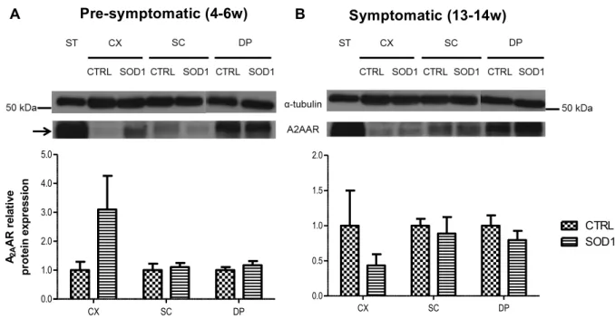
PNS through disease progression. However, there is an overexpression of A2A receptors in
the cortex of pre-symptomatic mice and a decrease in the symptomatic phase. The A2A
receptors are unaltered in the other tissues in both phases of the disease. The mRNA evaluation does not reveal significant alterations in both adenosine receptors during disease progression. Thus, regarding adenosine receptors in ALS, the first changes seem to occur early in the disease at the CNS in A2A receptors.
Key words: Amyotrophic Lateral Sclerosis (ALS); SOD1G93A mutation; A1 adenosine receptor,
Abbreviations list
A1R – A1 adenosine receptor
A2AR – A2A adenosine receptor
A2BR – A2B adenosine receptor
A3R – A3 adenosine receptors
ADP – Adenosine Diphosphate ALS – Amyothrophic Lateral Sclerosis ATP – Adenosine Triphosphate BSA – Bovine Serum Albumin CNS – Central Nervous System CTRL – Control (referring to Wild-type
endogenous SOD1 mouse model)
DEPC – Diethylpyrocarbonate
dNTP - Deoxynucleotide Triphosphates DTT – Dithiothreitol
ECL - Enhanced Chemiluminescence EDTA - Ethylenediaminetetracetic Acid ER – Endoplasmic Reticulum
FTD – Frontotemporal Dementia HEPES -
4-(2-hydroxyethyl)-1-piperazineethanesulfonic acid
LMN – Lower Motor Neuron MND – Motor Neuron Diseases
NADH - Nicotinamide Adenine Dinucleotide
Hidrogenase
NMJ – Neuromuscular Junction
NP-40 – Nonyl Phenoxypolyethoxylethanol PBS - Phosphate Buffered Saline
PD – Parkinson’s Disease
PMSF – Phenylmethanesulfonyl Fluoride PNS – Peripheral Nervous System PST – Pre-Symptomatic phase mice PVDF – Polyvinylidene Difluoride
RIPA - Radio-Immunoprecipitation Assay ROS – Reactive Oxygen Species
RPM - Revolutions Per Minute SDS - Sodium Dodecyl Sulfate
SOD1 – Superoxide Dismutase 1 (referring
to transgenic mouse model for human SOD1 with G93A mutation)
ST – Symptomatic phase mice TBS – Tris Buffered Saline
TBS-T - Tris-Buffered Saline with Tween TDB – Tail Digestion Buffer
INTRODUCTION
1. Amyotrophic Lateral Sclerosis
1.1. Historical background
It was in the latter half of the 19th century that the initial steps towards the unraveling of one of the most common motor neuron diseases (MND) were accomplished. Using clinical cases and autopsy material, a technique known as “anatomo-clinical method”, the famous French neurobiologist and physician Jean-Martin Charcot (Figure 1), showed that it could be possible to correlate anatomical lesions in the nervous system by the presence of clinical signs (Goetz et al., 1995; Goetz, 2000 and Rowland, 2001). In this context, his first major contribution was in 1865 (Charcot, 1865) when he presented a case of a young woman who developed profound weakness and showed increased muscle tone, with contractures of all extremities, despite she had no intellect or sensory abnormalities, and her urinary control was normal. At the autopsy study, Charcot found specific and isolated lateral column degeneration in the spinal cord:
“On careful examination of the surface of the spinal cord, on both sides in the lateral areas, there are two brownish-gray streak marks produced by sclerotic changes. These grayish bands begin outside the line of insertion of the posterior roots, and their anterior border approaches, but do not include, the entrance area of the anterior roots. They are visible throughout the thoracic region and continue, though greatly thinning out, up to the widening point of the cervical cord. Below, they are barely visible in the thoracolumbar region. Transverse sections taken at different levels allow one to see that the lateral columns have in their most superficial and posterior regions, a gray, semitransparent appearance, rather gelatinous.... At no point does the diseased tissue penetrate the gray matter which remains unaffected.” (Charcot, 1865).
In a second apparently unrelated observation (Charcot & Joffroy, 1869) with his colleague, Joffroy, they found pediatric cases of infantile paralysis in which the spinal cord lesions were systematically limited to the anterior horns of the grey matter. Thus raised the hypothesis that the spinal cord motor system was organized into two parts, and that lesions
Figure 1 | Jean-Martin Charcot (1825-1893). Jean-Martin Charcot was a French neurobiologist and physician
affecting each part cause different clinical signs. These conclusions became the pillars of modern neurology: when gray matter motor nuclei are damaged, weakness is associated with muscular atrophy in the body areas supplied by those cells, whereas when white lateral column damage occurs, weakness is associated with progressive contractures and spasticity. Charcot’s achievement to make sense of these evidences, led him for the first time in 1874 (Charcot, 1874) to use the term Amyotrophic Lateral Sclerosis to refer to this disorder and stated that:
“I do not think that elsewhere in medicine, in pulmonary or cardiac pathology, greater precision can be achieved. The diagnosis as well as the anatomy and physiology of the condition “amyotrophic lateral sclerosis” is one of the most completely understood conditions in the realm of clinical neurology.”
(Charcot, 1887).
ALS first became known as Charcot’s sclerosis but in North America the term “ALS” is used interchangeably with “Lou Gehrig’s disease” in memory of the famous baseball player who died of the disease in 1941. The word Amyotrophic comes from the Greek language. "A" means no, "Myo" refers to muscle, and "Trophic" means nourishment – "No muscle nourishment". When a muscle has no nourishment, it atrophies or wastes away. "Lateral" identifies the areas in a person's spinal cord where portions of the nerve cells that signal and control the muscles are located. As this area degenerates it leads to scarring or hardening ("sclerosis") in the region (The ALS Association, 2010).
1.2. Epidemiological and Clinical features of ALS
After 140 years, ALS is the most common adult-onset motor neuron disease. With a uniform worldwide incidence (frequency of new cases per year) of approximately 1-2 per 100 000 individuals and a prevalence (the proportion of affected individuals in the population) of 4-6 per 100 000. It affects people of all races and ethnic backgrounds and more commonly men than women (the male:female ratio is 3:2) (Kiernan et al., 2011). There are a few exceptions with higher frequency of cases, such as Guam (Reed et al., 1975), the Kii Peninsula of Japan (Kimura, 1965) and the southern lowlands of western New Guinea (Gajdusek & Salazar, 1982). Although 90-95% of cases have been classed as sporadic ALS (SALS) with no apparent genetic link, in the remaining 5-10% of instances the disease is inherited in an autosomal dominant manner, referred as familial ALS (FALS). The mean age of onset is 45-60 years in both forms of ALS. The primary hallmark is the degeneration of the upper motor neurons (UMN) of the motor cortex and of the lower motor neurons (LMN), which extend through the brainstem and spinal cord to innervate skeletal muscles. Clinical
presentation (figure 2) varies but most commonly consists of progressive muscle weakness, fasciculations (twitching muscles), atrophy and spasticity (the persistent contraction of certain muscles, which causes stiffness and interferes with gait, movement or speech). However ALS clearly spares cognitive ability, sensation, and autonomic nervous functions, like eye movement and control of urinary sphincters. It is less well recognized that at least 30% of small interneurons in the motor cortex and spinal cord also degenerate (Cleveland & Rothstein, 2001). Generally fatal within 1-5 years of onset, ALS culminates in death from respiratory failure because of denervation of respiratory muscles and diaphragm. The causes of almost all occurrences of the disease remain unknown (Pasinelli & Brown, 2006; Andersen & Al-Chalabi, 2011).
Regrettably there is no primary theraphy for this disorder and the single drug approved for use in ALS, Rilutek® (riluzole), acting through inhibition of pre-synaptic
glutamate release, only slightly prolongs survival for a few months (Bensimon et al., 1994). Symptomatic measures (for example, feeding tube and respiratory support) are the mainstay of management of this disorder in later stages of disease.
1.3. Superoxide dismutase 1 mutation
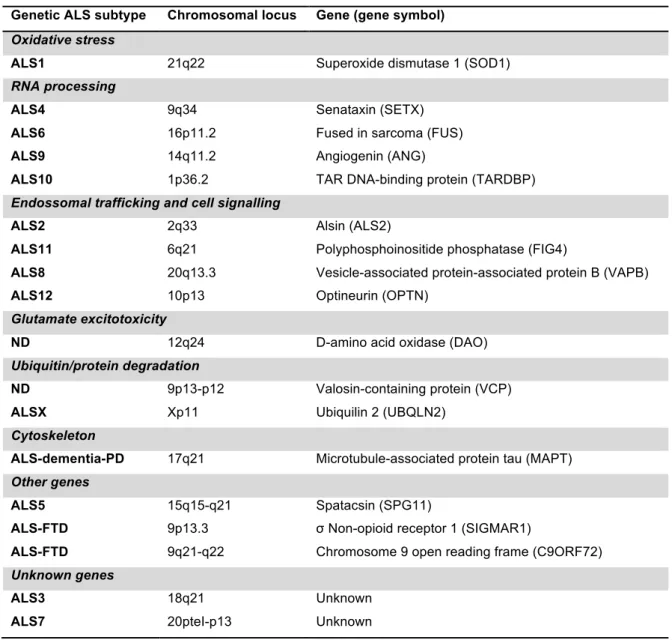
The identification of some of the genetic subtypes of ALS (table 1) has established key molecular and pathogenic mechanisms, which are applicable not only to the minority of cases that carry FALS mutations, but also to SALS more broadly. However, the discovery of Cu/Zn superoxide dismutase’s (SOD1) role in FALS (Rosen et al., 1993) offered the first insight to unravel ALS. The authors reported that mutations in this enzyme occur in an autosomal dominant manner in adult-onset ALS (ALS1) and account for 2-3% of ALS cases and about 15%-20% of instances of FALS.
Figure 2 | Clinical features of muscle wasting in patients with ALS. (A) Proximal and symmetrical
upper limb wasting results in an inability to lift arms against gravity. (B) Recessions above and below the scapular spine, indicating wasting of supraspinatus and infraspinatus muscles, as well as substantial loss of deltoid muscle. (C) Disproportionate wasting of the thenar muscles combined with the first dorsal interossei. (D) Substantial wasting of the tongue muscles. Note the absence of palatal elevation present on vocalisation. Difficulty in mouth opening and swallowing (extracted from Kiernan et al., 2011).
Table 1 | Reviewed genes associated with familial ALS. (adapted from Ferraiuolo et al., 2011).
SOD1 dismutates free oxygen radicals into O2 and hydrogen peroxide (H2O2). H2O2 is
then converted to H2O by either catalase or glutathione peroxidase (Nicholls & Ferguson,
2002). The SOD1 gene comprises five exons that encode 153 evolutionarily conserved amino acids which, together with a catalytic copper ion and a stabilizing zinc ion, form a subunit. Through covalent binding, pairs of these subunits form the SOD1 homodimers (Cleveland & Rothstein, 2001).
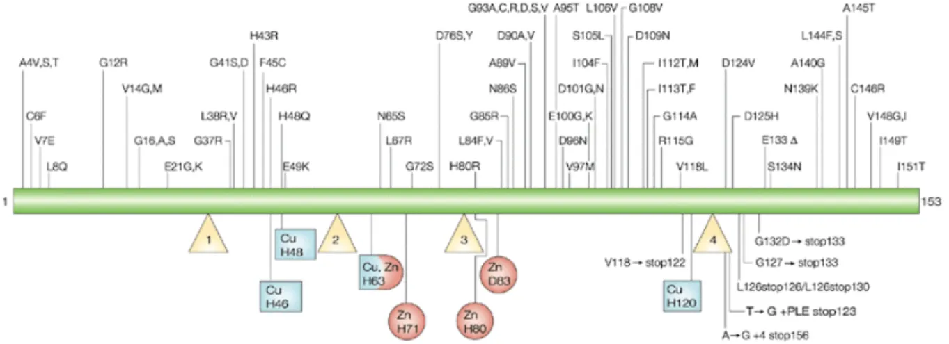
Following linkage analysis, in 1993, using modern genetic mapping methods and with DNAs from patients suffering from familial ALS, Rosen and colleagues (1993) identified 11 missense mutations in the SOD1 gene in 13 of 18 pedigrees with high-penetrance dominantly inherited FALS. Since then, 166 SOD1 mutations have been reported (figure 3) to be associated with ALS, plus 8 silent mutations and 9 intronic variants, presumed to be nonpathogenic. Of the 166 disease associated mutations, 147 are of the missense type. The remaining 19 mutations are nonsense and deletion mutations that result in a change in
Genetic ALS subtype Chromosomal locus Gene (gene symbol)
Oxidative stress
ALS1 21q22 Superoxide dismutase 1 (SOD1)
RNA processing
ALS4 9q34 Senataxin (SETX)
ALS6 16p11.2 Fused in sarcoma (FUS)
ALS9 14q11.2 Angiogenin (ANG)
ALS10 1p36.2 TAR DNA-binding protein (TARDBP)
Endossomal trafficking and cell signalling
ALS2 2q33 Alsin (ALS2)
ALS11 6q21 Polyphosphoinositide phosphatase (FIG4)
ALS8 20q13.3 Vesicle-associated protein-associated protein B (VAPB)
ALS12 10p13 Optineurin (OPTN)
Glutamate excitotoxicity
ND 12q24 D-amino acid oxidase (DAO)
Ubiquitin/protein degradation
ND 9p13-p12 Valosin-containing protein (VCP)
ALSX Xp11 Ubiquilin 2 (UBQLN2)
Cytoskeleton
ALS-dementia-PD 17q21 Microtubule-associated protein tau (MAPT)
Other genes
ALS5 15q15-q21 Spatacsin (SPG11)
ALS-FTD 9p13.3 σ Non-opioid receptor 1 (SIGMAR1)
ALS-FTD 9q21-q22 Chromosome 9 open reading frame (C9ORF72)
Unknown genes
ALS3 18q21 Unknown
length of the SOD1 polypeptide (Cleveland & Rothstein, 2001; Turner & Talbot, 2008). The pathological effects of SOD1 mutations are not thought to result from loss of dismutase activity but rather from gain-of-function effects through which the protein acquires one or more toxic properties. This theory is supported by several lines of evidence, including the absence of motor neuron degeneration in hSOD1-null mice and its occurrence in transgenic mice overexpressing mutant forms of SOD1, irrespective of residual dismutase activity (Gurney et al., 1994).
1.4. SOD1 mouse models
1.4.1. SOD1 overexpressing and knockout models
Mice deficient for SOD1 were generated by targeted gene deletion. Homozygote SOD1 knockout mice were viable and appeared to develop without any obvious motor abnormalities (Ho et al., 1998; Reaume et al., 1996). Hence, disruption of SOD1 alone appeared to be insufficient to cause spontaneous motor neuron degeneration in mice without injury or challenge. SOD1 knockouts are repeatedly reported to be normal or healthy which is interpreted as a major defeat for a loss-of-activity hypothesis for SOD1 mutations. However, SOD1 null mice develop chronic age-related peripheral axonopathy, denervation muscle atrophy and accelerated sarcopenia which confers significant locomotor deficits (Flood et al., 1999; Shefner et al., 1999; Muller et al., 2006). It seems that a loss-of-function cannot be completely excluded from a pathogenic mechanism of all SOD1 mutants.
Similarly with other diseases, increased dosage of SOD1 was also tested. Transgenic mice overexpressing human SOD1WT were generated (Epstein et al., 1987). This model was
characterized by hypotonia, hindlimb neuromuscular pathology (Avraham et al., 1988, Avraham et al., 1991; Rando et al., 1998), muscular dystrophy, vacuolar pathology, axonal
Figure 3 | Mutations causing ALS. So far, 166 mutations have been linked to ALS throughout the 153 SOD1
loss and motor neuron degeneration were described in spinal cords of aged animals (Dal Canto & Gurney, 1995; Jaarsma et al., 2000). No lines of transgenic human SOD1WT mice
have succumbed to ALS symptoms to date, although animals appear to undergo prolonged subclinical motor neuron degeneration.
1.4.2. SOD1 mutant transgenic model
In ALS research, the mainstay has been a mouse that bears the human gene for the known mutation of SOD1 associated with familial ALS. The mouse bearing this kind of mutated gene was the first laboratory model clearly linked to ALS based on a known cause of the disease. The interpretation of this particular model requires some consideration of wild-type SOD1 overexpressing and knockout mice, described above (Turner & Talbot, 2008). The discovery of SOD1 mutations in FALS was promptly followed by the generation of transgenic mice constitutively expressing mutant human SOD1 genes (Gurney et al., 1994). These transgenic constructs typically involve 12-15 kb human genomic fragments encoding SOD1 (harboring a 93 Gly – Ala substitution, from which SOD1G93A designation derives)
driven by the human endogenous promoter and regulatory sequences. Despite vast differences in transgene copy number, steady-state transcript and protein levels, dismutase activity and neuropathology, the mutations induce fatal symptoms strongly indicative of ALS with different disease latencies and progression rates. Crucially, the disease phenotype of transgenic mice expressing hSOD1 mutants on a background of endogenous enzyme argued for a dominant gain-of-function mechanism in toxicity (Gurney et al., 1994). Transgenic SOD1G93A mice are principally used in ALS research because of abundant
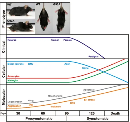
expression, stability and activity in the CNS. Mice develop hindlimb tremor and weakness at around 3 months detected by locomotor deficits progressing to hyper-reflexia, paralysis and premature death after 4 months (Gurney et al., 1994). Pathologically, neuromuscular junctions degenerate around 47 days which appears selective for fast-fatiguable axons (Pun
et al., 2006). Proximal axonal loss is prominent by 80 days coinciding with motor impairment
and a severe (50%) dropout of lower motor neurons, is evident at 100 days (Fisher et al., 2004). This retrograde sequence of neurodegeneration has led to an attractive proposal that ALS may be a distal axonopathy, described later.
At present, 12 different human SOD1 mutants have been expressed in mice. These include nine missense and three C-terminally truncated variants (Turner & Talbot, 2008).
1.5. Pathogenic mechanisms of ALS
Despite the fact that a number of genes have now been linked to ALS, the exact pathogenic mechanisms are still largely unclear. Our current understanding of the pathology of ALS is largely based on studies of ALS-associated gene mutations. Because the clinical and pathological profiles of sporadic and familial ALS are similar, it can be predicted that insights from studies of ALS-causing gene mutations apply to sporadic ALS. The mechanisms underlying neurodegeneration in ALS are multifactorial and operate through inter-related molecular and genetic pathways (figure 5). Specifically, neurodegeneration in ALS might result from a complex interaction of several factors including: cytoplasmic misfolded protein aggregates, glutamate excitotoxicity, mitochondrial dysfunction, oxidative stress, disruption of axonal transport process, endoplasmic reticulum stress and neuroinflammation. Other factors equally important, which are not going to be described in detail, are endossomal trafficking dysregulation, transcription and RNA processing
Figure 4 | Time course of clinical and neuropathological events in the high copy number transgenic SOD1G93A mice. Mice develop hindlimb tremor, weakness and locomotor deficits at about 3 months which is
preceded by distal synaptic and axonal degeneration. This progresses into fatal paralysis about 1 month later concomitant with spinal motor neuron loss and reactive gliosis. A sequence of mutant SOD1 aggregation into insoluble protein complexes (IPC), inclusion bodies modified by the ubiquitin-proteasome system (UPS) and subcellular degeneration in motor neurons may underlie the phenotype. (extracted from Turner & Talbot, 2008).
impairements and the role of non-neuronal cells (Boillée et al., 2006; Dion et al., 2009; Redler & Dokholyan, 2012).
1.5.1. Protein misfolding and aggregation
Protein misfolding and aggregation are prominent features of ALS. Aspects of toxicity can arise either through aberrant chemistry, mediated by the misfolded aggregated mutants, or through loss or sequestration of essential cellular components; for example, by saturating the protein-folding chaperones and/or the protein-degradation machinery. Consistent with the latter, the aggregates are intensely immunoreactive with antibodies to ubiquitin, a feature common not only to all instances of disease in mice, but also to many human examples (Clement et al., 2003; Henkel et al., 2006; Cassina et al., 2008). Partial inhibition of the proteasome is sufficient to provoke large aggregates in non-neuronal cells that express SOD1 mutants, leading to the proposal that proteasome activity could be limiting by combating such aggregates and moreover, that undue proteasomal attention to aberrantly folded forms of SOD1 could compromise the removal of even more important components (Guo et al., 2003).
Figure 5 | Schematic evolution of motor neuron degeneration during the course of SOD1 mutant ALS disease. Four stages are defined (normal, early phase, symptomatic, and end stage). Toxicity is
non-cell-autonomous, produced by a combination of damage incurred directly within motor neurons that is central to disease initiation and damage within non-neuronal neighbors, including astrocytes and microglia, whose actions amplify the initial damage and drive disease progression and spread. Selective vulnerability of motor neurons to ubiquitously expressed mutant SOD1 is determined by the unique functional properties of motor neurons (e.g., they are very large cells with large biosynthetic loads, high rates of firing, and respond to glutamate inputs) and damage to their supporting cells in the neighborhood. (extracted from Boillée et al., 2006).
1.5.2. Mitochondrial dysfunction and oxidative stress
Mitochondria, have developed defenses to detoxify superoxide (O2•-) generated by the
respiratory chain, a highly reactive molecule that contributes to oxidative stress and has been implicated in a number of diseases and aging (Turrens,1997; Barja, 1999). Multiple studies have shown that oxidative stress interacts with, and potentially exacerbates, other pathophysiological processes that contribute to motor neuron injury, including excitotoxicity (Rao & Weiss, 2004), mitochondrial impairment (Duffy et al., 2011), protein aggregation (Wood et al., 2003), endoplasmic reticulum stress (Kanekura et al., 2009), and alterations in signaling from astrocytes and microglia (Sargsyan et al., 2005; Blackburn et al., 2009). The most important are the Cu/Zn-superoxide dismutase (SOD1) and the manganese superoxide dismutase (Mn-SOD or SOD2). Age-related diseases, like neurodegenerative diseases, are associated with increased mitochondrial production of O2•- and H2O2. In ALS, there are
evidences that these two reactive oxygen species (ROS) can generate highly reactive radicals, like OH•, which will modify all kinds of macromolecules including lipids (Shibata et
al., 2001), proteins (Shaw et al., 1995), nuclear and mitochondrial DNA (Fitzmaurice et al.,
1996), and RNA species (Beal, 2005; Chang et al., 2008). Defective respiratory chain function associated with oxidative stress has also been found in tissue from patients with ALS at earlier stages of the disease. Dysfunction of components of the mitochondrial respiratory chain is also evident in the spinal cord of SOD1G93A transgenic mice at disease
end stage (Mattiazzi et al., 2002). Furthermore, mutant SOD1 insoluble aggregates could directly damage the mitochondrion through: swelling, with expansion and increased permeability of the outer membrane and intermembrane space, leading to release of cytochrome c and caspase activation; inhibition of the translocator outer membrane (TOM) complex, preventing mitochondrial protein import; and aberrant interactions with mitochondrial proteins such as the anti-apoptotic BCL2 (Pasinelli & Brown, 2006; Ferraiuolo
et al., 2011).
1.5.3. Excitotoxicity
Excitotoxicity, results from excessive influx of calcium cations through the over-stimulation of post-synaptic glutamate receptors which may be caused by increased synaptic levels of glutamate, or by increased sensitivity of the post-synaptic neuron energy homeostasis or glutamate receptor expression. This increase of calcium can activate enzymes such as phosphatases, proteases, lipases and endonucleases, causing protein and lipid alterations in cell membranes, generation of ROS, and mitochondrial damage and dysfunction (Corona et al., 2007). Decreased levels of the glutamate transporter EAAT2 (excitatory aminoacid transporter 2) are found in both human patients and in mutant SOD1
transgenic rodents (Rothstein et al., 1995). Thus, excitotoxicity may be involved in modulation of disease progression.
1.5.4. Impaired axonal transport
Motor neurons are highly polarized cells with long axons, and axonal transport is required for delivery of essential components, such as RNA, proteins and organelles, to the distal axonal compartment, which includes synaptic structures at the neuromuscular junction (NMJ). The main machinery for axonal transport uses microtubule-dependent kinesin and cytoplasmic dynein molecular motors, which mediate transport towards the NMJ (anterograde transport) and towards cell body (retrograde transport), respectively. Defects in either supply or clearance of material within an axon can lead to neuronal death. Axonal transport becomes impaired due to neurofilament disorganization via activation of protein kinases that phosphorylate neurofilament proteins (Pasinelli & Brown, 2006).
1.5.5. Endoplasmic reticulum stress
ER stress is an important pathway to cell death in ALS (Atkin et al., 2006; Atkin et al., 2008), and is triggered very early in SOD1G93A transgenic mice (Saxena et al., 2009). ER
stress is triggered when misfolded proteins accumulate within the ER lumen, inducing the unfolded protein response (UPR). Although the initial phases of the UPR aim to promote cell
survival, prolonged or severe ER stress triggers the apoptotic phase of the UPR. Up- -regulation of the three UPR sensor proteins, PERK, ATF6 and IRE1, have been observed
both at the symptom onset and at disease end stage of SOD1G93A transgenic mice, implying
the involvement of ER stress in disease mechanisms (Atkin et al., 2006; kikuchi et al., 2006). The ER chaperone, protein disulphide isomerase (PDI), was found to co-localize with mutant SOD1 inclusions in both cellular and animal models of ALS and overexpression of PDI decreased mutant SOD1 aggregation, ER stress, and apoptosis (Walker et al., 2010).
1.5.6. Neuroinflamation
Neuroinflammation is characterized in ALS by the appearance of reactive microglial and astroglial cells (Neusch et al., 2007; Van de Bosch et al., 2008), suggesting a non-cell autonomous process (Clement et al., 2003). In ALS, reactive astrocytes produce nitric oxide and peroxynitrite, and trigger mitochondrial damage and apoptosis in motor neurons (Cassina et al., 2008). Astrocytes may also contribute to damage motor neurons through excitotocicity. Furthermore, microglial cells are reported to be activated in the brain and spinal cord of patients with ALS, as well as mutant SOD1 transgenic mice. In fact activated
microglia were detected before motor neuron loss (Henkel et al., 2006). Damage within motor neurons is enhanced by injury from microglial cells via an inflammatory response that accelerates disease progression (Barbeito et al., 2004).
1.6. Where does ALS begin?
Despite Charcot’s initial observation of concomitant UMN and LMN pathological changes in ALS, the question of where ALS begins has not been established. Resolution of this question might enhance the understanding of the pathophysiology of ALS and has diagnostic and therapeutic importance (Meininger, 2011). Two theories have been proposed to explain which are the initial steps of ALS disease but more importantly where do they take place. The “dying-forward” hypothesis proposes that ALS is mainly a disorder of corticomotoneurons mediating anterograde degeneration of anterior horn cells. Support to this hypothesis includes:
- Transneuronal degeneration in ALS is an active excitotoxic process in which live but dysfunctional corticomotoneurons, originating in the primary motor cortex, drive the anterior horn cell into metabolic deficit. When this is marked, it will result in more rapid and widespread loss of lower motor neurons (reviewed here Eisen & Weber, 2001).
- Expression of SOD1G93A mutation induces energy dysfunction in discrete CNS motor
regions long before motor neuron degeneration occurs (Browne et al., 2006).
- Asymptomatic 60 day-old mice have lost approximately 9%, 12% and 14% of their corticospinal, bulbospinal and rubrospinal projections, respectively. 90 day-old mice that display the first clinical signs have lost approximately 30%, 33% and 33% of their corticospinal, bulbospinal and rubrospinal projections, respectively. Mice aged 110 days that have severe clinical signs, have lost approximately 53%, 41% and 43% of their corticospinal, bulbospinal and rubrospinal projections, respectively. (Zang & Cheema, 2002).
The prevailing and best documented proposal however, is the “dying-back” hypothesis in which motor neuron loss in ALS involves retrograde degeneration within the muscle cells or at the NMJ. Support for the dying-back hypothesis includes:
- A quantitative analysis demonstrating denervation at the NMJ by day 47, followed by severe loss of motor severe loss of motor axons (approximately 60%) from ventral roots between days 47 and 80, and loss of α-cell bodies from the lumbar spinal cord after day 80 (Fisher et al., 2004).
- Transgenic mice with skeletal muscle-restricted expression of hSOD1 gene develop neurologic and histopathologic phenotypes consistent with ALS. Muscle restricted expression is sufficient to cause dismantlement of NMJ and distal axonopathy (Dobrowolny et al., 2008, Wong & Martin, 2010).
- Magnetic Resonance Imaging (MRI) studies showed a significant reduction in muscle mass that parallels reduction in fiber diameter and muscle atrophy from week 8. Evidences of neurodegeneration in the brainstem detected only from week 10 (Marcuzzo et al., 2011).
2. The motor nervous system
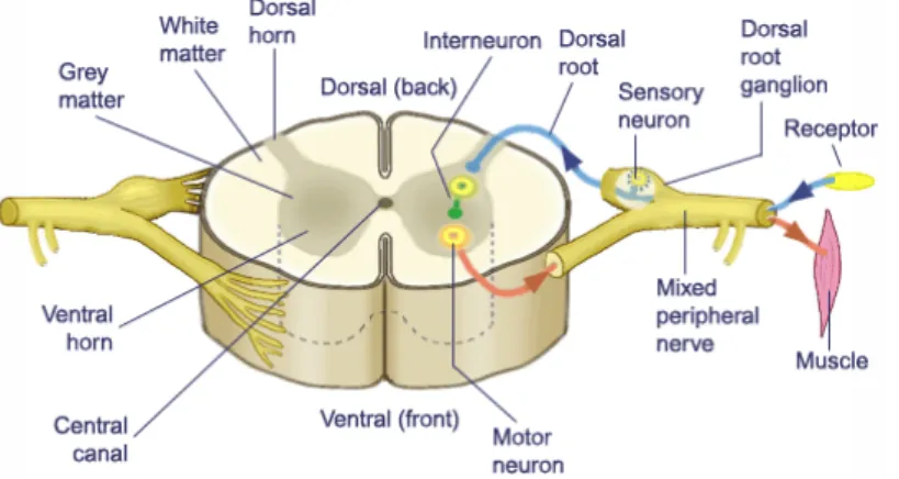
The somatic portion of the nervous system is composed of two major types of nerve cells which connect the spinal cord to the periphery (figure 6). These are primary sensory neurons (or afferent neurons), which relay input from the periphery to the spinal cord, and spinal cord motor neurons (or efferent neurons) which convey motor outflow from the spinal cord to the periphery. Motor neurons can be divided in two groups: UMN, which originate in the motor region of the cerebral cortex or the brain stem and carry motor information down to the final common pathway, and LMN, connecting the brainstem and spinal cord to muscle fibers. Axons of spinal cord motor neurons pass to the periphery to innervate striated muscle. Even for most other reflexes, such as the withdrawal reflex, there is at least one other neuron (an interneuron - Renshaw cells) interposed in the circuit (Kandel et al., 2000; Marieb & Hoehn, 2007).
Skeletal muscle is a form of striated muscle tissue existing under the control of the somatic nervous system. Some examples of this type of muscle are respiratory muscles and the diaphragm, severely targeted in patients with ALS disease (Marieb & Hoehn, 2007).
2.1. Motor neurons and neuromuscular synaptic transmission
The phenomenon of neuronal cross-talk is often termed neurotransmission and it is mediated by neurotransmitters. Neurotransmitters are endogenous chemicals that transmit signals from a neuron to a target cell across a synapse allowing the brain to communicate with the rest of the body (Marieb & Hoehn, 2007).
(http://davidsciencestuff.tripod.com/id1.html)
Figure 6 | Somatic Component of the Peripheral Nervous System. The
peripheral nervous system can be subdivided into somatic and autonomic components. The somatic nervous system contains motor nerves and sensory nerves innervating skin and muscle. The soma (cell bodies) of motor nerves and sensory nerves are located in the gray matter of the anterior horn of the spinal cord and in the dorsal root ganglia, respectively.
The nerve terminal is responsible for neurotransmitter release and stores it in small, uniformly sized vesicles. Synapses between motor neurons typically use glutamate or GABA as their neurotransmitters, while the NMJ uses acetylcholine exclusively. Glycine is also present in the interneurons of the spinal cord. Arrival of an action potential at the motor neuron ending leads to an instant opening of voltage-gated Ca2+ channels with a subsequent
abrupt increase in intracellular calcium concentration (Fagerlund & Eriksson, 2009; Martyn et
al., 2009). This increased calcium concentration triggers a cascade of intracellular signaling
events leading neurotransmitter-containing vesicles to migrate, dock, fuse to the surface of the nerve, rupture and discharge the specific neurotransmitter through the synaptic cleft to the receptive post-synaptic component, either a neuron or the NMJ. The energy required for these processes is generated by a large population of mitochondria present in the cytoplasm. At the NMJ (figure 7) the nicotinic acetylcholine receptors (nAChRs) in the sarcolemma, activated by the released acetylcholine, respond by opening their channels for influx of sodium ions into the muscle to depolarize it (Hughes et al., 2006).
3. Adenosine
Purinergic research has been demonstrated to be potentially fruitful on neurodegenerative disorders such as ischemia, neuropatic pain, multiple sclerosis, Parkinson’s, Alzheimer’s and Huntington’s diseases (Burnstock, 2008a,b) and might accordingly provide novel clues also for ALS. The validation that purinergic research could indeed meet ALS comes from the general notions that 1) microglia, astrocytes and degenerating neurons, commonly release and promptly respond to both ATP and adenosine; 2) extracellular ATP secreted at high concentrations is toxic to neurons and activates microglia and astrocytes, being thereafter degraded to adenosine creating neuroinflammation; 3) neuron toxicity, microglia and astrocyte activation are all common
(adapted from http://faculty.pasadena.edu)
Figure 7 | Structure of the Neuromuscular Junction. Schematic representation of the adult NMJ
with the three main components: pre-synaptic nerve terminal, synaptic cleft and post-synaptic membrane. Stimulation of a motor nerve results in the release of acetylcholine from vesicles at the pre-synaptic membrane; acetylcholine diffuses and binds to post-synaptic receptors, producing depolarization of the sarcolemma and leading to an action potential.
features to ALS (Volonté et al., 2011). Adenosine is a ubiquitous nucleoside, present and being released from apparently all cells, including neurons and glia. It comprises a molecule of adenine attached by a glycosidic bound to a ribose sugar molecule. Perhaps as a result of their ubiquitous nature, purines have also evolved as important molecules for both intracellular and extracellular signaling, roles that are distinct from their activity related to energetic metabolism, as adenosine diphosphate (ADP) and adenosine triphosphate (ATP), and synthesis of nucleic acids (Khakh and Burnstock, 2009).
Unlike ATP, which may function as a neurotransmitter in some brain areas, adenosine is neither stored nor released as a classical neurotransmitter. It does not accumulate in synaptic vesicles, being released from the cytoplasm into the extracellular space in a calcium independent process through a nucleoside transporter. The adenosine transporters also mediate adenosine reuptake, being the direction of the transport dependent on the concentration gradient at both sides of the membrane. As it is not exocytotically released, adenosine behaves as an extracellular signalling molecule that modulates synaptic transmission. Using G-protein coupled mechanisms, that not only lead to changes in second messenger levels but also to regulation of ion channels, such as calcium and potassium channels, adenosine modulates neuronal activity, pre-synaptically by inhibiting or facilitating transmitter release, post-synaptically by affecting the action of other neurotransmitters and non-synaptically by hyperpolarizing or depolarizing neurons and/or exerting non-synaptic effects (e.g. on glial cells). Adenosine, therefore, belongs to the group of neuromodulators, endogenous substances released at the synaptic cleft that influence the release (pre-synaptic modulation) or the action (post-(pre-synaptic modulation) of the neurotransmitters (Sebastião & Ribeiro, 2000; Ribeiro & Sebastião, 2010).
3.1. Adenosine receptors
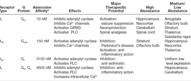
Adenosine receptors are a class of specific purinergic receptors with adenosine as the endogenous ligand. There are four adenosine receptor subtypes among vertebrates, which have been cloned and characterized to date (table 2): adenosine A1, A2A, A2B and A3
receptors that belong to the G-protein coupled receptors (GPCRs) family. (Fredholm et al., 1994, Fredholm et al., 2001). These receptors are also known as P1 receptors, from the P1 (adenosine selective)/P2 (ATP selective) nomenclature (Burnstock, 1978). A1R and A3R are
coupled to Gi/o inhibitory proteins while A2ARand A2BR are coupled to Gs excitatory proteins
(Linden, 2001; Ribeiro et al., 2002). Neuromodulation by adenosine is exerted through activation of high-affinity adenosine receptors (A1 and A2A) which are probably of
physiological importance, and of low-affinity adenosine receptors (A2B), which might be
low density in most tissues (Ribeiro & Sebastião, 2010). However, much of the data on coupling to other G-proteins are from transfection experiments and it is not known if such coupling is physiologically important. There are evidences that A2AR may be coupled to
different G-proteins in different areas (Kull et al., 2000). Other authors (Auchampach et al., 1997) found that one adenosine receptor may also be coupled with more than one G-protein.
3.2. Adenosine receptors distribution and interactions
Within the CNS, the A1R has the highest expression in the brain cortex, cerebellum,
hippocampus and dorsal horn of spinal cord, coupled to activation of K+ channels (Trussell &
Jackson, 1985) and inhibition of Ca2+ channels (Macdonald et. al., 1986), both of which would inhibit neuronal activity. The A2AR is expressed at high levels in only a few regions of
the brain (striatum has the higher expression) and is primarily linked to the activation of adenylate cyclase (Sebastião & Ribeiro, 1996). The A2BR, which also activates adenylate
cyclase, is thought to be fairly ubiquitous in the brain (Dixon et al., 1996), but it has been difficult to link this receptor to specific physiological or behavioral responses (Feoktistov & Biaggioni, 1997). The A3R is also somewhat poorly characterized, but apparently has
intermediate levels of expression in the human cerebellum and hippocampus and low levels in most of the brain (Fredholm et al., 2001). It has been reported to uncouple A1 and
metabotropic glutamate receptors via a protein kinase C–dependent mechanism (Dunwiddie
et al., 1997; Macek et al., 1998), and thus, one of its functions may be to modulate the
activity of other receptors. Both A1 and A2A receptors are predominantly, but not exclusively,
Table 2 | Adenosine receptors in the central nervous system and their properties (Dunwiddie & Masino,
located pre-synaptically (Rebola et al., 2003; Baxter et al., 2005; Rebola et al., 2005a; Rebola et al., 2005b). Previous evidence indicates that A1R and A2AR may be co-localized in
the same nerve terminals (Correia-de-Sá et al., 1991; Lopes et al., 1999a; Rebola et al., 2005b; Pousinha et al., 2010). Moreover, both receptors were shown to form a heteromeric complex in co-transfected cultured cells (Ciruela et al., 2006). It is believed that A1R or A2AR
are preferentially activated as a function of the source and amount of adenosine (Sebastião & Ribeiro, 2009). A1R are preferentially activated by adenosine generated intracellularly,
released through adenosine transporters, and A2AR are activated preferentially by adenosine
generated extracellularly (Cunha et al., 1996), due to the action of ectonucleotidases upon ATP release. The relative density of A1R or A2AR adenosine receptors in sub-regions of the
same brain area may differ. Whenever the two receptors co-exist, we can ask about their relative importance, i. e. the hierarchy of one receptor with respect to the other. This may change with neuronal activity, the age and even on other molecules that are in the vicinity of the site of action and that may be relevant for the production or inactivation of the ligand (Sebastião & Ribeiro, 2009). High frequency of neuronal firing favours ATP release (Cunha
et al., 1996) and adenosine formed from released adenine nucleosides seems to prefer A2AR
activation (Cunha et al., 1996) which may be due to the geographical distribution of ecto-5-nucleotidases and A2AR. A2AR activate adenosine transport, which in the case of high
neuronal activity and ATP release is in the inward direction. This induces a decrease in extracellular adenosine levels and a reduced ability of A1R to be activated by endogenous
extracellular adenosine. By themselves, A2AR are able to attenuate A1AR activation (Cunha
et al., 1994) which may further contribute to a decreased activity of A1R receptors under high
frequency neuronal firing. The ability of adenosine receptors to inhibit synaptic transmission is attenuated by the protein kinase C (PKC) activation (Sebastião & Ribeiro, 1990) and a similar mechanism appears to be involved in the A2AR-mediated attenuation of A1R
responses (Lopes et al., 1999). Adenosine receptors are also present in the peripheral nervous system, either autonomic or somatic, especially at the motor nerve endings. There are reports that human and rat skeletal muscle express both mRNA and protein of adenosine receptors (Dixon et al., 1996; Lynge & Hellsten, 2000).
OBJECTIVES
The present work was designed to determine the effect of the SOD1G93A mutation on
the expression of A1 and A2A adenosine receptors of the ALS transgenic mouse model for the
human gene SOD1, through disease progression (pre-symptomatic and symptomatic phases). As described above ALS’s primary hallmark is the degeneration of the upper motor neurons of the motor cortex and of the lower motor neurons, which extend through the brainstem and spinal cord to innervate skeletal muscles at the neuromuscular junction level (Boilléee et al., 2006). Therefore, three different tissues were analyzed, i.e. the motor cortex, spinal cord and phrenic nerve-diaphragm (herein referred as diaphragm). This approach covered both CNS and PNS adenosine receptor expression. Adenosine is known to modulate various physiological functions of most tissues, including skeletal muscles (Hespel & Richter, 1998). This is the main reason why adenosine is the target of this work.
Therefore, to achieve this main goal I formulated three specific questions:
Aim 1: Are the protein levels of A1 and A2A receptor changed in the motor cortex, spinal
cord and the neuromuscular junction throughout disease progression? To answer this question the immunoblotting technique was used.
Aim 2: Are the mRNA levels of A1 and A2A receptor altered in the neuromuscular
junction throughout disease progression? To answer this question quantitative Real-Time PCR technique was used.
Aim 3: Is the primary pathological feature, regarding the expression of adenosine receptors, observed in the central or in the peripheral nervous system? Specifically, I tried to establish throughout disease progression, if the first event related to adenosine receptor alterations occurs in the motor cortex, spinal cord or at the neuromuscular junction level.
This work is part of a project (PTDC/SAU-NEU/101752/2008) funded by Fundação para a Ciência e a Tecnologia (FCT).
MATERIALS & METHODS
1. Breedings and housbandry
Transgenic B6SJL-Tg(SOD1-G93A)1Gur/J males and wild-type B6SJLF1/J females were purchased from Jackson Laboratory (USA; Stock No. 002726 and 100012, respectively) and a colony was established at the rodent facility (Instituto Medicina Molecular). Since transgenic female line has a very high incidence of non-productive matings (Leitner et al., 2009), mice were maintained on a background B6SJL by breeding transgenic males with non-transgenic females in a rotational scheme. Animals were handled according to European Community Guidelines and Portuguese Law on Animal Care (2010/63/UE). At time of weaning, littermates were identified through ear punching and separated in different cages according to their gender. This system is a permanent procedure that attributes to each hole a number and allows individual identification of mice. Moreover, this method does not require anesthesia, guarantee animal welfare, and the tissue removed by the ear punch can be used for DNA analysis, phasing out the requirement of an additional procedure for genotyping (Costa & Antunes, 2010). All animals were housed 4-5 mice per cage, under a 12h light/12h dark cycle, and received food and water ad libitum.
2. Mice genotyping
Using the tissue removed by ear punching, as described above, the mice DNA was isolated by adding TDB (50 mM KCl, 10 mM Tris-HCl pH=9.0, 0.1% Triton X-100, 0.15 mg/mL proteinase K) followed by an overnight incubation at 56⁰C. Additionally a heat
proteinase K inactivation was performed during 15 min at 95⁰C. After a 2 min centrifugation to
remove debris a PCR reaction was prepared. Primers against interleukin-2 precursor (internal positive control) and human SOD1 transgene were raised (see annexe II, table 7). For a total of 25 µL the following components were added per sample: 0,2 mM dNTP mix, 10X DreamTaq Buffer containing MgCl2, 1.5 U DreamTaq DNA polymerase (Fermentas®),
1.33 µM of the transgene SOD1 primer, 0.75 µM of the control primer, water and 200-500 ng DNA template (BioRad® C1000 Thermal Cycler, see annexe I, table 4 for PCR conditions).
Both PCR products and DNA ladder (1 kb gene ruler, Fermentas®) were loaded in a 2%
agarose gel and an electrophoretic migration took place. With a Red Safe dyed gel it was possible to inspect bands in a transiluminator (Molecular Imager® Gel Doc™ XR System) and
3. Tissue extraction and dissection
Male or female, wild-type and transgenic, F2 mice (4-6 and 13-14 weeks old) were anesthetized under isoflurane atmosphere before being decapitated. The brain was rapidly removed from the brain cavity and dissected free in ice-cold PBS 1X (137 mM NaCl, 2.7 mM KCl, 4.3 mM Na2HPO4, 1.47 mM KH2PO4, pH 7.4) in order to separate the motor cortex (rich
in A1R) from the striatum (rich in A2AR) to avoid contamination. Additionally, the spinal cord
and the diaphragm were extracted. The different tissues were either homogenized or separately frozen at -80⁰C until further use, as described below.
4. Protein quantification
4.1. Total protein homogenates
Frozen tissue (cortex, striatum, spinal cord and diaphragm) was placed in 800 µL of RIPA buffer (1 M Tris pH 8.0, 0.5 M EDTA pH 8.0, 5 M NaCl, 0.1% SDS, 10% NP-40, 50% Glycerol), supplemented with protease inhibitors (RocheTM tablet and PMSF were added just prior to use), and homogenized in a Potter-Elvehjem homogenizer with a Teflon piston. The samples were then centrifuged at 3.5 rpm (1000 x g) during 10 minutes at 4⁰C, the
supernatant was collected and corresponds to the whole tissue lysate. Protein was quantified using the BioRadTM Dc Protein assay Kit based on Lowry (1951), due to the high levels of
detergents in the lysis buffer, by measuring the absorbance at 750 nm.
4.2. Gel electrophoresis and immunoblotting
In order to assess the protein levels of A1 and A2A adenosine receptors in the motor
cortex, spinal cord and in the diaphragm throughout disease progression the immunoblotting technique was used. After homogenate preparation and protein quantification, the appropriate volume of each sample (corresponding to 30 µg and 50 µg of protein to A1R and
A2AR quantification, respectively) was diluted in water and loading buffer 5X (350 mM Tris
HCl, 30% Glycerol, 10% SDS, 600 mM DTT and 0.012% Bromophenol blue, pH 6.8) performing a total volume of 25 µL to apply per lane. The striatum (5 µg) was used as positive internal control for A2AR due to its high content in this adenosine receptor. The cortex
was used as positive control for A1R since it has high levels of this protein. As endogenous
control, α-tubulin was used because it proved to be a reliable reference control. Prior to loading, the samples were boiled at 95⁰C for 5 minutes. Under reducing and denaturing
samples, in a 10% resolving (and 5% stacking) gel concentration. A standard running buffer 1X (25 mM Tris Base, 190 mM Glycine and 0.1% SDS) was used. Submerged in transfer buffer 1X (25 mM Tris Base, 190 mM Glycine and 20% Methanol), the separated gel proteins were then transferred to PVDF membranes, through a 250 mA electrical current for 90 minutes. A Ponceau Red staining followed in order to check for success of transfer confirming that no air bubbles have formed between the gel and membrane. At this point membranes were cut into pieces separating above α-tubulin proteins from the receptor ones. Membranes were blocked with 5% non-fat dry milk for 1 hour, washed with TBS-T (200 nM Tris Base, 1.5 M NaCl and 0.1% Tween-20, pH 7.6) and incubated with primary antibodies overnight at 4⁰C. Primary antibodies were diluted in 3% BSA, 0.02% NaN3 and TBS-T. After
washing again for 30 minutes, the membranes were incubated with secondary antibody, for 1 hour at room temperature, and washed again (see table 1 for antibodies information). Finally, a chemoluminescent detection method was performed with ECL western blot detection reagent (GE HealthcareTM) using X-Ray films (FujifilmTM). Western blots densitometry was
determined with Image J software and normalized to the respective α-tubulin band density. ImageJ Gel analyzer options: Uncalibrated OD; Inverted Peaks; Plot lanes (ImageJ v.1.47c, Wayne Rasband, National Institutes of Health, USA) (according to Miller, 2010; McLean, 2011).
Table 3 | Antibodies used in this study. Primary and secondary antibodies and related conditions used in the
immunoblotting experiments for individual proteins. All primary antibodies were diluted in 3% BSA with 0.02% NaN3 and secondary antibodies in 5% non-fat dry milk.
5. mRNA quantification
5.1. Total RNA homogenates
Frozen tissue (diaphragm) was placed in 1000 µL QIAzol Lysis Reagent (according to QuiagenTM RNeasy Lipid Tissue Mini Kit protocol) and homogenized in a Potter-Elvehjem
homogenizer with a Teflon piston. The subsequent steps were followed from the same protocol and total RNA was eluted in 40 µL RNase-free water supplied. The concentration of total RNA was determined by measuring the absorbance at 260 nm in NanoDrop ND-1000 (Thermo ScientificTM).
Protein Predicted
protein size Primary antibody Animal Dilution
Secondary
antibody Dilution
A1R 37 kDa Thermo Scientific TM (PA1-041A) Rabbit 1:1000 Sta. Cruz BiotechnologyTM: goat anti-mouse (SC-2005); goat anti-rabbit (SC-2004) 1:10000 A2AR 42 kDa Upstate TM (05-717) Mouse 1:1000 1:10000
5.2. cDNA synthesis and qRT-PCR
Quantitative Real-Time PCR technique was used to evaluate the mRNA expression levels of A1 and A2A adenosine receptors in the diaphragm throughout disease progression.
cDNA was obtained using the SuperScriptTM First-Strand Synthesis System for RT-PCR (InvitrogenTM) according to manufacturer’s protocol. For a total volume of 20 µL in each
reaction tube, the following components were mixed: 0.5 mM dNTP, Random hexamears (50 ng), DEPC-treated water, 1X RT buffer, 5 mM MgCl2, 10 mM DTT, 25 U of SuperScriptTM II
reverse transcriptase and 2-5 µg RNA template. Minus RT controls were performed for each sample, in which DEPC-treated water was added instead of SuperScriptTM II reverse
transcriptase. The reverse transcription of all samples took place in Bio Rad C1000 Thermal Cycler (see annexe I, table 5). These PCR products were quantified through qRT-PCR in Rotorgene 6000 (QuiagenTM) using SYBR® Green master mix method (Applied
BiosystemsTM). Specific primers against A1R, A2AR and β-actin DNA sequence (see annexe I,
table 6) were used in this reaction. β-actin was used as a reference gene to normalize target gene results. For a total volume of 25 µL in each reaction tube, these components were added: 2x SYBR® Green master mix, 5 µM primer solution, water and cDNA template. Non
Template Controls (NTC) were performed for each primer, in which DEPC-treated water was added instead of cDNA templates.
In order to make valid comparisons between different samples it is important to determine the primer amplification efficiency. Ideally, amplification efficiencies for control and target primer should be roughly equal. However, the amplification efficiency for a specific pair of primers is affected by differences in primer binding sites, the sequence of the amplification product, and PCR product sizes, and thus should be determined experimentally. The efficiency equation is:
E = 10(!!/!)− 1
where E is the efficiency of the reaction and M refers to the slope of the plot of Ct value
versus the log of the input template amount. A slope between −3.6 and −3.1 corresponds to an efficiency between 90% to 110% (which corresponds to a value of E between 0.9 and 1.1) (Fraga et al., 2008). The Ct (cycle threshold) is defined as the number of cycles required for
the fluorescent signal to cross the threshold (i.e. exceeds background level). Ct levels are
inversely proportional to the amount of target nucleic acid in the sample (i.e. the lower the Ct
level the greater the amount of target nucleic acid in the sample). The results for β-actin and A2AR were calculated based in the efficiency obtained from calibration curve analysis (see
annexe III) and determined for each target gene using serial dilutions (1:5) of cDNA of pre-symptomatic diaphragm homogenate. The presented results are fold change values calculated with the Pfaffl equation (Pfaffl, 2001). Therefore, they reflect the difference in adenosine receptors mRNA expression between CTRL and SOD1 animals. Values equal to
1 (=1) are indicative of same CTRL and SOD1 expression; fold change above the unit (>1) mean that SOD1 has a higher expression in relation to the CTRL; and values below 1 (<1) mean that SOD1 has a lower expression when compared to CTRL.
The Pfaffl equation to calculate fold change values (Pfaffl, 2001) is:
𝒓𝐚𝐭𝐢𝐨 (𝐟𝐨𝐥𝐝 𝐜𝐡𝐚𝐧𝐠𝐞) = 𝐄𝐭𝐚𝐫𝐠𝐞𝐭
𝚫𝐂𝐭𝐭𝐚𝐫𝐠𝐞𝐭(𝐜𝐨𝐧𝐭𝐫𝐨𝐥!𝐬𝐚𝐦𝐩𝐥𝐞)
𝐄𝐫𝐞𝐟𝐞𝐫𝐞𝐧𝐜𝐞𝚫𝐂𝐭𝐫𝐞𝐟𝐞𝐫𝐞𝐧𝐜𝐞(𝐜𝐨𝐧𝐭𝐫𝐨𝐥!𝐬𝐚𝐦𝐩𝐥𝐞)
in which Ct reference corresponds to the number of amplification cycles obtained in the
qRT-PCR for β-actin triplicates, and Ct target for target gene triplicates, both belonging to the
same animal sample. β-actin fold change results were calculated considering the efficiency (E=1,01), slope (M=-3,288) and the correlation coefficient (R2=0,99) of the calibration curve. A2AR efficiency is 1,11, slope is -3,089 and correlation coefficient 0,99. For A1R, due to
insufficient levels of mRNA expression in the diaphragm required to obtain a calibration curve, data from spinal cord experiments (laboratory results, unpublished data) were used. Efficiency for A1R calibration curve is 1,0, slope is -3,351 and correlation coefficient 0,99.
Melting point analysis was always performed after each qRT-PCR run as a quality control step (Fraga et al., 2008). Melting point analysis is used to distinguish target amplicons from PCR artifacts such as primer-dimer or mis-primed products. Specificity is confirmed by the presence of a unique peak in the melting curve (see annexe III).
6. Statistics
For statistical evaluation of the data, Graphpad PRISM® 5 (San Diego, California, USA) software was used. To assess the significance for protein quantification between CTRL and SOD1 mice for each tissue, the Student’s t-test was used (with the Welch’s correction). The significance of diaphragm mRNA levels between the pre-symptomatic and suymptomatic phases were assessed with a Student’s t-test (with the Welch’s correction). The values presented, for protein and mRNA quantification, are mean ±SEM of n=5 experiments. Values of p<0.05 were considered to be statistically significant.

