UNIVERSIDADE DO ALGARVE
Nutritional modulation of innate immune parameters in
the epidermal mucus of Senegalese sole
(Solea senegalensis)
Influência da dieta nos parâmetros da resposta imunitária
inata do muco de linguado (Solea senegalensis)
Vera Lúcia Fernandes Rodrigues
Sob a supervisão de: Prof. Maria Teresa Dinis, Professor titular em Aquacultura da Universidade do Algarve Dr. Jorge Dias, Assistente de Investigação no Centro de Ciências Marinhas do Algarve Dr. Laura Ribeiro Investigador Pos‐Doc do Centro de Ciências Marinhas do Algarve Dissertação de mestrado em Biologia marinha: especialização em Aquacultura e pescas
Faro 2007/2008
This thesis is dedicated to my
parents without whom none
of this would have been
possible.
Vera Rodrigues
Fish are in constant interaction with their habitat and potential pathogens. Fish epidermis acts as the first line of defense, since mucus secreted by mucous cells contains innate immune parameters, such as enzymes and antimicrobial proteins. Knowledge about the mechanisms of immune defense at the skin and mucus level in fish is still scarce.
This study was undertaken to evaluate the role of nutritional factors as tools to enhance the immune parameters in the mucus of Senegalese sole (Solea senegalensis). Homogenous groups of Senegalese sole (initial body weight: 92.8 ± 1.52g) were fed: a control diet (CTRL); the CTRL diet supplemented with vitamin C (1000 mg/kg) (VIT C); and a diet identical to CTRL but in which 70% of the dietary fat was originated from coconut oil (COC), identified as a potential immunostimulant. After a period of 4 and 6 weeks of experimental feeding, fish mucus was sampled for quantification of lysozyme, alkaline phosphatase, proteases, lipid antioxidation (TBARS) and antimicrobial activity. Growth performance and lysozyme activity was not affected by dietary treatments, while the proteolytic activity and lipid oxidation was affected positively by both dietary treatments. Alkaline phosphatase activity was only affected by diets rich in coconut oil. Epidermal mucus of sole showed antibacterial activity against a series of marine pathogen bacteria. Highest inhibitory action was associated to mucus extracts derived from coconut oil fed fish.
The modulation of selected mucus immune parameters through dietary factors is possible in Senegalese sole. A four weeks period of feeding seems enough to induce such changes. Further research is needed to determine the processes associated to such modulation and evaluate to what extent these beneficial changes contribute to an enhanced immune response of sole.
Os peixes estão em constante interacção com o seu habitat, e potenciais agentes patogénicos. A epiderme dos peixes actua como primeira linha de defesa, uma vez que o muco secretado pelas células secretoras de muco contém factores da resposta imunitária inata, tais como enzimas e proteínas antimicrobianas. O conhecimento sobre os mecanismos da defesa imunológica ao nível da pele e do muco dos peixes é reduzido.
Este estudo foi realizado com o objectivo de avaliar o papel dos factores nutricionais como ferramentas para estimular os parâmetros imunológicos em muco de linguado (Solea
senegalensis). Grupos homogéneos de linguado (peso inicial: 92.8 ± 1.52g) foram alimentados:
com uma dieta controlo (CTRL); a dieta CTRL suplementada com vitamina C (1000 mg/kg) (VIT C); e uma dieta idêntica ao CTRL mas na qual 70% da gordura provinha de óleo de coco (COC), identificada como tendo um efeito imunoestimulante. Após um período de 4 e 6 semanas de alimentação experimental, foram recolhidas amostras de muco epidérmico e quantificadas a actividade da lisozima, fosfatase alcalina, proteases, oxidação lipídica (TBARS) e a actividade antibacteriana. O crescimento e a actividade da lisozima não foram afectados pelos tratamentos alimentares, enquanto que as proteases e a oxidação lipídica foram afectadas positivamente pelos dois tratamentos. No caso da actividade da fosfatase alcalina, verifica‐se apenas um aumento da sua actividade com a dieta suplementada em óleo de coco. O muco epidermal do linguado mostra actividade antibacteriana contra uma série de bactérias marinhas patogénicas, e o maior grau de inibição está associada ao extrato de muco derivado de peixes alimentados com a dieta com óleo de coco.
A modulação por via nutricional de alguns parâmetros imunitários no mucus do linguado é possível. Um período de quatro semanas parece ser suficiente para induzir tais mudanças. São necessários estudos complementares para determinar os processos associados a esta modulação e avaliar em que medida estas mudanças serão benéficas como contributos para o reforço da resistência do linguado a patologias.
Although the thesis has in its cover only the author’s name, none of this work would have been even possible to accomplish without the hefty support of some people, that I fear I may unintentionally forget here. If that is the case, please accept my sincere apologies. I sincerely hope that anyone who in anyway contributed and helped me get this far will share with me this particular moment of happiness and satisfaction which I am now enjoying.
First of all, I think of Prof. Dr. Mª Teresa Dinis, and wish to say how grateful I am. For the extraordinary example that she is to me. For showing me the fascinating world of aquaculture, and giving me the inspiration to work in it. Is an honor and a privilege to have her as my supervisor. I would like to express my deep appreciation to Dr. Jorge Dias, for supporting and offering me such an opportunity to work in this Masters, but also for all his patient and tireless guidance during the whole process, and for his incentive and support. Although I feel that I will never be able to express my full appreciation for what he has done for me, I wish to convey to him my eternal gratitude. Special thanks are also for Drª Laura Ribeiro, for being my tutor, for giving my first opportunity in the world of aquaculture, for bringing me to Aquagroup, for the rich conversations about life and work, and for her good advice. For all this and for always being present as a boss but more importantly as a friend.
I would also like to thank Drª Sónia Pedro, INRB‐IPIMAR, for the invaluable contribute in the antibacterial essays, without whom this work would have been poorer.
I’m also grateful to my dear friends in the lab. Wilson, Patricia, Elisabete, Andre, Mónica, Helena, my friends, colleagues, comrades. By their friendship, patience, laughs, help, for being always there… and because they are… beautiful. Dorinda, Violeta, Rita and François far but always present.
I also owe a special Word of gratitude to Carina and Débora, for always “being there”, offering unwavering support. Since the very beginning they were never too busy to provide a warmth encouragement to carry on. I spent many formative years at Algarve’s University, and gained so much from so many. I would like to take this opportunity and thank all academic Professors and BMP colleague friends. Finally and foremost, I am forever indebted to my family for the unconditional support they have provided throughout my entire life.
Mommy and Daddy I love you, for your encouragement, guidance, love, patience, confidence, caring, word of advice and continuous support, you were critically important for my success. For my dear grandmother, my second mother, and above all a great inspiration in life and a pillar in my family. My “godmother” Arminda and my “cousins” Lino e Elisabete, who most of all are my brothers of heart.
For my dog Golias goes a canine thanks. For being able to make me smile even in the worst moments.
Sofia, Davidinho, Cristiano are names that were always present during this master. Guardian angels.
Table of contents
Abstract I Acknowledgements Iv Table of Contents Vi List of figures Vii List of tables Viii 1. Introduction 1 1.1. The farming of Senegalese sole (Solea senegalensis) 2 1.2. The immune response in fish 3 1.3. Mucus as a natural immune barrier in fish 4 1.4. The potential of immunostimulants in fish 8 1.5. Objectives of the study 11 2. Materials and methods 13 2.1. Experimental diets 13 2.2. Experimental animals and rearing conditions 14 2.3. Mucus collection 14 2.4. Analytical methods 16 2.4.1. Protein 16 2.4.2. Lysozyme 17 2.4.3. Alkaline phosphatase 17 2.4.4. Proteases 18 2.4.5. Lipid oxidation (TBARS) 18 2.4.6. Antibacterial assay 19 2.4.7. Statistical analysis 20 3. Results 21 3.1. Growth performance 21 3.2. Immune parameters 22 3.3. Antibacterial activity 26 4. Discussion 27 5. Conclusion 38 6. References 39
List of figures
Fig. nr. Title Page nr.
1 The Senegalese sole (Solea senegalensis) 2
2 Non‐specific and specific immune responses and protection of fish against infection. Source: DSM Nutritional Products brochure “The effect of vitamin C on fish health”.
4
3 Section of Senegalese sole skin. (C, cuticle; E, epidermis; D, dermis; H, hypodermis; * fish scale (haematoxylin, H/VOF). Source: Arellano, 2004.
5
4 Schematic view of factors influencing the immune response in fish. Source: DSM Nutritional Products brochure “The effect of vitamin C on fish health”.
9
5 Mucus collection in Senegalese sole. 15
6 Transfer of mucus from the collecting bag to a 50 ml sterile
centrifuge tube. 15
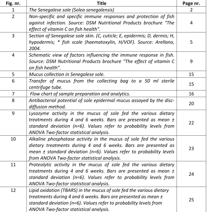
7 Flow chart of sample preparation and analytics. 16 8 Antibacterial potential of sole epidermal mucus assayed by the disc‐
diffusion method. 20
9 Lysozyme activity in the mucus of sole fed the various dietary treatments during 4 and 6 weeks. Bars are presented as mean ± standard deviation (n=6). Values refer to probability levels from ANOVA Two‐factor statistical analysis.
22
10 Alkaline phosphatase activity in the mucus of sole fed the various dietary treatments during 4 and 6 weeks. Bars are presented as mean ± standard deviation (n=6). Values refer to probability levels from ANOVA Two‐factor statistical analysis.
23
11 Proteolytic activity in the mucus of sole fed the various dietary treatments during 4 and 6 weeks. Bars are presented as mean ± standard deviation (n=6). Values refer to probability levels from ANOVA Two‐factor statistical analysis. 24 12 Lipid oxidation (TBARS) in the mucus of sole fed the various dietary treatments during 4 and 6 weeks. Bars are presented as mean ± standard deviation (n=6). Values refer to probability levels from ANOVA Two‐factor statistical analysis. 25
List of tables
Table. Nr. Title Page nr.
I Formulation and composition of the experimental diets. 13 II Characteristics of the bacterial strains tested. 19 III Growth performance of sole fed the various dietary treatments over 6 weeks. 21 IV Inhibition of bacterial growth by 100 μl of epidermal mucus of sole. 26
1. Introduction
Aquaculture is the farming of aquatic organisms including fish, mollusks, crustaceans and aquatic plants under controlled conditions. Aquaculture, probably the fastest growing food‐producing sector, now accounts for almost 50 percent of the world’s food fish supply and is perceived as having the greatest potential to meet the growing demand for aquatic food. Given the projected population growth over the next two decades, it is estimated that at least an additional 40 million tons of aquatic food will be required by 2030 to maintain the current per capita consumption (FAO, 2006). Aquaculture has evolved into an industrial activity. As a consequence higher productivity has been closely associated to an intensification of production conditions (e.g. higher rearing densities). As a result, production losses due to diseases have also increased, given the enhanced exposure of fish to pathogens, such as bacteria, parasites, or virus, throughout the production cycle (Laidler et al., 1999).
Infectious diseases are the major cause of economic losses in commercial aquaculture. Current methods for treating diseases in fish include a limited number of government‐approved antibiotics and chemotherapeutics that have in most cases limited effectiveness. As such, the aquaculture industry has begun to focus on prevention of disease rather than treatment. One undisputed concept behind such approach is the fact that to avoid disease epidemics in aquaculture, fish need to be reared in good environmental conditions and priority should be given to the optimization of their welfare status (Mozumder, 2005).
Fish welfare must be assured, by guaranteeing that the fish live as well as possible and express their natural behavior as much as possible, free from negative experiences (stressors). Given that fish are in direct contact with their environment through the large surface of their gills and skin, water quality (dissolved oxygen, CO2, ammonium and pH) and the presence of contaminants
(organics and inorganic pollutants) are some of the most critical aspects of the environment for fish welfare (Mellor & Stafford, 2001). Furthermore, under captivity, fish are also subjected to a
great number of other potential stress factors, such as husbandry and feeding practices.
1.1. The farming of Senegalese sole (Solea senegalensis)
Senegalese sole (Solea senegalensis Kaup, 1858) has longtime been considered a promising candidate species for marine aquaculture in the Mediterranean and South Atlantic costal areas (Figure 1). Senegalese sole is commonly raised in extensive polyculture (in earth ponds) in the South of Portugal and Spain, where it can achieve higher growth rates than European seabass (Dicentrarchus labrax), being second only to gilthead seabream (Sparus aurata). Sole’s high commercial value has motivated farmers to initiate its intensive farming. This interest has gain momentum in Southern Europe over recent years, as a result of the rapid increase in the production of European seabass and gilthead seabream leading to market saturation and a decline in profitability for these traditional species (Morais et al., 2006). Figure 1. The Senegalese sole (Solea senegalensis).
Significant progress on sole farming has been obtained on aspects related to spawning‐control under captivity, the development of feeding protocols, in particular during early life stages, and the estimation of nutritional requirements of larvae and juveniles. Over the last years, research effort has been put into optimizing larval rearing techniques for this species, since this was considered as the bottleneck, hampering the development of commercial scale culture. At present, the main problem associated to the large scale cultivation of sole it is the high sensitivity of the post‐larvae and juveniles to stressful situations commonly found in aquaculture activities, such as grading,
weighing or initial feeding with artificial diets. These stressful conditions cause not only sub‐ optimal growth, but also a high susceptibility to opportunistic pathogens, mainly Flexibacter, which may lead to mass mortalities. Being a new species to aquaculture, knowledge on the mechanisms associated to disease resistance and stress response in sole are extremely scarce. 1.2. The immune response in fish Fish are a diverse group of animals specialized for life in the aquatic environment. Their connection with their habitat is very close and so they are always in contact with bacteria, viruses and fungi that can appear in large concentrations, be pathogenic and cause damage to these organisms. However, under normal conditions the fish can maintain its well state, defending itself against potential invaders, because of a complex system of defense (Mozumder, 2005; Arellano et al., 2004).
The immune system is divided into innate (non‐specific) and acquired (specific) (Figure 2). The first is the only weapon of defense of invertebrates and a key defense mechanism of fish. It is of vital importance in resistance to diseases, especially given that the innate response generally precedes the adaptive response, activates and determines the nature of the adaptive response and co‐ operates in the maintenance of homeostasis (Magnadóttir, 2006). The specific immune mechanisms in fish are slow and limited by temperature constrains on their metabolism. So, fish are likely to rely highly on their innate immune mechanisms for protection against invading pathogens (Bly & Clem, 1991; Ellis, 2001; Ingram, 1980).
Figure 2. Non‐specific and specific immune responses and protection of fish against infection. ure “The effect of vitamin C on fish health”. .3. Mucus as a natural immune barrier in fish nt of contact between ost and potential pathogen (Mozumder, 2005).
In the skin of Senegalese sole, four morphologically distinct layers were identified: cuticle, epidermis, dermis and hypodermis (Figure 3). The epidermis is composed of stratified epithelium, containing three cellular layers: the outermost or mucosa layer, the middle or fusiform layer and the stratum germinativum or the basal layer. In the mucosa, two mucous cell types are
Source: DSM Nutritional Products broch
The innate system is divided in physical barriers (scales, mucus surfaces of skin, gills, gut and the epidermis which act as the first barriers against infection), cellular (phagocytic cells and the non‐ specific cytotoxic cells) and humoral components (growth inhibitors, various lytic enzymes and components of the complement pathways, agglutinins and precipitins, natural antibodies, cytokines, chemokines and antibacterial peptides) (Magnadóttir, 2006; Subramanian et al., 2007).
1
The epidermis is a metabolically very active border tissue containing mucous, sensory, chloride and club cells, and is covered by a mucous layer that forms an additional external barrier containing enzymes (Arellano et al., 2004). This mechanical barrier impedes the entry of the majority of microorganisms into the body and the lining of the alimentary, respiratory, and urogenital tracts as well as total epidermis. Together, the skin and mucus represent the first poi
differentiated: type‐A cells containing several round vesicles of different electron density and type‐ B cells containing mucosomes of uniform electron density (Arellano, 2004). Figure 3. Section of Senegalese sole skin. epidermis; D, dermis; H, hypodermis; * fish scale (haematoxylin, H/VOF) Source: Arellano, 2004. The epidermal mucus is produced primarily by epidermal goblet or mucus cells and is composed mainly of water and gel‐forming macromolecules, including mucins and other glycoproteins (Shephard, 1993), and presents a diversity of functions in fish, including: protection against and resistance to disease, respiration, ion
(C, cuticle; E,
ic and osmotic regulation, excretion, communication and eding (Ingram, 1980; Shephard, 1994; Arellano et al., 2004). In recent years it has been fe demonstrated that mucus plays an important role in the prevention of colonization by parasites, bacteria and fungi (Mozunder, 2005). The information available on the antimicrobial function of epidermal mucus is restricted to a few fish species. For example, Austin & McIntosh (1988) demonstrated the existence of antimicrobial property in the epidermal mucus of rainbow trout (Oncorhynchus mykiss); in ayu (Plecoglossus
them with Listonella anguillarum resulted in increased mortality (Kanno et al., 1989, Fouz et al., 1990). Lemaître et al. (1996) observed in their work in carp (Cyprinus carpio) that loss of epidermal mucus increased susceptibility to bacterial infection. Antimicrobial activity of epidermal mucus xtracts against a broad range of microbial pathogens was observed by Hellio et al. (2002). All
and ious other antibacterial proteins and peptides (Subramanian et al., 2008; Palaksha et al., 2008; s that in the mucus yer are present many antimicrobial enzymes and proteins, which are involved in innate immunity n 19 p t e
these experiments support the hypothesis that the epidermal mucus plays a protective function against microbial infection in fish (Subramanian et al., 2008).
The antimicrobial function of epidermal mucus appears to result from its mechanical and biochemical properties (Pickering, 1974). As an element of the innate immune mechanism, mucus has a dual function: presents a continuous production, thereby preventing the adherence of pathogens, and also acts as a repository of several innate immune factors: lysozymes, immunoglobulins, complement proteins, lectins, C‐reactive proteins, proteolytic enzymes var
Fast et al., 2002; Ross et al., 2000; Cole et al., 1997). Altered expression of these different non‐ specific factors was associated with variations in resistance between species (Fast et al., 2002). Little is known about the role of enzymes in the epidermal mucus in the innate immune system of some species of fish (Subramanian et al., 2008), but there are many indication la of fish (Mozunder, 2005). Innate immune parameters such as lysosyme, proteases, and cathepsins have been used as indicators of disease resistance of fish (Magnadóttir, 2006). Lysosyme is the best know bacteriolytic in non‐specific defense in animals and fish, aiding in the preventio of bacterial infection (Aranishi, 99). It is resent in mucus, lymphoid issue, plasma and other body fluids in most species of fish (Magnadóttir, 2006). Also referred as N‐ acetylmuramide glycanohydrolase or muramidase, it functions by hydrolyzing the ß‐(1‐4) linkage between N‐acetylmuramic acid and 3‐acetyl amino‐2‐deoxy‐D‐glucose residues of mucopolysaccharide found in bacterial cell walls (Magnadóttir, 2006, Subramanian et al., 2007). It acts directly on the walls of Gram‐positive bacterial cells, causing lysis of its outermost
peptidoglycan layer, and in Gram‐negative bacterial cells acting subsequently to the degradation of the outer membrane, by complementing other enzymes that expose the peptidoglycan layer (Yano, 1996). This enzyme has been reported in the mucus of many species such as Japanese flounder (Paralichthys olivaceus), coho salmon (O. kisutch), koi carp (Cyprinus carpio), haddock
lanogrammus aeglefinus), Atlantic cod (Gadus morhua), hagfish (Myxine glutinosa), Arctic char
as a otential stress indicator in the epidermal mucus of Atlantic salmon (S. salar). It is also thought to
asotus), has been shown to activate the production of the antimicrobial peptide
arasin I (Cho et al., 2002). Moreover, cathepsins B and L showed bacteriolytic activity against the sh pathogens Edwardsiella tarda, Flavobacterium columnare and L. anguillarum (Aranishi, 1999). (Me (Salvelinus alpinus), brook trout (S. fontinalis), striped bass (Morone saxatilis) and others (Hikima et al., 1997; Schrock et al., 2001; Subramanian et al., 2007).
Alkaline phosphatase (AP), another enzyme present in the mucus, has been established p
act in a protective role in the initial stage of wound healing in carp and as an antibacterial agent because of its hydrolytic activity (Ross et al., 2000; Fast et al, 2002; Subramanian et al., 2007).
Proteases have a significant role in the innate immune mechanisms (Ingram, 1980). They can cleave bacterial proteins thus directly damaging the pathogen. They also function indirectly by activating and enhancing the production of various immunological components such as complement, immunoglobulin and antimicrobial peptides. Proteases are classified into serine, cysteine, aspartic and metalloproteases based on the chemical nature of the groups responsible for catalysis. Fish mucus has been found to have serine proteases (trypsin); cysteine proteases (cathepsins B and L); aspartic proteases (cathepsin D) and metalloproteases (Subramanian et al. 2007). Several proteases that have been observed in fish have been correlated with one or more of the above mentioned activities. For instance, a cathepsin D found in the mucus of catfish (Parasilurus p fi
Oxidative stress is an inevitable result of life in an oxygen‐rich environment. For aerobic organisms, oxygen is paradoxically both vital and dangerous. The so‐called “oxygen paradox” derives from the chemical nature of oxygen, which in its atomic form (O) is a free radical and in its molecular form (O2) is a bi‐radical (Davies, 2000). Aerobic tissues continuously generate superoxide radicals and
hydrogen peroxide (H2O2) as by‐products of oxidative metabolism (Passi et al., 2004). Oxidative
stress occurs when reactive oxygen species (ROS) generation exceeds its removal. The deleterious effects include oxidation of proteins, DNA, steroid components, as well as peroxidation of unsatured lipids in cell membranes (Martínez‐Álvarez et al., 2005). In particular, ROS are able to start the lipid peroxidation process, which is potentially dangerous in fish, since they contain a high percentage of polyunsatured fatty acids. Lipid peroxidation is involved in a variety of disorders and pathological conditions (Nakamura et al., 1998). Intrinsic factors, such as age, phylogenetic osition, nutritional status and environmental factors such as dissolved oxygen, temperature, ites, can either enhance or decrease antioxidant in fish (Martínez‐Álvarez et al., 2005).
outbreak of diseases in fish farming can be a major concern. The high susceptibility f fish to stress and the rapid spread of diseases in water have forced fish farmers to concentrate
the use of immunostimulants such as antioxidant p
toxins present in the water, pathologies, paras defences 1.4. The potential of immunostimulants in fish The goals of the aquaculture industry are to optimize growth and to produce high‐quality fish. As in all farming, the o
their efforts on maintaining fish in good health in order to achieve sustainable economic performances.
Growing healthy fish requires them to be able to develop strong defense mechanisms against pathogen invasion. Intensively raised fish may be exposed to stressful situations which often result in a depressed immune status. Good management practices reduce stress and therefore help to maintain healthy animals. However, since not all stressing situations can be avoided, fish with enhanced defense mechanisms will be better prepared to combat the negative effects of stress. The nutritional quality of the feed is a major factor in sustaining healthy fish. It has been shown that the immune system can be enhanced by
vitamins, carotenoids and other feed additives (Vadstein, 1997). The combination of good management, and improve growth in ional factors as munostimulants, and hormones/cytokines. These compounds may directly initiate activation of vaccination and nutritional prophylaxis will insure higher survival rates intensive farming systems (Figure 4). Figure 4. Schematic view of factors influencing the immune response in fish. Source: DSM Nutritional Products brochure “The effect of vitamin C on fish health”.
Bricknell & Dalmo (2005) define immunostimulant as “a natural occurring compound that modulates the immune system by increasing the host’s resistance against diseases that in most circumstances are caused by pathogens”. Sakai (1990) divided the immunostimulants into several groups depending on their sources: bacterial, algae‐derived, animal‐derived, nutrit
im
the innate defense mechanisms acting on receptors and triggering intracellular gene activation that may result in production of antimicrobial molecules (Bricknell & Dalmo, 2005).
Nutritional status is considered one of the important factors that determine the ability of fish to resist diseases. Nutritional modulation of resistance to infectious diseases, based upon the information on fish and terrestrial animals, is divided into four major groups. In the first category,
one must consider a proper balance of macro‐ and micronutrients, including amino acids, polyunsaturated fatty acids (PUFA), vitamins and trace elements, which are essential for the development of immune system starting at the larval stage. Deficiencies in these nutrients may impact several development events including the proper development of lymphoid organs. Marginal deficiencies may negatively affect the immune system at later stages of life. Severe deficiencies will increase susceptibility to disease and may result in the death of the animal. The second point is to consider that adequate nutrition is essential for cells of the immune system to divide and synthesize effector molecules. The diet supplies the immune system with the amino acids, PUFA, enzyme co‐factors and energy necessary to support lymphocyte proliferation and the synthesis of effector (e.g. immunoglobulins, lysozyme and complement) and communication molecules (e.g. cytokines and eicosanoids). The quantitative need for nutrients to maintain a normal immune function is relatively small compared to the requirements for growth and reproduction. In the third category, it is important to consider that some nutrients provide essential substrates for the proliferation of pathogens (e.g. iron) and their presence at low oncentrations in body fluids may limit the growth of pathogens within the fish. The fourth echanism may include the indirect regulatory effects of diets on the immune system that are ediated through the endocrine system (Lall, 2000). c m m
1.5. Objectives of the study This study was undertaken to evaluate the role of nutritional factors as tools to enhance selected mune parameters in the mucus of Senegalese sole. dies have also shown positive effects of vitamin C on everal non‐specific immunological parameters, such as lysosyme, respiratory burst, and resistance es (Verlhac et al., 1996; Sahoo & Mukherjee, 2003; Ai et al. 2004; Lin & Shiau, im The nutritional variables chosen were: Vitamin C level Vitamin C (ascorbic acid, AA) is an essential vitamin for normal growth, physiological functions and plays an important role in immunity in animals including fish (Lin & Shiau, 2005; Ren et al, 2007). It is the most important water‐soluble antioxidant and is a cofactor in many hidroxylating reactions (Kumari & Sahoo, 2005). The influence of AA on the immune response may be related to its antioxidant activity as a free radical scavenger, protecting cells from auto‐oxidation and maintaining their integrity for an optimal functioning of the immune system. Since ascorbic acid is involved in several enzymatic processes, it is difficult to determine the exact mechanism of action behind the stimulation of the immune response (Kumari & Sahoo, 2005). Dietary supplemental levels of vitamin C have been shown to enhance immune status and disease resistance, in a wide variety of species, such as channel catfish (Ictalurus punctatus), Indian major carp (Labeo rohita), rainbow trout, Atlantic salmon, Japanese seabass (Lateolabrax japonicus) and large yellow croaker (Pseudosciaena crocea) (Sahoo & Mukherjee, 2003; Ai et al., 2004; Lin & Shiau, 2005; Kumari & Sahoo, 2005; Ai et al., 2006). A number of stu s to stress and diseas 2005; Kumari & Sahoo, 2005; Ai et al; 2006). Coconut oil Recent studies show that fatty acids are important effectors in the immune functions (Puertullano et al., 2004). Dietary lipids may modulate a great number of immune parameters, such as lymphocyte proliferation, cytokine synthesis, natural killer cell activity, phagocytes and others
ction rojan et al., 1994), and another antimicrobial effect in viruses is due to lauric acid interference ith virus assembly and viral maturation (Hornung et al., 1994; Isaacs et al., 1992). To our nowledge the potential of coconut oil as an immune effector has never been studied in fish.
(Pablo & Cienfueros, 2000). Coconut oil is constituted by a unique blend of several fatty acids (50% lauric acid and 7% capric acid), which show potent antiviral, antibacterial and antiprotozoal functions. In general, it is reported that fatty acids and monoglycerides produce their killing/inactivating effect by lysing the plasma membrane of lipid bilayer. The antiviral action attributed to monolaurin is that of solubilizing the lipids and phospholipids in the envelope of the virus, causing its disintegration. However, there is evidence from recent studies that one antimicrobial effect in bacteria is related to monolaurin interference with signal transdu (P w k
2. Materials and Methods
2.1. Experimental diets
Three experimental diets were formulated to fulfill the nutritional requirements of Senegalese sole. A control diet (CTRL diet) was a practical formulation currently used for sole farming, with 150 mg/kg of vitamin C. The control formula was supplemented with vitamin C at 1000 mg/kg (VIT C diet) and a third formula, identical to control but in which 70% of the original dietary fat source (fish oil) was replaced by coconut oil (COC diet). The formulation in terms of ingredients and the proximate composition of the experimental diets are presented in Table I.
Table I. Formulation and composition of the experimental diets.
Ingredients (%) CTRL COC VIT C
Fishmeal LT 35.00 35.00 35.00 Fish protein concentrate (CPSP G) 12.50 12.50 12.50 Soybean meal 48 16.00 16.00 16.00 Corn gluten 8.00 8.00 8.00 Wheat meal 21.00 21.00 20.73 Coconut oil 0.00 5.00 0.00 Fish oil 7.00 2.00 7.00 Choline chloride 50% 0.10 0.10 0.10 Vitamin C (Lutavit C35) 0.03 0.03 0.30 Vitamin E (Lutavit E50) 0.05 0.05 0.05 MIN & Vit Mixture 0.25 0.25 0.25 Betaine 0.07 0.07 0.07 Proximate composition Dry matter (DM) (%) 92.47 91.25 92.33 Crude protein (%DM) 46.21 46.21 46.19 Crude fat (%DM) 11.93 11.93 11.93 Gross Energy (kJ/g DM) 20.79 20.79 20.75 Vitamin C (ascorbic acid) (mg/kg DM) 141.00 144.00 1093.00
Ingredients were finely ground, mixed in an horizontal helix ribbon mixer (model Mano, 100 L capacity, CPM, San Francisco, USA) for 15 min and pelleted dry using a steamless pelleting machine (model 3000, CPM, San Francisco, USA) fitted with a die of 3.0 mm in diameter. Pellets were dried
at room temperature and subsequently stored at 4ºC. Diets were manufactured at the Zootechnical Departement of the University of Trás‐os‐Montes e Alto Douro.
2.2. Experimental animals and rearing conditions
Three homogeneous groups of 20 Senegalese sole (initial body weight: 92.8 ± 1.52 g) were maintained at the Experimental Station of Ramalhete (University of Algarve), in 3 rectangular PVC tanks (31 x 55 x 94 cm); volume: 160 L; water‐flow rate: 3.6 L∙min‐1), supplied with recirculated seawater (constant temperature: 19.2 ± 1.6ºC; salinity: 36.2 ± 1‰; dissolved oxygen: 94.1 ± 17.6 mg∙L‐1). A photoperiod of 13/11 fluorescent light/dark cycle was adopted, with a light intensity of 90 Lux. Fish were fed with one of the three experimental diets by means of automated feeders for 12 hours (10.00 till 22.00) during 6 weeks. 2.3. Mucus collection Fish used in this study were individually measured and weighed at the beginning and end of the experiment. After 4 and 6 weeks of experimental feeding, the epidermal mucus was collected in individual fish according to the method initially described by Ross et al. (2000) and slightly modified by Subramanian et al. (2007). The fish were starved for 24 hours prior to sampling. The mucus samples were obtained from 18 fish per tank and aggregated in pools of 3 fish. The fish were anaesthetized with a sub‐lethal dose (150 mg∙L‐1) of 2‐phenoxy‐ethanol (Sigma‐Aldrich). Each individual fish was then placed in a plastic bag with zip (Figure 5) containing 5 ml of 100 mM NaCl. The bags were then gently shaken by hand during 1‐2 min and afterwards fish were carefully placed in a recovery tank.
Figure 5. Mucus collection in Senegalese sole. The mucus sample in the bag was transferred to a 50 ml sterile centrifuge tube and placed in ice. The mucus remaining in the bag was rinsed with additional 5 ml of NaCl and pooled in the same tube (Figure 6). Figure 6. Transfer of mucus from the collecting bag to a 50 ml sterile centrifuge tube.
The mucus samples were immediately transported to the laboratory, where they were centrifuged at 2000 x g for 15 min at 4ºC. The pellet was eliminated and the supernatant was homogenized with a sonicator for 20 seconds, aliquoted in 2 ml eppendorf tubes and stored at ‐80ºC until subsequent assay of total protein, lysozyme activity and lipid oxidation (TBARS). Prior to these assays, the thawed samples were further centrifuged at 8850 x g for 2 min at 4ºC. Only this last supernatant was used for assays. For alkaline phosphatase (AP) and protease activity the initial supernatant was aliquoted in 1 ml samples, freeze dried and stored at ‐80 ºC until further use. Prior to the assay, samples were reconstituted to 1 ml with the respective enzyme assay buffers, centrifuged at 8850 x g for 2 min at 4ºC. Only this last supernatant was used for assays (Figure 7). Figure 7. Flow chart of sample preparation and analytics. 2.4. Analytical methods 2.4.1. Protein
Protein concentration of mucus samples was determined by spectrophometry measurement of protein‐dye binding according to the Bradford method (Bradford, 1976). The total protein concentration determined was then used to calculate the specific activity for the various assays. The absorbance for various assays was read using a microplate reader BioRad Benchmark.
2.4.2. Lysozyme
Lysozyme activity was determined using a turbidimetric assay adapted from Ross et al. (2000), with some modifications. Twenty‐one mg of lyophilized Micrococcus lysodeikticus bacterial cells (Sigma) were suspended in 100 ml of 0.05 M Na‐phosphate buffer (50 ml sodium phosphate dibasic dehydrate and 200 ml potassium phosphate monobasic), adjusted to pH 6.2 with HCl. This solution was made fresh everyday. The Micrococcus lysodeikticus suspension was incubated in a water bath at 30 ºC under gentle stirring for not less than 30 minutes. 50 µl of both buffer (for blank) and sample were pipetted into a 96‐well microplate in triplicate. 150 µl of the bacterial solution was rapidly added using a multi‐dispenser pipette and absorbance was read after 5 and 20 min at 450 nm. One unit of lysozyme activity was defined as the amount of enzyme that catalyzed a decrease in absorbance of 0.001 min‐1. Activity units were expressed per mg of protein (specific activity). 2.4.3. Alkaline phosphatase Alkaline phosphatase (AP) activity was determined according to Ross et al. (2000). Fifty µl of mucus reconstituted in 100 mM ammonium bicarbonate, 1 mM magnesium chloride hexahydrate, pH 7.8 buffer were incubated in a 96‐well plate Thermo Shaker (Grant) at 30 ºC for 15 min. 150 µl of 4 mM p‐nitrophenol phosphate substrate was rapidly added using a multi‐dispenser pipette. Absorbance was read at 405 nm after 20 and 25 minutes of reaction. One unit of activity was defined as the amount of enzyme required to release 1 nmol of p‐nitrophenol in 1 min. The extinction coefficient of p‐nitrophenol in the microplate wells was experimentally determined using a standard curve with concentrations ranging 0‐50 µM. Activity units were expressed per mg of protein (specific activity).
2.4.4. Proteases
Protease activity was determined using the azocasein hydrolysis assay according to Firth et al., (2000). Azocasein hydrolysis was assayed by incubating 50 μl of the mucus sample re‐suspended in 100 mM ammonium bicarbonate, pH 7.8, with 50 μl azocasein substrate 0.25% (w/v) in the same buffer for 19 h at 30 °C. The reaction was stopped by adding 50 μl of 20% (w/v) trichloroacetic acid followed by a 5 min centrifugation at 15400 × g. Equal volumes (100 μl) of the resultant supernatant and 0.5 M NaOH were added to a 96‐well plate and the absorbance measured at 405 nm. One unit of activity was defined as the amount of enzyme that caused a change in absorbance of 0.001 min‐1. Activity units were expressed per mg of protein (specific activity). 2.4.5. Lipid oxidation (TBARS)
Oxidation of lipids was determined in mucus by measuring the thiobarbituric acid reactive substances (TBARS) according to the method adapted from Burk et al. (1980). TBARS was assayed by incubating 750 µl of the mucus samples with 1500 µl of trichloroacetic acid 20% and 50 µl 1% butylated hydroxytoluene (BHT) in methanol. A standard curve with concentrations ranging from 0 to 5000 nmol of MDA solution (1,1,3,3‐tetraethoxypropane) was also assayed. Both samples and standards were incubated with 2950 µl of 2‐thiobarbituric acid 50 mM in a 90 ºC water bath for 20 min. After cooling and centrifugation at 2000 x g for 20 min at 4 ºC, supernatants (200 µl) were transferred to a 96‐well plate and absorbance read at 540 nm. TBARS concentration was calculated by correspondence to the standard curve, and expressed per mg of protein.
2.4.6. Antibacterial assay The antibacterial assays were done by disc‐assay method (Chabbert, 1963). Mucus extracts (100 ul) were impregnated in sterile filter paper discs (6 mm in diameter, Schleicher and Schuell, Inc.). Discs were allowed to evaporate and then placed on Zobell’s agar test plates (9 cm diameter) inoculated with 18 hours culture of the test pathogens (107 bacteria /ml) in 15‰ NaCl TSA broth. The plates were then incubated for 48 h at 22.8 ºC before reading the inhibition zone diameter. The antibacterial tests were performed in triplicate against five Gram‐negative bacteria Aeromonas
salmonicida, Aeromonas hydrophila, Yersinia ruckeri, Vibrio alginolyticus and Listonella anguillarum. The test bacterial pathogen cultures were obtained from stock cultures maintained at
the Microbiology Laboratory of the Unity of Upgrading of Fishery and Aquaculture Products, from the National Institute of Biological Resources (Table II).
Table II. Characteristics of the bacterial strains tested.
Name Origin Characteristics
Aeromonas hydrophila INRB – IPIMAR Isolated from trout
Aeromonas salmonicida IFREMER
Listonella anguillarum IFREMER Serotype I
Vibrio alginolyticus INRB – IPIMAR ATCC 17749
Yersinia ruckeri Institute Pasteur Collection CIP: 8280T
The zone of inhibition of bacteria around the disc was recorded using Vernier calipers (Figure 8). The assay was scored positive (+) if it was < 2 mm, doubly positive (++) if the zone was ≥ 2 mm, triple positive (+++) if the zone of inhibition was ≥7 mm, and negative (‐) if there was no inhibition of microbial growth (Thompson et al., 1985). The bacterial assay results were compared with those obtained when challenging the bacterial pathogens with two reference antibiotics (i.e. discs containing chloramphenicol 30 mg, oxytetracycline 30 UI from Sanofi Diagnostics Pasteur discs).
Figure 8. Antibacterial potential of sole epidermal mucus assayed by the disc‐diffusion method.
2.4.7. Statistical analysis
Data are presented as means ± standard deviation. Data were subjected to a two‐way analysis of variance, considering dietary treatment and duration of feeding as variables, and when appropriate, means were compared by the Newman‐Keuls test. Parameters expressed as percentages were subjected to arcsin square root transformation. Statistical significance was tested at 0.05 probability level. All statistical tests were performed using the STAGRAPHICS Centurion XV, version 15.1.02 software.
3. Results
3.1. Growth performance
Data on growth performance of sole fed the different experimental diets during 48 days are reported in Table III. Specific growth rate (SGR) values ranged from 0.38 to 0.63 %/day and neither weight gain nor growth rates were significantly affected (P>0.05) by vitamin C supplementation or coconut oil inclusion. However it is worth mentioning a slight trend to a growth enhancement observed in fish fed the coconut oil diet (COC). Still, the high weight dispersion that generally characterizes sole rearing makes it difficult to attain statistical significance. Similarly to what is observed in terms of weight, the condition factor (K) of fish was not affected by the various dietary treatments. Moreover, K values were not altered between the beginning and the end of the trial. Table III. Growth performance of sole fed the various dietary treatments over 6 weeks. Dietary treatments CTRL COC VIT C Initial body weight, g 92.5 ± 21.9 94.5 ± 19.1 91.5 ± 13.8 Final body weight, g 113.3 ± 26.0 130.0 ± 28.6 116.4 ± 16.6 Weight gain, %IBW/d 0.42 ± 0.25 0.76 ± 0.30 0.61 ± 0.34 SGR, %/d 0.38 ± 0.20 0.63 ± 0.22 0.52 ± 0.27 Initial total length, cm 17.5 ± 1.5 17.7 ± 1.5 17.9 ± 0.9 Final total length, cm 18.9 ± 1.5 19.6 ± 1.4 19.3 ± 0.9 K 1.66 ± 0.19 1.71 ± 0.24 1.61 ± 0.16 Values are means ± sd. Specific growth rate: SGR = 100 x (Ln final body weight ‐ Ln initial body weight) / 48 days. Condition factor: K = (100 x total weigh) / total length3
3.2. Immune parameters
The presence of lysozyme, alkaline phosphatase and protease was confirmed for the first time in the mucus of the Senegalese sole. The activity of lysozyme ranged from 0.05 to 0.08 U/mg protein at the 4 weeks sampling and 0.05 to 0.06 U/mg protein at the end of the trial. Lysozyme activity was not significantly affected (P>0.05) by the various dietary treatments or by the duration of feeding (Figure 9).
0.00
0.02
0.04
0.06
0.08
0.10
0.12
0.14
CTRL
COC
VIT C
Dietary treatments
U/
m
g
pr
ot
e
in
4 weeks 6 weeks ANOVA Two‐factors Statistical analysis95% confidence level Diet Time Interaction
Lysozyme 0.211 0.050 0.059 Figure 9. Lysozyme activity in the mucus of sole fed the various dietary treatments during 4 and 6 weeks. Bars are presented as mean ± standard deviation (n=6). Values refer to probability levels from ANOVA Two‐factor statistical analysis.
Despite the extremely low levels of alkaline phosphatase (AP) found in the mucus of Senegalese sole, results show that AP activity in fish fed coconut oil and vitamin C rich diets was significantly higher (P<0.05) than that found in fish fed the control diet (Figure 10). After 4 weeks of experimental feeding, alkaline phosphatase levels for the CTRL treatment (0.001 U/mg protein)
were six‐fold increased in both COC and VIT C treatments. At the end of the trial (6 weeks of experimental feeding), this situation was maintained, but such increase on AP activity seen in fish fed diets COC and VIT C was only three‐fold in relation to CTRL diet. The duration of feeding (either 4 or 6 weeks) had no significant effect (P>0.05) on alkaline phosphatase activity. Similarly, the interaction between the two variables (dietary treatment and duration of feeding) did not prove statistical significant at a 95% confidence level.
0.0000
0.0002
0.0004
0.0006
0.0008
0.0010
0.0012
CTRL
COC
VIT C
Dietary treatments
U/
m
g
pr
ot
e
in
4 weeks 6 weeks ANOVA Two‐factors Statistical analysis95% confidence level Diet Time Interaction
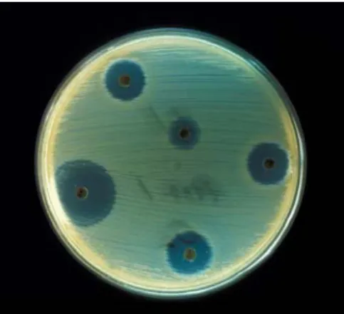
Alkaline phosphatase 0.000 0.507 0.685 Means comparison by Student‐Newman‐Keuls test Diet a b b Figure 10. Alkaline phosphatase activity in the mucus of sole fed the various dietary treatments during 4 and 6 weeks. Bars are presented as mean ± standard deviation (n=6). Values refer to probability levels from ANOVA Two‐factor statistical analysis. At both 4 and 6 weeks of experimental feeding, the proteolytic activity in the mucus of Senegalese sole fed the CTRL diet was 0.05 U/mg protein (Figure 11). The activity of proteases in the mucus of
fish fed the VIT C supplemented diet ranged from 0.05 to 0.06 U/mg protein and were not significantly affected by either dietary treatment or duration of feeding. However, sole fed the diet incorporating coconut oil (COC) showed a significant increase (P<0.05) of proteolytic activity in their mucus. Both the duration of feeding (either 4 or 6 weeks) and its interaction with the dietary treatment had no significant effect (P>0.05) on mucus proteolytic activity.
0.00
0.02
0.04
0.06
0.08
0.10
0.12
CTRL
COC
VIT C
Dietary treatments
U/
m
g
pr
ot
e
in
4 weeks 6 weeks ANOVA Two‐factors Statistical analysis95% confidence level Diet Time Interaction
Protease activity 0.000 0.653 0.645
Means comparison by Student‐Newman‐Keuls test
Diet a b a
Figure 11. Proteolytic activity in the mucus of sole fed the various dietary treatments during 4 and 6 weeks. Bars are presented as mean ± standard deviation (n=6). Values refer to probability levels
from ANOVA Two‐factor statistical analysis.
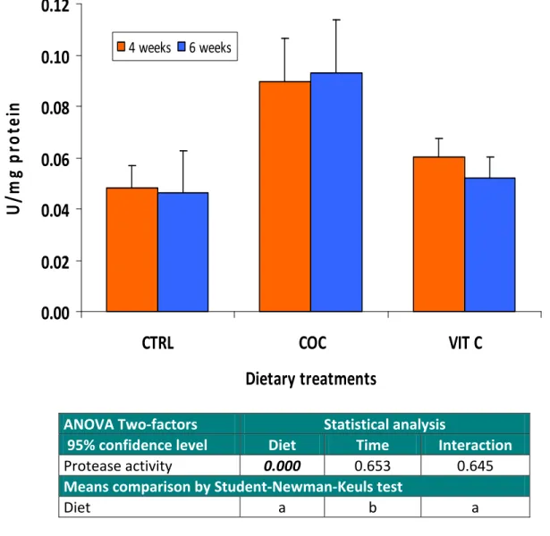
Data regarding the lipid oxidation potential (measured as TBARS) in mucus of Senegalese sole fed the various experimental diets during 4 and 6 weeks are presented in Figure 12. After 4 weeks of feeding, TBARS levels varied between 0.74 and 1.03 nanomoles MDA/mg protein, with the lowest values being found in fish fed the CTRL diet. However, after 6 weeks of feeding and if comparing to the CTRL treatment, feeding sole with either coconut oil or a high dose of vitamin C significantly reduced (P<0.05) mucus susceptibility to lipid oxidation. Despite the fact that the duration of experimental feeding has not affected significantly TBARS, its interaction with the dietary treatments was highly significant statistically.
0.00
0.50
1.00
1.50
2.00
2.50
CTRL
COC
VIT C
Dietary treatments
na
no
M
MD
A
/m
g
pr
ot
e
in
4 weeks 6 weeks ANOVA Two‐factors Statistical analysis95% confidence level Diet Time Interaction
Lipid oxidation (TBARS) 0.026 0.093 0.000
Means comparison by Student‐Newman‐Keuls test
Diet b a a
Figure 12. Lipid oxidation (TBARS) in the mucus of sole fed the various dietary treatments during 4 and 6 weeks. Bars are presented as mean ± standard deviation (n=6). Values refer to probability
3.3. Antibacterial activity
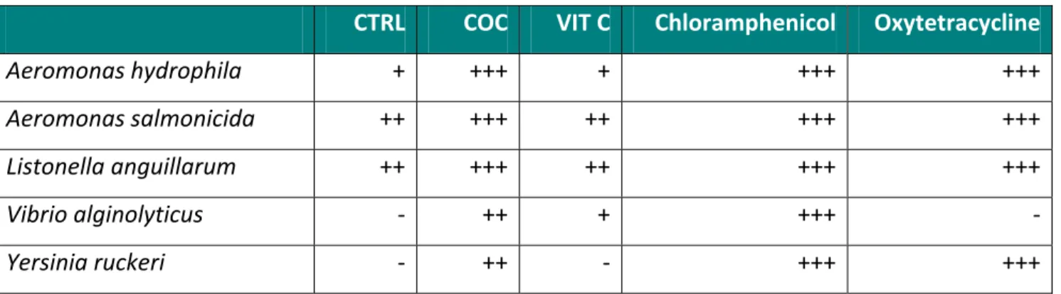
The growth inhibitory effects of experimental mucus extracts against selected pathogens commonly occurring in a marine environment are presented in Table IV. The epidermal mucus extracts of Senegalese sole showed antibacterial activity against a series of known marine pathogen bacteria. A moderate inhibitory effect was observed against Aeromonas hydrophila by mucus of fish fed the CTRL and the VIT C treatments. However, the mucus from fish fed the coconut oil diet (COC) showed a potent antibacterial effect against Aeromonas hydrophila. For both Aeromonas salmonicida and Listonella anguillarum, mucus from all dietary treatments showed an important growth inhibitory effect, but again highest inhibitory potential was observed in the mucus of fish fed the coconut oil diet. Vibrio alginolyticus was not inhibited by the mucus of the CTRL treatment, moderately by the VIT C treatment and strongly by the COC treatment. The mucus from fish fed coconut oil was the only extract that showed and inhibition effect on Yersinia ruckeri. Overall, results show that mucus derived from fish fed the coconut oil based diet showed the highest antibacterial effect on the selected marine pathogens. Table IV. Inhibition of bacterial growth by 100 μl of epidermal mucus of sole.
CTRL COC VIT C Chloramphenicol Oxytetracycline
Aeromonas hydrophila + +++ + +++ +++ Aeromonas salmonicida ++ +++ ++ +++ +++ Listonella anguillarum ++ +++ ++ +++ +++ Vibrio alginolyticus ‐ ++ + +++ ‐ Yersinia ruckeri ‐ ++ ‐ +++ +++ (‐): no activity; (+): 0 < D < 2 mm; (++): 2 ≤ D < 7 mm; (+++): D ≥ 7 mm. D: Diameter of the inhibition zone in millimeters not including the diameter of the disc.
4. Discussion
The aquatic environment is a complex ecosystem, which obscures the distinction between health, sub‐optimal performance and disease. During disease outbreaks, the underlying cause is often difficult to ascertain and is usually the end result of a series of linked events involving environmental factors, health condition of the stocks, presence of an infectious agent and/or poor husbandry and management practices. The whole aquatic production environment, including ecological processes, must be taken into consideration. Therefore, an aquatic system health management approach needs to be developed to replace the more traditional pathogen‐focused approach applied traditionally to disease diagnosis.
Fish are in constant interaction with their aquatic environment, which contains a large range of non‐pathogenic and pathogenic microorganisms. The epidermal mucus secretions and epidermis act as the first biological barrier between fish and the potential pathogens present in the environment (Shephard, 1994). Several studies suggest the protective role of the mucus and its components in various fish species (Aranishi, 1999; Palaksha et al., 2008). The presence in the mucus of certain hydrolytic enzymes including lysozyme, alkaline phosphatase and proteases has been studied in a number of fish species (Ross et al., 2000; Fast et al., 2002; Subramanian et al., 2007), but to date no published information was available on the presence of such factors in the mucus of Senegalese sole.
Presently, the main factor hampering the successful implementation of large scale cultivation of sole it is the high sensitivity of the post‐larvae and juveniles to stressful situations commonly found in aquaculture activities, such as grading, weighing or initial feeding with artificial diets. Such stressful conditions are often associated to mass mortalities due to a high susceptibility to opportunistic pathogens, mainly Flexibacter. As a benthonic species, sole lives in direct contact with the bottom substrate. It has been suggested that by this fact, benthonic species, such as hagfish, may present higher levels of innate immune factors in skin mucus (Subramanian et al., 2007). It was therefore our objective to characterize the epidermal mucus of sole in terms of its
content of selected immune‐associated factors. Furthermore, this study also evaluated the role of certain nutrients (vitamin C and coconut oil as a source of lauric and capric fatty acids) as modulators of the activity of some mucus immune factors. In the present study, mucus samples were collected by placing individual sole in plastic bags and gently shaken the fish with NaCl 100 mM. An alternative methodology, by means of skin scrapping of recently killed fish is described by Mozumder (2005). However, we have adopted the previous methodology given that Subramanian et al. (2007) suggested that such method of mucus collection yielded samples which accurately represented what was naturally present in the fish skin mucus at the time of sampling. Growth performance
Specific growth rate values ranged from 0.38 to 0.63 %/day and neither weight gain nor growth rates were significantly affected (P>0.05) by vitamin C supplementation or coconut oil inclusion. Li et al. (2007) found also no relationship between vitamin C supplemented diets (33 to 424 mg/kg feed) and the growth rates of juvenile Atlantic salmon. This work is in accordance with our results in the vitamin C treatment. Similarly to our results with sole, no effect on weight gain or growth was observed in climbing perch (Anabas testudineus) fed diets containing 20% coconut oil. However, given the relatively short duration of the experimental period, it was not a primary objective of this study to induce a growth enhancement in fish. Furthermore, the usually high weight dispersion found in sole rearing makes it hard to infer statistical significance from our data. Immune factors As a component of the innate immune mechanism, the epidermal mucus plays a dual role in fish. First, by being continuously produced and sloughed off it prevents pathogen adherence (Pickering, 1974). Secondly, it also serves as a repository of numerous innate immune factors such as lysozyme, immunoglobulins, complement proteins, lectins, C‐reactive protein, proteolytic enzymes