Maio de 2015
Henrique Pedro Pereira da Silva Amaral
Machado
The mechanisms underlying
Mycobacterium tuberculosis
virulence
granted by the nrp gene
Mecanismos subjacentes à virulência do
Mycobacterium tuberculosis conferidos pelo
gene nrp.
Dissertação de Mestrado
Mestrado em Ciências da Saúde
Trabalho efetuado sob a orientação de
Doutora Margarida Saraiva
ii
DECLARAÇÃO
Nome: Henrique Pedro Pereira da Silva Amaral Machado Endereço electrónico: [email protected] Telefone: +351 961083628
Número do Bilhete de Identidade: 14146246
Título da dissertação: The mechanisms underlying Mycobacterium tuberculosis virulence granted by the nrp gene.
Orientadores:
Doutora Margarida Saraiva Doutor Egídio Torrado
Ano de conclusão: 2015
Designação Ramo de Conhecimento do Mestrado: Ciências da Saúde
DE ACORDO COM A LEGISLAÇÃO EM VIGOR, NÃO É PERMITIDIDA A REPRODUÇÃO DE QUALQUER PARTE DESTA TESE.
Universidade do Minho, 27 de Maio de 2015
iii
The work presented in this dissertation was done in the Microbiology and Infection Research Domain of the Life and Health Sciences Research Institute (ICVS), School of Health Sciences, University of Minho, Braga, Portugal (ICVS/3B’s – PT Government Associate Laboratory, Braga/Guimarães, Portugal).
v
A
GRADECIMENTOS
Começo por agradecer à Doutora Margarida Saraiva. As palavras aqui escritas dificilmente conseguiriam fazer justiça à importancia que teve no meu desenvolvimente ciêntifico e pessoal ao longo dos ultimos dois anos. Sem duvida a qualidade trabalho por mim, e muitos outros, desenvolvido nos ultimos anos não teria sido atingida sem o seu esforço, dedicação e profissionalismo. Por tudo isto lhe fico imensamente grato, e desejo apenas que os próximos anos tragam ainda maior sucesso.
Quero expressar a minha gratidão ao Doutor Egídio Torrado por todo o seu empenho neste projecto. Sem dúvida o seu contributo tanto a nível científico como técnico foi essencial para levar este trabalho a bom porto. Obrigado também por todas as discussões científicas que tivemos ao longo desde último ano, sem dúvida ajudaram-me a analisar a “ciência” de forma mais critica!
Agradeço ao Doutor Apoorva Bhatt por ter disponibilizado as estirpes utilizadas neste trabalho e por ter partilhado os seus resultados preliminares. Fico também grato pela hospitalidade e prontidão com que nos recebeu em Birmingham para discutir este projecto.
Ao Professor Gil Castro, uma das primeiras pessoas que me acolheu neste instituto, queria também expressar a minha gratidão por todas as vezes que partilhou o seu vasto conhecimento cientifico comigo. Sem duvida que a sua experiência é uma mais valida para todos aqueles que trabalham à sua volta. Gostaria também de manifestar a minha gratidão ao Professor Fernando Rodrigues por me ajudar a perceber o meu próprio trabalho de outra perspectiva, a da mycobactéria.
Ao Diogo, obrigado por tudo! Foste a primeira pessoa que me acolheu no laboratório, e sinceramente acho que nesse ponto não podia ter tido maior sorte! Ajudaste-me a dar os primeiros passos no mundo da investigação, algo que aliás te vejo a fazer ano após ano com novos estudantes sempre com o mesmo empenho. Como bem sabes este percurso nem sempre é facil, mas não tenho qualquer dúvida que ultrapassarás todos os obstáculos que encontrares. Por tudo isto, e pela constante companhia neste últimos 2 anos, muito obrigado!
À Isabel quero também expressar a minha mas sincera gratidão! Ajudaste-me em muito adquirir as competências necessárias para desenvolver este tipo de trabalho. Quantas vezes as 2 horas dentro do P3
vi
se tornaram em 4 ou em 6? Tanto melhor, em boa companhia o tempo voa! Espero que o futuro seja repleto de alegrias, tanto profissionais como. Muito obrigado!
Hélder, não podia de forma nenhuma deixar de ter agradecer! Obrigado pela oportunidade de trabalhar no teu projecto, foi e ainda é uma aventura não só pelo valor do trabalho em si como também pela tua constante boa disposição e vontade de levar tudo a bom porto. Tenho a certeza que o futuro será brilhante!
Quero também agradecer ao Jeremy, tanto pelo trabalho que desenvolvemos juntos como pela frequente companhia dentro do P3. Muito obrigado!
Aos membros do I3.02 que fazem deste ICVS mais do que um local de trabalho, muito obrigado! Quero agradecer à Joana, Bruno, Ana Cardoso, Alice, Cláudia, Alexandra, Gabriela, Ritinha, Carine, Teresa, Ana Ribeiro, Inês, Patrícia, e Ana Belinha, algumas de vós companhias mais antigas, outras mais recentes mas sem dúvidas todas sempre prestáveis e optimas companhias! Ao Nuno, frequentemente companhia de horas tardias, um obrigado. À Flávia, Vânia e Joana Gouveia queria também deixar aqui o meu apreço. Apesar de agora trilharem diferentes percursos, ficam sempre as memórias dos bons momentos e os conselhos que por aqui deixaram, muito obrigado.
Aos meus pais posso apenas expressar a minha eterna gratidão, sendo que sem os seus esforços e por vezes privações nada disto teria sido possível. Farei tudo ao meu alcance para fazer com que todos estes anos de esforços tenham valido a pena, muito obrigado. Quero deixar também um agradecimento muito especial ao meu irmão, que desde sempre me acompanhou e apoiou. Obrigado Carlos!
Finalmente, e apesar de implícito, quero expressar a minha gratidão à Filipa. Certamente serei das pessoas mais afortunadas por ter a tua companhia neste percurso nem sempre fácil. Para mim, o teu valor é inestimável. Ajudaste-me constantemente a superar os meus próprios limites, e foste presença constante quando as coisas correram menos bem, já para não falar na ajuda prática neste mesmo trabalho! Por tudo, obrigado.
vii
A
BSTRACT
Mycobacterium tuberculosis (Mtb), the etiological agent of tuberculosis (TB), is a highly complex pathogen estimated to have caused more deaths than any other infectious agent throughout human existence. Despite global efforts to eradicate TB, this is still a leading cause of death by infectious disease, second only to the human immunodeficiency virus (HIV). Although public health measures have greatly diminished TB burden, the reality is that little has changed in anti-TB therapy and vaccination over the past decades. As an obligatory intracellular pathogen Mtb has co-evolved with its human host for over 70 thousand years. This timeframe has given Mtb ample opportunity to evolve its virulence mechanisms in order to thrive in the harsh conditions imposed by the immune system and eventually allowing its transmission between hosts. In light of this ancient history between host and pathogen, a greater understanding of Mtb and its interactions with the host will be required to develop more efficient anti-TB drugs and vaccines. In this work we have focused on the role of the Mtb protein Rv0101, a non-ribosomal peptide synthetase predicted to be involved in lipid metabolism and cell wall biogenesis, during infection. We found that mice infected with a Mtb mutant deficient for Rv0101 have a transient lower lung bacterial burden during the early stages of infection. Additionally, in mice infected with this strain the dissemination of the bacterium to the spleen was delayed and remained lower up to 90 days post-infection. These differences were accompanied by a diminished recruitment of activated CD4+ T cells, inflammatory monocytes and an overall lower immunopathology. Furthermore, mice with severely compromised or absent adaptive immune response were able to survive infection with this mutant strain at least twice as long as animals infected with the complemented strain. In summary, we have shown that Rv0101 is a virulence factor required by Mtb to optimally overcome the innate immune response of the host, in addition to being a potent inducer of immunopathology. Characterizing the role of key Mtb molecules in infection is urgently needed to understand how the pathogen overcomes and modulates the host immune response to allow for its persistence and transmission. In turn, this knowledge may provide good drug targets and a better rational for vaccine design.
ix
R
ESUMO
O Mycobacterium tuberculosis (Mtb), agente etiológico da tuberculose (TB), é um agente patogénico altamente complexo sendo estimado que tenha levado a mais mortes que qualquer outro agente infeccioso ao longo da existência humana. Apesar de esforços globais para erradicar a TB, ela continua a liderar como uma das causas de morte por doença infecciosa, sendo ultrapassada apenas pelo vírus da imunodeficiência humana (HIV). Apesar de medidas de saúde pública terem diminuído o fardo da TB, a realidade é que nas ultimas décadas pouco mudou em termos da terapia anti-TB e vacinação. Como um patógeno intracelular obrigatório, o Mtb co-evoluiu com o seu hospedeiro humano durante mais de 70 mil anos. Esta janela temporal conferiu ao Mtb amplas oportunidades para evoluir os seus mecanismos de virulência de forma a prosperar no ambiente hostil imposto pelo sistema imune inato, eventualmente permitindo a sua transmissão entre hospedeiros. À luz deste história milenar entre hospedeiro e patógeno, uma maior compreensão do Mtb e das suas interacções com o hospedeiro serão necessárias ao desenvolvimento de antibióticos e vacinas mais eficientes. Neste trabalho focámo-nos no papel durante a infecção do Rv0101 do Mtb, uma sintetase peptídica não ribossomal previsivelmente envolvida em metabolismo lipídico e na biogénese da parede celular. Mostramos que ratinhos infectados com um mutante deficiente para o Rv0101 apresentam uma carga bacteriana temporariamente inferior durante o estágio inicial da infecção. Além disso, esses mesmos ratinhos apresentaram um aparecimento tardio da bactéria no baço e controlaram-na melhor pelo menos até ao dia 90 após a infecção. Estas diferenças foram acompanhadas por um recrutamento diminuído de células T CD4+, monócitos inflamatórios e em geral uma menor imunopatologia. Adicionalmente, ratinhos sem sistema imune adaptativo, ou com o mesmo severamente comprometido, infectados com com esta estirpe mutante conseguiram sobreviver pelo menos o dobro do tempo que aqueles infectados com a estirpe complementada. Resumidamente, mostramos que o Rv0101 é um factor de virulência do Mtb, necessário para uma óptima superação da resposta imune inata do hospedeiro, sendo também capaz de induzir diferenças imunopatológicas duradouras. A caracterização do papel de moléculas do Mtb durante a infecção será necessário para compreender como o patógeno supera e modula a resposta imunitária do hospedeiro, permitindo a sua persistência e transmissão. Por sua vez, este conhecimento pode providenciar bons alvos para novas drogas e assistir no desenho de novas vacinas.
xi
T
ABLE OF CONTENTS
ABSTRACT ... vii
RESUMO ... ix
FIGURE INDEX ... xiii
LIST OF ABBREVIATIONS ... xv
INTRODUCTION ... 1
1. Epidemiology ... 3
2. Early events in the immune response to Mtb ... 4
2.1. Transmission... 4
2.2. Innate immune response ... 4
2.2.1. Recognition ... 4
2.2.2. Inflammatory mediators ... 4
2.2.2.1. IL-12p40 - homodimers and heterodimers ... 5
2.2.2.2. IL-6 ... 6
2.2.2.3. TNF-α ... 6
2.2.2.4 The IL-1β – Eicosanoid – Type-1 IFN network ... 6
2.2.2.5. Chemokines ... 8
2.3. Adaptive immune response... 9
3. Host-pathogen interactions... 12
3.1. The cell wall of Mtb and its impact in host-pathogen interactions ... 12
3.1.1. TDM ... 14
3.1.2. PDIMs ... 14
3.1.3. SLs ... 15
3.1.4. Other cell wall lipids ... 15
4. The role of Rv0101 in Mtb virulence ... 16
AIMS ... 17
METHODS ... 21
1. Bacteria and Infection ... 23
2. Animals ... 23
3. Differentiation and Infection of Bone Marrow-Derived Macrophages (BMDM) and Peritoneal Macrophages.. 23
xii
5. Survival ... 25
6. Bacterial Burden Determination ... 25
7. Histology ... 25
8. Flow Cytometry ... 25
9. Mtb growth curves ... 26
10. Cytokine concentration by Enzyme-Linked Immunosorbent Assay (ELISA) ... 27
11. RNA extraction and quantification ... 27
12. Reverse transcriptase polymerase chain reaction (RT-PCR) ... 27
13. Quantitative Real-Time PCR (qRT-PCR) ... 27
14. Statistical Analysis ... 28
RESULTS ... 29
1. Initial considerations ... 31
2. In vivo infection... 32
2.1. Nrp is required for optimal establishment of infection ... 32
2.2. Nrp induces lung immunopathology ... 33
2.3. Immune response characterization ... 35
2.3.1. Nrp modulates the cellular immune response ... 36
2.3.2. Cytokine and chemokine expression is modulated by nrp ... 40
2.3.3. Nrp is required for full virulence in mouse models of deficient adaptive immunity ... 43
2.3.4. Innate immune deficiencies ... 46
2.3.4.1. Nrp-dependent virulence is not mediated by TLR-2 or TLR-4 activation ... 46
2.3.4.2. Nrp-dependent virulence is not mediated by the eicosanoid pathway ... 47
3. in vitro studies ... 48
DISCUSSION AND FUTURE PERSPECTIVES ... 53
xiii
F
IGURE INDEX
Figure 1 The balanced IL-1β – eicosanoid – type-I IFN network. Page 8
Figure 2 The cellular immune response to Mtb. Page 11
Figure 3 Schematic representation of the cell envelope of Mtb. Page 13
Figure 4 Cytometry analysis. Page 26
Figure 5 nrp-complemented and ∆nrp-mutant strains growth kinetics in liquid media.
Page 31
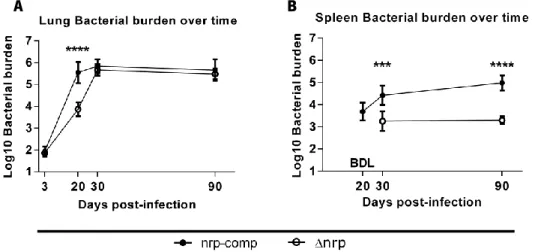
Figure 6 Animals infected with the ∆nrp-mutant or the nrp-complemented strain presented different bacterial burdens over time in the lungs and spleen.
Page 32
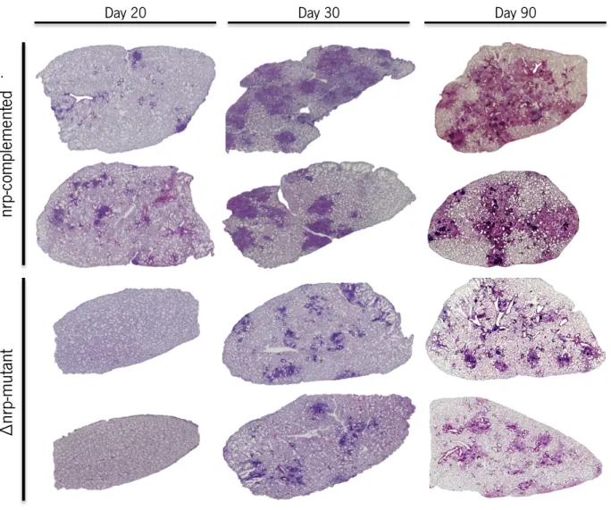
Figure 7 Animals infected with the ∆nrp-mutant or the nrp-complemented strain presented different immunopathology in the lungs.
Page 33
Figure 8 Characterization of H&E preparations from the lungs of animals infected with the ∆nrp-mutant or the nrp-complemented strain revealed
differences in immunopathology.
Page 34
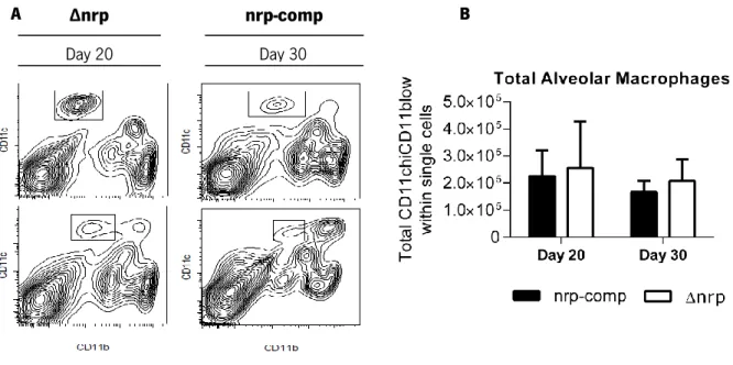
Figure 9 The ∆nrp-mutant and nrp-complemented strain induced different myeloid cell recruitment in the lungs of WT mice.
Page 37
Figure 10 Total number of alveolar macrophages was independent of infecting strain.
Page 38
Figure 11 The ∆nrp-mutant and nrp-complemented strains present different CD4+ T cell features in the lungs of WT mice.
Page 39
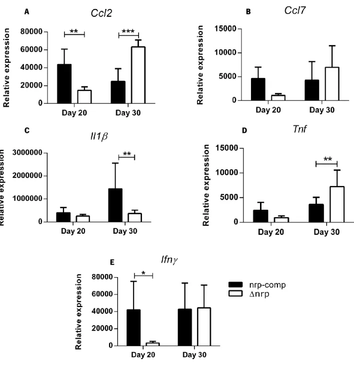
Figure 12 The ∆nrp-mutant and nrp-complemented strain induced differential cytokine/chemokine expression in the lungs of WT mice.
Page 42
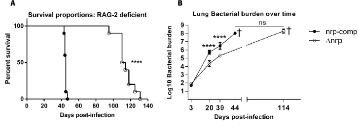
Figure 13 The ∆nrp-mutant presented reduced virulence in RAG-2 deficient mice. Page 44
Figure 14 The ∆nrp-mutant presented reduced virulence in IFN-γ deficient mice. Page 45
Figure 15 nrp does not appear to modulate the recognition of Mtb by immune cells through TLR-2 and TLR-4.
xiv
Figure 16 Shifting the eicosanoid pathway towards PGE2 production did not seem to impact control of the ∆nrp-mutant.
Page 47
Figure 17 The eicosanoid pathway was not modulated by nrp. Page 48
Figure 18 nrp was required for optimal Mtb growth inside macrophages but did not protect against IFN-γ dependent mechanisms.
Page 49
Figure 19 The ∆mutant induced higher nitric oxide production than nrp-complemented Mtb.
Page 50
Figure 20 The ∆nrp-mutant induced higher production of TNF-α, but not IL-1β or IL-10, than the nrp-complemented Mtb.
Page 51
xv
L
IST OF ABBREVIATIONS
AMP Antimicrobial peptide
AIDS Acquired immune deficiency syndrome
ALOX Arachidonate lypoxigenase
AMPh Adenosine monophosphate
ANOVA Analysis of variance
APC Antigen-presenting cell
BCG Bacillus Calmette-Guérin
BDL Below detection level
BMDM Bone marrow-derived macrophages
CCL C-C chemokine ligand
CR Complement receptor
cDNA Complementary DNA
CCR C-C chemokine receptor
CFU Colony-forming unit
CLR C-type lectin receptor
DCs Dendritic cells
ELISA Enzyme-Linked Immunosorbent Assay
FBS Fetal bovine serum
GMM Glucose monomycolate
H&E Hematoxylin-eosin
HIV Human immunodeficiency virus
IFN Interferon
xvi LAM Lipoarabinomannans LCCM L929-cell-conditioned medium LM Lipomannans LOS Lipooligosaccharides LTB4 Leukotriene B4 LXA4 Lipoxin A4 MDR Multidrug resistant
MOI Multiplicity of infection
MHC Major histocompatibility complex
MMP Matrix metalloproteinase
mRNA Messenger RNA
Mtb Mycobacterium tuberculosis
MyD88 Myeloid differentiation primary-response protein 88
NOD2 Nucleotide-binding oligomerization domain-containing protein 2
NOS2 Nitric oxide synthase 2
Nrp Non-ribosomal peptide synthetase
OADC Oleic acid/albumin/dextrose/catalase
PAMP Pathogen-associated molecular patterns
PB Proskauer Beck
PBS Phosphate-buffered saline
PDIM Phthiocerol dimycocerosate
PGE2 Prostaglandin E2
PIM Phosphatidylinositolmannoside
PPE Proline-Proline-Glutamic acid
PRR Pattern recognition receptors
xvii RORγt Retinoic acid receptor-related orphan receptor-γt
RAG Recombination activating gene
ROS Reactive oxygen species
SL Sulfolipids
SR Scavenger receptor
TB Tuberculosis
TGF-β Transforming growth factor-β
TRIF TIR-domain-containing adapter-inducing interferon-β
TDM Trehalose dimycolate
Th T helper
TLR Toll-like receptor
TNF Tumor necrosis factor
WHO World Health Organization
1
3
1. Epidemiology
Tuberculosis (TB) is an infectious disease caused by Mycobacterium tuberculosis (Mtb). As a predominantly lung disease, some of its main clinical features include sputum producing chronic cough, appetite and weight loss, night sweats, fever, hemoptysis and ultimately mortal respiratory failure in case treatment fails1. To the furthest extent of our knowledge, TB has killed more people than any other infectious disease, and it is estimated to have killed more than smallpox, malaria, plague, influenza, cholera and acquired immune deficiency syndrome (AIDS) combined in the past two hundred years2. Despite major global efforts towards prevention and treatment, TB remains one of the leading causes of death due to infectious disease, second only to human immunodeficiency virus (HIV)3. In is estimated that 2 billion people are affected by a latent form of this disease and in 2013 alone 9 million new cases of active TB and 1.5 million deaths were registered3. Although the numbers of new TB cases and its death toll are still a matter of grievous concern, they are in drastic contrast to the reality observed during the first half of the twentieth century2,3. Since the beginning of the twenty first century, there has been an average 1.5% decrease of TB incidence per year, accompanied by an overall decrease of disease prevalence and mortality, and a stabilization of TB incidence among HIV-positive patients3. Even though these data are overall positive, a drastic decrease in the drop of TB incidence rate was observed between 2012 and 2013, registering a drop of 0.6%, less than half of the average since 20002,3. To reach the milestone of less than a TB case per million individuals set by the World Health Organization (WHO) for 2050, numerous roadblocks will have to be overcome. Among these, the most notorious are the absence of an efficient vaccine, the lack of fast and reliable diagnostic tools and the requirement for more efficient anti-mycobacterial drugs. Furthermore, the large number of TB/HIV co-infections and the increased incidence of infections with multidrug resistant (MDR) Mtb strains further prevent the success of this milestone. Indeed, the current strategy to stave off TB is based on public health measures, a 50 years old multidrug regimen for at least six months and the inconsistent protection of the Bacillus Calmette-Guérin (BCG) vaccine3,4. It is clear that a greater understanding regarding the mechanisms underlying the success Mtb as a pathogen is required to develop more efficient vaccines and drugs. In the past few decades, efforts have been made to close this gap of knowledge, both regarding the immune system, the pathogen and their interactions, a topic that will be further developed in this thesis.
4
2. Early events in the immune response to Mtb
2.1. Transmission
It is thought that transmission of Mtb occurs when an infected individual with an advanced form of TB, characterized by high bacterial burden coughs and disrupted lesions with access to the airways, coughs and releases small bacteria-containing droplets into the atmosphere, which will subsequently be inhaled by individuals in close proximity5. Although transmission models of TB have not yet been fully established, it is though that mainly the smaller infectious droplets, containing fewer bacteria, will make it past the upper respiratory track, and into the relatively sterile environment of the distal alveoli6–8.
2.2. Innate immune response
2.2.1. Recognition
Once in the distal alveoli, recognition and phagocytosis of the bacteria occurs mainly by alveolar macrophages, but also by dendritic cells (DCs), monocytes, neutrophils and even by epithelial cells 9–14. Several receptors expressed in phagocytic cells are known to bind pathogen-associated molecular patterns (PAMPs) present in Mtb. These receptors include toll-like receptors (TLRs), nucleotide-binding oligomerization domain-containing protein 2 (NOD2), C-type lectin receptors (CLRs), scavenger receptors (SRs) and complement receptors (CRs)15,16. Although in vitro studies have revealed a specific role for many of these receptors in response to Mtb, most in vivo works using animals deficient for a single receptor revealed only a minor or no phenotype16. These studies suggest that recognition of Mtb by innate immune cells is a complex event requiring multiple receptors, with some of them probably sharing some degree of redundancy16.
2.2.2. Inflammatory mediators
Upon recognition and internalization, a vast array of inflammatory mediators is released to the vicinity of the infected cell in response to stimulation of pattern-recognition receptors (PRRs) by Mtb PAMPs17,18. These mediators include the interleukin (IL)- 12 subunit 12p40, tumor necrosis factor α (TNF-α), 6 and IL-1β, eicosanoids, type-I interferons (IFN) and chemokines18. Together these molecules shape the immune response to Mtb.
5
2.2.2.1. IL-12p40 - homodimers and heterodimers
Overall, the relevance of IL-12p40 derived cytokines is highlighted by the fact that humans with mutations on the IL-12β1 receptor present higher susceptibility to TB resulting in increased incidence, severity and disease dissemination19–25.
IL-12p40 dimerizes either with another IL-12p40 molecule, IL-12p35 or IL23-p19, forming IL-12p80, IL-12 (also known as IL-12p70) and IL-23, respectively18,26. IL-12p80 is readily formed after infection and is essential to induce a migratory phenotype in DCs, allowing them to travel to the lymph node where naïve T cells will be activated5,14,18,27, thus triggering and adaptive immune response. This IL-12p80 dependent mechanism is believed to be enhanced through the expression of a splice variant of the IL-12β1 receptor (which binds to IL-12p40) by DCs early after infection28. Although IL-12p80 is important in the early events leading to the adaptive immune response, it is unable to trigger the production of IFN-γ, a critical cytokine required for the control of Mtb29.
IL-12p40 also dimerizes with IL-12p35. Unlike IL-12p40, IL-12p35 is not significantly expressed in the early stages after Mtb infection29. Accordingly, the critical role of IL-12p70 does not involve the initial triggering of an adaptive immune response to Mtb, as is the case of IL-12p8019,30. Instead, IL-12p70 is essential in the induction and maintenance of IFN-γ-producing cells, which are protective in later stages of infection19,30. Thus, IL-12p70 is essential for long lasting protection against Mtb, as mice lacking this cytokine are unable to control bacterial proliferation19,29.
The dimerization of IL-12p40 with IL-23p19 leads to the formation of IL-23 which, in contrast to IL-12p70, is not essential to control Mtb infection14. Indeed, mice lacking IL-23p19 infected by a low-dose aerosol are equally able to control Mtb as WT mice30. However, IL-23 is required for optimal differentiation of an IL-17-producing adaptive immune response30. The relevance of this type of adaptive immune response is discussed later in this work. Furthermore, IL-23 is known to share some functions with IL-12p70. Specifically, IL-12p35 deficient mice are able to generate an IFN-γ-producing adaptive immune response through an IL-23p19 dependent mechanism30. However, this mechanism does not fully compensate the absence of IL-12p70, as the adaptive immune response triggered is not sufficient to control Mtb30.
6
2.2.2.2. IL-6
Although IL-6 is rapidly produced by phagocytes after Mtb challenge, its relevance in the context of infection is still a matter of debate18. It is currently known that IL-6 is required for the induction of an optimal T cell response31, even though after low dose aerosol infection IL-6 deficient mice are able to control Mtb with only a slight increase in lung bacterial burden32. On the other hand, when these mice are exposed to a high-dose aerosol they rapidly succumb to infection33, clearly pointing for a role for this cytokine as a necessary response factor against high-burden challenges.
2.2.2.3. TNF-α
Unlike IL-6,it is well established that TNF-α is essential to control Mtb, as TNF-α deficient mice are unable to control the growth of Mtb and quickly succumb to infection34–36. The importance of this cytokine is further emphasized by human studies. Indeed, rheumatoid arthritis and inflammatory bowel disease patients undergoing the standard anti-TNF therapy have an increased chance to reactivate a latent case of TB37–39. TNF-α exerts its main function in controlling Mtb by activating microbicidal mechanisms in macrophages, thus allowing for the elimination of Mtb10. In addition to this critical function during the earlier stages of TB, TNF-α plays a role in the formation of the granuloma and is essential to maintain its structural integrity during infection, allowing a better control of Mtb during the chronic stage of infection40,41.
2.2.2.4 The IL-1β – Eicosanoid – Type-1 IFN network
The exact role of IL-1β in TB has not yet been fully characterized, but it is clear that mice lacking this cytokine or its receptor are highly susceptible to Mtb, and succumb shortly after infection42,43. Furthermore, this cytokine requires a tight regulation, as its overproduction leads to extreme immunopathological features associated with poor survival44. Even though IL-1β exerts a protective role against Mtb, it does so independently of TNF-α, IFN-γ, nitric oxide synthase 2 (NOS2) and IL-12p4044,45. Indeed, the levels of these molecules were not diminished in mice deficient for IL-1β signaling when compared to WT animals44,45. More recently however, the role of IL-1β has been partially attributed to the regulation of the eicosanoid pathway46.
7
The eicosanoid pathway has only recently been implicated in the immune response to Mtb47,48. The eicosanoids are immunomodulatory lipids derived from arachidonic acid and are divided into 3 distinct classes: prostaglandins (PGs), leukotrienes (LTs) and lipoxins (LXs)49. All these classes of eicosanoids are produced by infected macrophages and it has been proposed that the ratio between each individual class may significantly impact the outcome of infection14. Specifically, PGE2 is known to induce apoptosis of infected cells leading to phagocytosis of apoptotic cells by phagocytes in their vicinity (efferocytosis), a mechanism associated with protection against Mtb47,50. Indeed, arachidonate 5-lypoxigenase (ALOX5) deficient mice, which are unable to produce LTB4 and LXA4 thus shifting the metabolic pathway towards PGE2 production, are more resistant to Mtb51. In contrast, LXA4 induces a necrotic phenotype in infected cells, which favors bacterial proliferation and infection of bystander cells, thus enabling the formation of new infectious foci14,47,50. The extent of the role of eicosanoids is not yet fully understood, but human studies highlight their relevance in TB. Specifically, polymorphisms in Alox5 and Lta4h (involved in LTA4 synthesis) increase susceptibility to TB52,53. From an immunological standpoint, PGE2 production has been shown to be induced by IL-1β, and in addition to its direct effects in the control of Mtb, it also plays a role in regulating the crosstalk between IL-1β and type-I IFN46,54,55.
Type-I IFNs comprise IFN-α and IFN-β and have been associated with detrimental effects to the host during Mtb infection, both in humans and in the mouse model. Specifically, high severity of active TB correlates with high levels of circulating type-I IFNs and high levels of expression of genes induced by these cytokines46,56. Accordingly, mice deficient in the receptor for these cytokines show increased survival and lower bacterial burdens upon Mtb infection57,58. It has been suggested that type-I IFNs exert their detrimental role in TB by downregulating the production of 1β and 12, partially due to an increase in 10 and IL-1β antagonist production45,54,55,59. A schematic of the crosstalk between these cytokines is proposed in Figure 1.
8
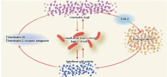
Figure 1: The balanced IL-1β – eicosanoid – type-I IFN network. Mtb induces IL-1α/β and type-I IFN production. IL-1β
activates cyclooxygenase-2 (COX-2) resulting in PGE2 production. PGE2 inhibits type-I IFN activity, favoring control of Mtb. Type-I IFN induces IL-10 and IL-1R antagonist, blocking IL-1β activity and favoring Mtb growth. Originally from Jon S. Friedland, Clinical Implications of Basic Research, The New England Journal of Medicine, 2014.
The relevance of the immune network formed by IL-1β, eicosanoids and type-I IFNs has just begun to be explored, but it already presents itself as a good target for anti-TB therapy, with the possibility to be used in a patient-specific manner54,55.
2.2.2.5. Chemokines
In addition to cytokines, chemokines play a major role in building the inflammatory environment encountered in TB. These chemokines are predominantly composed by two distinct classes, defined by the presence of a C-C or C-X-C motif and a single receptor may be able to bind multiple chemokines60. This family of proteins is responsible for orchestrating the recruitment of leukocytes to the site of infection and subsequent migration to different organs60. In the mouse model, there are at least 34 chemokines reported to be differential expressed throughout Mtb infection60, attesting the complexity of the cell-recruitment dynamics encountered during infection. Resorting to gene deficient mice, a vast effort has been placed towards characterizing the role of individual chemokines and their receptors in the context of Mtb infection. Few of these studies have reported differences regarding bacterial burden, and when these differences were reported they are predominantly in mice deficient for a chemokine receptor60. Indeed, only few studies reported a role for individual chemokines, suggesting a redundant role for these molecules60–63. Specifically, these studies have reported that mice deficient in C-C chemokine ligand 2 (CCL2), a CCR2 ligand, have a
9
transient increase in lung bacterial burden after low-dose aerosol infection, accompanied by a decreased macrophage influx and accumulation of protective IFN-γ CD4+ T cells64. These findings are in line with the increased risk of developing TB presented by humans with a Ccl2 polymorphism65. A similar phenotype was observed in mice deficient in CCL5 (CCR1/CCR5 ligand), although in this case the transiently increased lung bacterial burden was associated with a delayed recruitment of CD11c+ myeloid cells and T cells66. Other studies have shown that mice deficient for CCR7 or its known ligands (CCL19/CCL21) have an impaired DC migration to the lymph node, and are consequently impaired in T cell induction, translating into a diminished control of Mtb67–70. Interestingly, the action of chemokines is not restricted to cell-migration between organs, as they also play a role in the location of immune cells within the inflammatory lesion. Specifically, mice deficient in CXCL13 (CXCR5 ligand) have been shown to be more susceptible to Mtb, not due to an overall change in the number of a specific cell type, but instead due to the location of T cells within the granuloma69. In this case, in the absence of CXCL13, T cells were unable to locate in close proximity to infected macrophages, thus leading to insufficient macrophage activation and subsequent Mtb growth69.
Taken together, these studies point for a complex role of chemokines during infection, controlling temporal and spatial features of the formation and action of both innate and adaptive immunity.
2.3. Adaptive immune response
The activation of the innate immune response is crucial for the establishment of an adaptive immune response. It is known that 8-10 days after infection, antigen-presenting cells (APCs) carrying Mtb or Mtb molecules, mainly DCs, arrive at the lymph nodes14. Once in the lymph node, these cells will present Mtb molecules to naïve T cells, driving their differentiation and proliferation, and up to 5 days later these Mtb-specific T cells reach the site of infection5,14. It has been extensively described in humans and mice that adaptive immune responses are absolutely required to control Mtb. Specifically, HIV patients with low CD4+ T cell counts quickly succumb to Mtb infection if not rapidly treated4,71,72. A similar outcome is observed in recombination activating gene (RAG) deficient mice, which are unable to produce B and T lymphocytes and quickly succumb to Mtb shortly after infection73.
There are two main CD4+ adaptive immune responses involved in TB, the T helper 1 (Th1) and Th17 responses. These responses are generated when APCs present Mtb peptides in the context of a major
10
histocompatibility complex class 2 (MHC-II) to naïve CD4+ T cells in the presence of a specific cytokine milieu. For a CD4+ naïve T cell to acquire a Th1 phenotype it requires the expression of the transcription factor T-bet and IL-12 and IFN-γ as polarizing cytokines26,29,74,75. One the other hand, for optimal Th17 polarization expression of the transcription factor retinoic acid receptor-related orphan receptor-γt (RORγt) is required, as well as the presence of IL-6, transforming growth factor-β (TGF-β), IL-1β and IL-2314,74,75. The currently accepted paradigm is that a Th1 cell response must be induced for the host to control an otherwise lethal Mtb infection10,11,76. The Th1 cell subset is mainly characterized by the production of IFN-γ, which activates macrophages leading to the production of reactive nitrogen and oxygen species (RNS and ROS) 10,11,14,77–80. In the mouse model, the onset of a Th1 cell adaptive immune response leads to the stabilization of bacterial burden10,11,14. Indeed, mice deficient for IFN-γ, NOS2 or MHC-II are unable to control bacterial growth and rapidly succumb to infection77,81,82. Although the Th1 cell subset is clearly essential for the host’s protection, it is not able to sterilize the infected, even after boosting this response with the currently available vaccine.
To a lesser extent, the Th17 cell subset also plays a role in the immune response to Mtb. This subset secretes mainly IL-17 which is known to modulate granuloma formation and is required for BCG vaccine-dependent accelerated recruitment of IFN-γ producing T cells to the site of infection83,84. However, it has been shown that repeated BCG exposure of Mtb-infected mice leads to increased Th17 cell responses accompanied by neutrophil/granulocyte recruitment and consequently increased immunopathology due to enhanced neutrophilia85. Thus, although the induction of a Th17 response is largely benign in the context of TB, a tight regulation is required to avoid immunopathological consequences to the host83.
During the immune response in TB, both Th1 and Th17 cell subsets are active and it is likely that a fine balance between them is required for a better outcome of TB. In this regard, it is known that Th1 cells are able to act as a negative regulator of the Th17 response by limiting neutrophil recruitment and downregulating IL-17 expression73, thus limiting excessive immunopathology. On the other hand, IL-17 is able to anticipate the recruitment of Th1 cells, thus improving resistance to infection86. The mechanisms through which the host and Mtb regulate these different subsets of Th cells are not fully understood. It is possible that the key to a more efficient vaccine lies in finding the right balance between Th1 and Th17 cells.
11
The immune response in TB is a vast field of study, and although numerous works have helped to shed light on the matter, we are still unaware of the full spectrum of the mechanisms that govern this response and most importantly if there is an immune response capable of completely eliminating the pathogen. A simplified version of current knowledge regarding the cellular immune response to Mtb after low-dose aerosol infection is depicted in Figure 2.
Figure 2: The cellular immune response to Mtb. After aerosol infection, Mtb can be phagocytosed by DCs, alveolar
macrophages and neutrophils. Alveolar macrophages secrete inflammatory mediators including IL-1, IL-12p40, IL-6 and chemokines and together with neutrophils may die an apoptotic or necrotic cell death. DCs migrate to the draining lymph-node 8-12 days after infection, under the influence of CCL2, CCL19, CCL21, IL-12p80 (IL-12(p40)2) and IL-12P70. Th1 cells are generated upon antigen presentation in the presence of IL-12p70. Th1 cells migrate to the lung 14-17 days after infection under the influence of CCL2, CCL19 and CCL21. Upon arrival to the lung, Th1 cells required IL-12p70 for maintenance and activate macrophages by secreting IFN-γ. These macrophages increase in microbicidal activity by upregulating NOS2 (iNOS), TNF-α and IL-12p40 production. Adapted from Anne O’Garra, et al., The Immune Response in Tuberculosis, Annual Reviews of Immunology, 2013.
12
3. Host-pathogen interactions
The events that take place in the immune response during TB are the result of a complex interaction between the host and Mtb. A great portion of the work performed on deciphering this immune response has focused on characterizing host factors, often disregarding how pathogen factors modulate this response. Indeed, our knowledge on how Mtb is able to subvert and thrive under the seemingly unfavorable conditions found within the host is still unclear. To fully understand the intricate relationship between Mtb and humans, the only host known for Mtb, we must consider that both have been coevolving since the appearance of anatomically modern humans about 70 000 years ago87. This time frame has given both host and pathogen ample opportunity to fine tune their defense and virulence mechanisms, respectively, to one another87. Indeed, the fact that TB presents a wide range of outcomes, ranging from clearance after infection, to establishment of subclinical latent disease and ultimately to active disease attests to the complexity of TB1,4,14. Remarkably, for extended periods of time in active TB, the balance between a protective and detrimental immune response allows the host to remain ambulatory, thus increasing the opportunity for Mtb to be transmitted5. These different outcomes may be largely defined by the result of the initial interaction between Mtb and innate immune cells1. Accordingly, it has been shown that different strains of Mtb induce different inflammatory profiles after infection of these cell types88–91.
3.1. The cell wall of Mtb and its impact in host-pathogen interactions
The initial interactions between Mtb and the host are predominantly dependent of recognition of antigens present of the cell wall of Mtb by the PRRs on innate immune cells. This cell wall is an extremely complex structure, composed by 4 distinct layers: the innermost polysaccharide layer composed by peptidoglycans and arabinogalactan; the inner mycomembrane composed of long chain mycolic acids covalently attached to the innermost proteins; the outer mycomembrane composed many non-covalently lipids, including phthiocerol dimycocerosates (PDIMs); trehalose dimycolate (TDM); sulfolipids (SLs); phosphatidylinositolmannosides (PIMs) modified into lipomannans (LM) and lipoarabinomannans (LAM); glucose monomycolate (GMM); lipooligosaccharides (LOS); and the outermost capsular layer containing poorly characterized glycans, lipids and proteins92,93. A simplified schematic representation of the cell envelope of Mtb is depicted below in Figure 3.
13
Figure 3: Schematic representation of the cell envelope of Mtb. The cell wall is mainly composed by a large complex
that contains three different covalently linked structures (peptidoglycan (grey), arabinogalactan (blue) and mycolic acids (green)). The outer mycomembrane contains various free lipids non-covalently linked to mycolic acids. These free lipids include TDM, PDIMs, SLs, LOS, GMMs, PIMs and its glycoconjugates. The capsule contains poorly characterized glycans, lipids and proteins. Adapted from Abdallah M. Abdallah, et al., Type-VII secretion – mycobacteria show the way, Nature Reviews Microbiology, 200794.
These lipid components of the outer mycomembrane and capsular layer are thought to be in direct contact with the host’s cells and therefore their study is important to understand how their recognition modulates the immune response16.
As a result of the availability of the Mtb genome, and upon the development of efficient techniques to genetically manipulate this pathogen95,96, the employment of gene-deficient Mtb has allowed us to assess the role of specific Mtb molecules during infection. Particularly, this has allowed us to gain insight on the role of the relevance the Mtb cell wall lipids in infection.
Mycolic acid layer (Inner Mycomembrane) Capsule Peptidoglycan and Arabinogalactan Inner membrane Free lipids (Outer Mycomembrane)
14
3.1.1. TDM
Among Mtb cell wall lipids, TDM is one of the most studied. This lipid is recognized by Mincle (a C-type lectin) and has been consistently associated as a virulence factor o Mtb15. Specifically, WT mice infected with a Mtb strain deficient for an enzyme responsible for adding distal-oxygen groups to mycolates presented increased survival when compared to mice infected with the WT strain97. In subsequent studies, this phenotype was attributed to an increased induction of IL-12p40 and TNF-α by the mutant strain98–100. Indeed, mice deficient for IL-12p40 presented the same survival time, regardless of the infecting Mtb strain98.
3.1.2. PDIMs
PDIMs were first reported to be a virulence factor upon the observation that Mtb clinical isolates deficient for PDIMs were unable to induce a severe disease in the ginea pigs101. More recently, it has been reported that PDIM-deficient Mtb induces a hyper inflammatory profile in macrophages, putatively due to higher exposure of PAMPs8. Additional, studies have shown that PDIM-deficient Mtb has an impaired growth inside resting macrophages, although these studies reported opposing results regarding the role of PDIMs on phagosomal acidification102,103. Recently, work performed with Mycobacterium marinum in the zebrafish larvae model has elegantly shown that PDIMs exert a role in modulating the inflammatory environment towards a bacterium-permissive one8. Specifically, it was shown that by avoiding TLR detection, and without myeloid differentiation primary-response protein 88 (MyD88) activation, the bacterium is able to induce the recruitment of permissive macrophages to the site of infection8. These macrophages show a reduced NOS2 activation and are therefore a better environment for bacterial replication8. Unfortunately, this mechanism cannot fully explain the role of PDIMs in Mtb, as an independent study using the mouse model of infection was unable to rescue the phenotype observed with PDIM-deficient Mtb, even when infecting IFN-γ, NOS2, MyD88 and CCR2 deficient mice104. This particular study has shown that the lower bacterial burden, observed in PDIM-deficient Mtb infected animals, is due to an increased bacterial death, and not a deficit in replication, early during infection104. The question of whether PDIMs grant virulence by subverting any specific immune mechanism, or whether it does so by granting sheer cell wall integrity remains to be elucidated.
15
3.1.3. SLs
The role of SLs during infection has not yet been clearly defined, but they are suggested to relevant during infection since the cell wall composition of Mtb extracted from infected humans or animals is enriched in SLs when compared to the composition found in Mtb under culture conditions105. SLs are recognized by NOD2 and class A SRs15,106, and as an anionic molecule it has been shown that SL-deficient Mtb has decreased susceptibility to LL-37, a human cationic antimicrobial peptide (AMP), by reducing Mtb-AMP interactions107. Despite this, no discernible phenotype has been observed between mice and murine macrophages infected with either WT or SL-deficient Mtb. Despite the fact that no differences regarding bacterial burden were observed, mice and guinea pigs infected with the SL-deficient Mtb presented a longer survival time than those infected with WT strain108,109. Interestingly, it has been reported that SLs are preferentially uptaken by neutrophils leading to high ROS production110, a microbicidal mechanism that confers limited protection against Mtb. Whether SLs induce Mtb phagocytosis by neutrophils, a cell-type considered to be a permissive niche for mycobacterial growth14, has not yet been assessed. One possible factor that may have hindered the attempt to define a clear role to SLs in TB is that the Mtb mutants employed so far were deficient exclusively for the SL-1 family. However, it has recently been proposed that the SL-2 family actually accounts for the highest quantity of SLs in Mtb111, thus it is possible that by removing this predominant SL family more pronounced phenotypes may be discovered.
3.1.4. Other cell wall lipids
Although in vitro studies have provided compelling evidence that PIMs and its glycoconjugates are able to modulate recognition and phagosome maturation in macrophages112–114, only limited in vivo work performed with Mtb mutants engineered for these structures has been published. Specifically, only one study using the zebrafish larvae model reported that a Mycobacterium marinum strain deficient for an enzyme required for LAM/LM branching, resulted in impaired growth of the bacterium when compared to the WT strain115. Hence, the role for these molecules during infection remains to be elucidated. Similarly, this Mtb-mutant strategy is yet to be employed for the study of GMMs and LOS.
It is now clear that Mtb has developed a cell wall capable of modulating the immune response of the host in its favor. The fact that these lipidic cell wall molecules are subject to modifications in saturation, chain length, cyclization among others with unpredictable effects92,93, further affirms the complexity of this
16
pathogen. The study of these host-pathogen interactions has just begun to scratch the surface of the intricacy beyond this timeless disease, but the employment of genetically engineered Mtb may prove a powerful tool to further advance this field of study.
4. The role of Rv0101 in Mtb virulence
The study of Mtb cell wall components has yielded valuable information on how host and pathogen interact, allowing for the choice of better targets for the future development of antimycobacterial drugs. However, there are still many unexplored Mtb molecules whose role during infection remains uncharacterized. Our knowledge on how the pathogen is able to thrive in the hostile conditions encountered inside the host could greatly benefit from the study of these molecules in the context of infection. Therefore, to advance this knowledge, we proposed to characterize a non-ribosomal peptide synthetase (nrp) encoded by Mtb, whose role during infection has not yet been defined.
Nrp is coded by Rv0101, and is part of a larger operon comprising other five genes: Rv0096, a member of the Proline-Proline-Glutamic acid (PPE) family; Rv0097, an oxidoreductase; Rv0098, a type III thioesterase; Rv0099, a fatty acid adenosine monophosphate (AMPh) ligase and Rv0100, which encodes an acyl carrier protein116. Nrp is predicted to interact with Rv0099 and Rv0101 to form a multimodal enzyme 117 and is an ubiquitous protein, present in cytosol, cell membrane and cell wall fractions of H37Rv 118. Sequence analysis of the nrp gene predicts that this enzyme is involved in lipid metabolism 119. The function of nrp in lipid metabolism together with its localization in the cell wall, are good indications that this enzyme takes part in cell wall synthesis.
Although so far no study has directly addressed the role of this enzyme, it has been mentioned multiple times in the literature as a potential Mtb virulence factor. Specifically, it was reported as a non-essential gene for in vitro growth by transposon mutagenesis, but was on the other hand required for growth in the spleen of C57BL/6J mice 119,120. Interestingly, nrp was reported to be the most abundant Mtb protein in the lungs of infected guinea pigs by day 30 post infection, while being undetected at 90 days post infection 121, suggesting that Mtb regulates the expression of this protein throughout the course of infection. Overall these reports point for a strong role of nrp during infection.
Taking these data in consideration, in this present work we propose to characterize the relevance of nrp during infection, and how it impacts the interactions between Mtb and the host.
17
19
Mtb is arguably the most successful pathogen known to mankind, with its existence spanning as long as that of mankind. To once and for all eradicate TB, a greater understanding of the fine mechanisms by which the pathogen is able to survive and thrive inside its human host is required. Many potentially pivotal Mtb virulence factors remain either unknown or uncharacterized, and their study could benefit greatly the development of new vaccines and anti-mycobacterial therapies.
The main goals of this thesis are to define the impact of nrp on: The growth of Mtb under optimal culture conditions.
The interactions of Mtb with macrophages in an in vitro setting. The course of Mtb infection in the mouse model.
21
23
1. Bacteria and Infection
Mtb ∆nrp-mutant and nrp-complemented strains, were generated on a H37Rv background by Dr Apoorva Bhatt (School of Biosciences – University of Birmingham). Bacteria were grown in Middlebrook 7H9 liquid media (BD Biosciences) for 7–10 days and then sub-cultured in Proskauer Beck (PB) medium, supplemented with 0.05% Tween 80 and 2% glycerol, to the mid-log phase. Bacterial stocks were aliquoted and frozen at –80°C. To determine the concentration of Mtb aliquots, 6 frozen aliquots were serial diluted and plated in Middlebrook 7H11 (BD Biosciences) agar plates supplemented with 10% oleic acid/albumin/dextrose/catalase (OADC) and 0.5% glycerol. Viable bacteria were determined by counting colony-forming units (CFUs) after 19-21 days of incubation at 37°C.
Mice were infected via aerosol route using an inhalation exposure system (Glas-Col). Briefly, bacterial clumps were disrupted by forcing them through a 26G needle 6 times and diluted in water (Aqua B. Braun) to a concentration of 2x10⁶ CFU/mL. Mice were exposed to the infectious aerosol for 40 minutes, resulting in the delivery of 100-200 viable bacteria to the lungs. Assessment of initial bacterial burden was performed 3 days post infection by growing viable bacteria from the whole lung homogenate of 5 mice.
2. Animals
C57BL/6 mice were purchased from Charles River Laboratory (Barcelona, Spain). RAG-2 and IFN-γ (generated on a C57BL/6 background) deficient mice were generously provided by Dr. Margarida Correia-Neves (ICVS) and Dr. Susana Roque (ICVS), respectively. IL-10 (generated on a C57BL/6 background) deficient mice were a kind gift from Dr. Anne O’Garra (MRC-NIMR, London). ALOX-5, ALOX-15, TLR-2 and TLR-4 (generated on a C57BL/6 background) deficient mice were also purchased from Charles River Laboratory. All mice were kept and bred under the same conditions in the Life and Health Sciences Research Institute (ICVS) animal housing facilities, at the school of Health Sciences, University of Minho. All mouse protocols were performed according to the European Union Directive 2010/63/EU, and previously approved by Direcção Geral de Alimentação e Veterinária.
3. Differentiation and Infection of Bone Marrow-Derived Macrophages (BMDM)
and Peritoneal Macrophages
Bone marrow cells were flushed from the tibiae and femurs of mice with complete Modified Eagle Medium (cDMEM, DMEM supplemented with 10% of heat-inactivated fetal bovine serum (FBS), 1% of HEPES 1M, 1% L-glutamine and 1% sodium pyruvate 100mM (all from GIBCO)). Bone marrow cells were counted and
24
cultured in 8mL cDMEM supplemented with 20% L929-cell-conditioned medium (LCCM) at a concentration of 0.5 x 10⁶ cells/mL in 8-cm diameter plastic petri dishes (Sterilin) for 7 days under 37°C and 5% CO2 conditions. On day 4 of differentiation, 10mL of cDMEM supplemented with 20% LCCM were added to the cultures. At day 7, adherent cells were mechanically detached and seeded in 24-well plates (Nunc) at a concentration of 1x10⁶/well and incubated at 37°C. Cells were infected with Mtb at a multiplicity of infection (MOI) of 1:1 (bacteria/macrophage ratio) and incubated at 37°C with 5% CO2. Four hours after infection, cells were washed 4 times with phosphate-buffered saline (PBS) (GIBCO) to remove extracellular bacteria. Washed cells were resuspended in 1mL of cDMEM and either incubated at 37°C for 48/96 hours in the presence or absence of 100 U/mL of IFN-γ or used to determine bacterial internalization. For that, 0.1% saponin (Sigma-Aldrich) in PBS was added to the wells and the cells were incubated at RT for 10 minutes to release intracellular bacteria. The number of viable bacteria was determined by plating 10-fold serial dilutions of the saponin treated cell suspensions in supplemented Middlebrook 7H11, as previously mentioned.
Peritoneal macrophages were obtained by intraperitoneally injecting 1 mL of thioglycollate in wild-type (WT) C57BL/6 mice. After 4 days, mice were euthanized and the peritoneum washed with PBS. The number of cells of the obtained peritoneal macrophage-enriched cell suspension was counted and processed for infection as described above for BMDM.
Supernatants from these cultures were used to determine nitric oxide concentration by the Griess method and cytokine concentration by ELISA.
4. Sample Collection and Preparation of Single Cell Suspensions
At selected time-points post-infection, mice were euthanized by CO2 asphyxiation and the organs aseptically excised. Lungs were perfused with 10 mL PBS through the right ventricle of the heart to flush blood cells, sliced with 2 sterile scalpels and incubated at 37ºC with collagenase IX (0.7mg/mL, Sigma-Aldrich) for 30 minutes. Single cell suspensions from the spleen and digested lung were obtained by homogenized by sieving them through a 40-µm-pore-size nylon cell strainer (BD Biosciences) with a syringe plunger. Lung and spleen cells were treated with erythrocyte lysis buffer (0.87% of NH4Cl solution and 5% of PBS in water) to lyse red blood cells and resuspended in cDMEM to use for bacterial burden determination, flow cytometry analysis and RNA extraction. Single cell suspensions were counted using a Countess® Automated Cell Counter (Life Technologies).
25
5. Survival
RAG-2 and IFN-γ deficient mice infected with Mtb weight was determined before infection and every 48 hours from day 30 post-infection onward. Mice were euthanized when they lost 20% weight or upon losing responsiveness to physical stimulation. Whenever possible, the lungs of moribund animals were harvested for histology and bacterial burden assessment.
6. Bacterial Burden Determination
To determine bacterial burdens, lung and spleen single cell suspensions were incubated with 0.1% saponin (Sigma-Aldrich) for 10 min to release intracellular bacteria. The number of CFUs was determined by plating 10-fold serial dilutions of saponin-treated cell suspensions in Middlebrook 7H11 agar plates supplemented as described above. BBL™ MGIT™ PANTA™ antibiotic mixture (BD Bioscience) was added to prevent contaminations of lung samples. Viable Mtb colonies were counted after 19-21 days of incubation at 37ºC.
7. Histology
The right upper lobe of the lung was inflated with 5 mL of PBS containing 3.7% formaldehyde and kept for 1 week in this solution. Afterwards, these lobes were embedded in paraffin, sectioned in 2-3μm thickness slices, and stained with hematoxylin- eosin (H&E).
8. Flow Cytometry
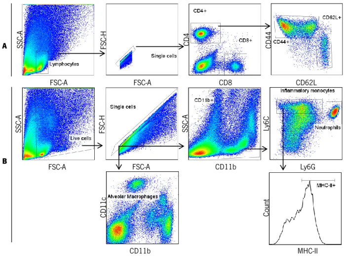
For surface staining 1-2x10⁶ cells were washed with FACS buffer (PBS containing 2% FBS and 0.01% azide) and stained for surface antigens for 30 minutes at 4°C. Cells were washed with FACS buffer and PBS and then fixed overnight (ON) in PBS containing 2% paraformaldehyde. The following antibodies were used: CD4-PB (clone RM4-5, eBioscience), CD8-FITC (clone 5H10-1, Biolegend), CD44-PerCPCy5.5 (clone 1M7, eBioscience), CD62L-PECy7 (clone MEL-14, Biolegend, Ly6G-APC (clone 1A8, Biolegend), MHC II-FITC (clone AMS-32.1,BD Pharmingen), Ly6C-PerCPCy5.5 (clone AL-21,BD Pharmingen), CD11b-PE (clone M1/70 from Biolegend), CD11c-PB (clone N418, Biolegend). Samples were acquired on a LSRII flow cytometer with Diva Software. All data were analyzed using FlowJo version 7.6.5 software. A schematic of the gating strategy performed is depicted below in Figure 4.
26
Figure 4: Cytometry analysis. Gating strategy for A) lymphoid cells and B) myeloid cells.
The total number of cells in each gate was calculated using the total number of cells determined by Countess® Automated Cell Counter.
9. Mtb growth curves
H37Rv WT, nrp-complemented and ∆nrp mutants were inoculated in 40mL of either 7H9 with 10% OADC or PB medium at an initial OD of 0.002 at 570nm. The cultures were grown in ventilated Erlenmeyers (Cole-Parmer) at 37°C and 120 rpm. Every 48 hours a sample of each culture was used for OD 570nm readings.
FSC-A SSC -A FSC-A FS C-H SSC -A CD11b Ly 6C Ly6G CD1 1c CD11b Co un t MHC-II FSC-A FSC-A CD8 CD62L SSC -A FS C-H CD4 CD4 4 A B
27
10. Cytokine concentration by Enzyme-Linked Immunosorbent Assay (ELISA)
The concentration of TNF-α, IL-1β and IL-10 in the supernatants was determined by using the commercially available ELISA kit for IL-10 (88-7104), IL-1β (88-7013) and TNF-α (88-7324), all from eBioscience, according to the manufacturer’s instructions.
11. RNA extraction and quantification
Total RNA from cell suspensions was extracted by using TRIzol® Reagent (Invitrogen, San Diego, CA) according to the manufacturer’s instructions. Briefly, glycogen (20μg/μl from Roche) was added to each sample and incubated for 5 minutes at RT. After incubation, 50μl of chloroform (Sigma-Aldrich) were added and the samples were mixed by vortexing and incubated on ice for 15 minutes. Afterwards, samples were centrifuged at 13000 rpm for 15 minutes at 4°C, and the upper aqueous phase was carefully recovered, and mixed with an equal volume of isopropyl alcohol (Sigma-Aldrich) to precipitate the RNA. Samples were incubated ON at -20°C and centrifuged at 13000 rpm for 15 minutes at 4°C. The supernatant was removed and the pellet washed with 800μl of 70% ethanol (Carlo Erba reagents). Ethanol was completely removed after centrifugation at 9000 rpm for 5 minutes and the dried RNA pellet was resuspended in RNase/DNase-free water (Gibco). RNA concentration was measured at 260nm (Nanodrop ND-1000 Spectrophotometer) and the purity assessed through the A260/A280 and A260/A230 ratios.
12. Reverse transcriptase polymerase chain reaction (RT-PCR)
Complementary DNA (cDNA) was synthesized using the GRS cDNA Synthesis Mastermix Kit (GRISP) according to the manufacturer’s instructions. Briefly, reaction mix was composed by 10μl of GRS RT Mastermix, 1μl of oligo(dT)20 and 9μl of RNA sample mixed with nuclease-free water. The cDNA synthesis reaction was performed in thermocycler (MyCycler, Bio-Rad) with the following program: 25°C for 10 minutes followed by 10 minutes at 42°C and 5 minutes for 85°C. The resultant cDNA template was used for quantification of target genes expression by real-time PCR (RT-PCR) analysis using SYBR green or TaqMan detection systems.
13. Quantitative Real-Time PCR (qRT-PCR)
For SYBR Green reactions 1µL of each cDNA sample was mixed with 9µL of reaction mix containing 3µL of water, 1µL of 0.4µM forward and reverse specific primer and 5µL of SYBR green qPCR Master Mix (GRISP). RT-PCR was performed in CF*96TM Real-time system (Bio-Rad) using the following program: 95°C for 15 minutes, followed by 40 amplification cycles of 95°C for 3 seconds, 60°C for 20 seconds and
28
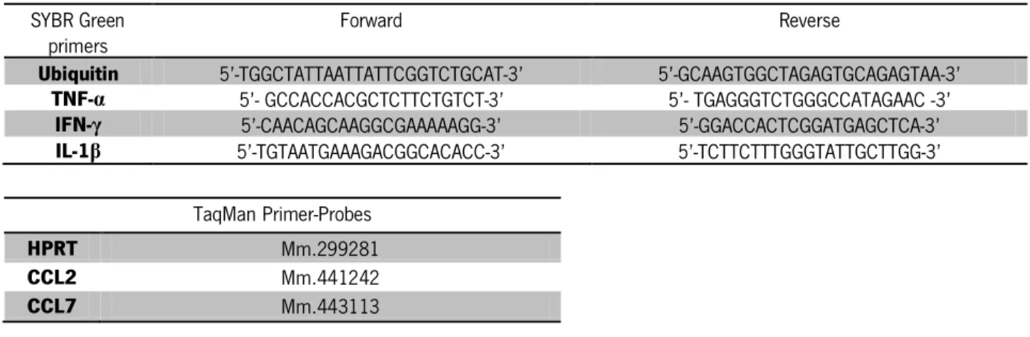
70ºC for 15 seconds, for melting curve analysis. For TaqMan reactions 1µL of each cDNA sample was mixed with 9µL of reaction mix containg 3.5µL of water, 0.5µL of specific primer-probes and 5µL of TaqMan Gene Expression Master Mix (Applied Biosystems). The RT-PCR program used was 50°C for 2 minutes, 95ºC for 10 minutes, 40 cycles of 95°C for 15 seconds and 60°C for 1 minute. Relative mRNA expression of the target gene was normalized to the levels of the housekeeping gene using the ∆Ct method: 1.8^(Housekeeping gene mRNA expression – Target gene mRNA expression) x 100000. The sequences of primers and references of the TaqMan primer-probe sets used in RT-PCR are listed in Table 1.
Table 1: Sequences of SYBR Green primers and TaqMan primer-probes used. SYBR Green
primers Forward Reverse
Ubiquitin 5’-TGGCTATTAATTATTCGGTCTGCAT-3’ 5’-GCAAGTGGCTAGAGTGCAGAGTAA-3’
TNF-α 5’- GCCACCACGCTCTTCTGTCT-3’ 5’- TGAGGGTCTGGGCCATAGAAC -3’
IFN-γ 5’-CAACAGCAAGGCGAAAAAGG-3’ 5’-GGACCACTCGGATGAGCTCA-3’
IL-1β 5’-TGTAATGAAAGACGGCACACC-3’ 5’-TCTTCTTTGGGTATTGCTTGG-3’ TaqMan Primer-Probes
HPRT Mm.299281
CCL2 Mm.441242
CCL7 Mm.443113
14. Statistical Analysis
Data were analyzed using GraphPad Prism 6. Depending on the nature of the dataset, differences between groups were analyzed using Student’s t-Test or Two-way ANOVA using Sidak’s test for multiple comparisons. Survival curves were compared using the Log-rank (Mantel-Cox) test. Differences were considered significant for p≤0.05 and represented as follows: *p≤0.05;**p≤0.01;***p≤0.001 and ****p≤0.0001.
29
31
1. Initial considerations
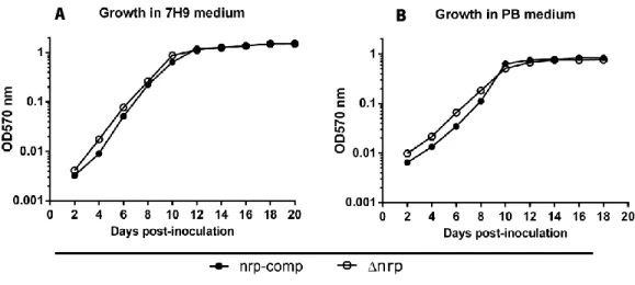
The ∆nrp mutant and the nrp-complemented strain were generated on a H37Rv background by our collaborator Dr. Apoorva Bhatt (School of Biosciences, University of Birmingham). Our collaborator is currently characterizing the biochemical and functional properties of nrp. From a microbiological standpoint, we have only assessed whether nrp was required for optimal Mtb growth under normal culture conditions. We found no significant differences in growth kinetics between Mtb strains in either PB or 7H9 liquid medium (Figure 5).
Figure 5: nrp-complemented and ∆nrp-mutant strains growth kinetics in liquid media. Biomass over time measured
by optic density at 570nm in A) 7H9 and B) PB inoculated with either nrp-complemented or ∆nrp-mutant strains. Culture media were inoculated with either Mtb strain at a theoretical OD570nm value of 0.002. Average doubling time in hours (nrp-complemented vs ∆nrp-mutant) was 25.15 vs 24.79 in 7H9 medium and 34.92 vs 34.08 in PB medium.
Thus, we focused on characterizing the relevance of this protein in the context of infection. To achieve this goal we took advantage of the mouse model infected via the aerosol route previously established in our laboratory. From these animals we assessed survival and bacterial burdens, gene expression as well as immunopathology in specific organs throughout the course of infection. We have also resorted to infecting macrophages from different origins to further elucidate innate interactions between host cells and pathogen. Overall this experimental setup has allowed us to better understand the role of nrp in the context of infection.
32
2.
In vivo
infection
2.1. Nrp is required for optimal establishment of infection
To assess the relevance of nrp during infection, we started by infecting via the aerosol route WT C57BL/6 mice with either ∆nrp-mutant or nrp-complemented strains. Bacterial burdens in the lung and spleen were analyzed at different time-points throughout infection (Figure 6A and 6B). Our data showed a difference regarding lung bacterial burden by day 20 post-infection between Mtb strains expressing or not Nrp (Figure 6A). Specifically, animals infected with the ∆nrp-mutant strain presented on average a 48-fold lower bacterial burden (1.683 Log10 average difference) when compared to those infected with the nrp-complemented strain.
We have also observed differences regarding bacterial burdens in the spleen of infected animals (Figure 6B). In this organ we were unable to detect Mtb before day 30 post-infection in animals infected with the ∆nrp-mutant. Moreover, the bacterial burden shown at day 30 post-infection in these animals remained stable (≈ 3.25 Log10) throughout the time period assessed, significantly lower than the one observed in animals infected with the nrp-complemented strain.
Figure 6: Animals infected with the ∆nrp-mutant or the nrp-complemented strain presented different bacterial burdens over time in the lungs and spleen. A) Lung bacterial burden differs between Mtb strains at day 20 between ∆nrp
and complemented strain (average 3.867 vs 5.550 Log10 bacterial burden, respectively). B) Spleen bacterial burden in
∆nrp-mutant infected mice was lower for every time-point assessed when compared to mice infected with the nrp-complemented strain. Data analyzed performing a Two-way ANOVA using Sidak’s test for multiple comparisons. BDL – Below detection level.**** p<0.0001 *** p<0.001; **p<0.01. Each time-point contains pooled data from 2 separate experiments with 5 animals each. Initial bacterial burden for animals infected with the ∆nrp-mutant and the nrp-complemented strain were 1.860±0.0446 Log10 and 1.931±0.0822 Log10, respectively.