TAINÁ GARCIA DA FONSECA
ENVIRONMENTAL RISK ASSESSMENT AND TOXICITY OF
PHARMACEUTICALS IN COASTAL TROPICAL AND
TEMPERATE ORGANISMS
Faculdade de Ciências e Tecnologia
Centro de Investigação Marinha e Ambiental
TAINÁ GARCIA DA FONSECA
ENVIRONMENTAL RISK ASSESSMENT AND TOXICITY OF
PHARMACEUTICALS IN COASTAL TROPICAL AND
TEMPERATE ORGANISMS
Doutoramento em Ciências do Mar, Terra e Ambiente Especialidade em Ecotoxicologia
Trabalho efetuado sob a orientação da Professora Doutora Maria João da Anunciação Franco Bebianno
Faculdade de Ciências e Tecnologia
Centro de Investigação Marinha e Ambiental
Environmental Risk Assessment and toxicity of pharmaceuticals in coastal tropical and temperate organisms
Declaração de autoria de trabalho
Declaro ser a autora deste trabalho, que é original e inédito. Autores e trabalhos consultados estão devidamente citados no texto e constam da listagem de referências incluída.
___________________________
©Tainá Garcia da Fonseca
A Universidade do Algarve tem o direito perpétuo e sem limites geográficos, de arquivar e publicitar este trabalho através de exemplares impressos reproduzidos em papel ou de forma digital, ou por qualquer outro meio conhecido ou que venha a ser inventado, de o divulgar através de repositórios científicos e de admitir a sua cópia e distribuição com objetivos educacionais ou de investigação, não comerciais, desde que seja dado crédito ao autor e editor.
The present thesis was financed and supported by the Conselho
Nacional de Desenvolvimento Científico e Tecnológico (CNPq - Brazil),
through the Ciência Sem Fronteiras Program (202360/2014-8).
I
A
CKNOWLEDGEMENTS
Firstly, my sincere gratitude to Prof. Maria João Bebianno for providing me the opportunity to develop the present research, overseas, filled of support and advices that helped me to move forward and grow in Science, guiding me towards the final objective.
To the Conselho Nacional de Desenvolvimento Científico e Tecnológico (CNPq - Brazil) for the financial support throughout my PhD course, through the Ciência sem Fronteiras Program (202360/2014-8).
A special gratitude to my co-supervisor Prof. Denis Abessa, who believed that I could do it, and that have inspired and encouraged me to pursue on making the difference.
To all my co-workers and friends involved at any step of this work and its progress, with determinant help, advices, patience and support “from the polychaete in the mud to the final writing”: Thiago Rocha, Nélia Mestre, Cátia Cardoso, Manon Auguste, Karyna Pereira, Francisca Ribeiro, Elna Fernandes, Tânia Carriço and Sarit O´Donovon.
To Luciane Maranho (Lu), for the valuable recommendations and knowledge shared about the studied polychaete.
To my brazilian friends and co-workers from NEPEA-UNESP for receiving me back with love and assistance: Guacira, Gabi, Carol, Itaquera, Roberta, Ana Lívia, Luiza, Ney, Lucas and Gian.
To my friends, that from close or far, helped to make it simpler: Manon, Tixa, Malária, Gabi, Roberta, Bis, Bru, Mel, Cátia, Sofia, Mônica and Renata.
To my best friend and companion, Luciano, for sharing with me the ups and downs throughout this journey, filled with daily encouragement, understanding and patience.
To my blessed and beloved family, for the endless care, motivation and support in all the steps to reach this point, and to whom I dedicate these 4 years of work.
II
G
ENERAL
A
BSTRACT
Demographic attributes of the urban society have prompted a milestone shift towards structural aging of global population and increase of life expectancy, crucial to the prominence of cancer as one of the non-communicable diseases leading mortality. Currently, cancer diseases were accountable for about 9.6 million deaths in 2018, with concerning increasing projections of cancer incidence and mortality, by 2030, at a greater proportion in developing countries. As a result, there is an increasing trend of production and consumption of pharmaceuticals applied in cancer treatments. Once administered and metabolized, such drugs are excreted into waterways following to waste water treatment plants (WWTPs), ending up in freshwater and coastal ecosystems, where they can be found at a sub ng to ng L-1 range. Anticancer drugs are designed to damage DNA and disrupt mechanisms of its transcription, replication and synthesis, as well as suppress the cell´s defense, ultimately causing cell death. In cancer therapy, drugs are typically administered in a combinatory cocktail, that covers different molecular targets, reducing the risk of clonal selection based upon cell resistance to a single drug. However, these chemicals may bring potential toxicity if discharged into the aquatic environment, both in temperate and tropical zones, particularly in the benthic compartment, where they are expected to accumulate.
In this sense, the present thesis aimed to assess the effects of anticancer agents of different classes, namely the cytotoxic platinum-based cisplatin (CisPt), the alkylating agent cyclophosphamide (CP) and the endocrine disruptor tamoxifen (TAM), on non-target benthic marine species under realistic environmental concentrations, at individual and combined exposures. For this purpose, a multi-biomarker approach was applied including the assessment of behavioural responses, oxidative stress, biotransformation, neurotoxicity, lipid peroxidation and genotoxicity in the temperate polychaete Nereis diversicolor, in addition to the evaluation of acute and chronic effects of drugs in tropical and representative benthic organisms: the sea urchin Echinometra lucunter, the polychaete Scolelepis squamata and the amphipod Tiburonella viscana from the Brazilian coast.
The main results revealed that single drug exposures caused alteration of AChE activity, oxidative stress and oxidative damage and, ultimately, DNA damage in N.
III
diversicolor. Besides, alterations in its burrowing behaviour also occurred, as an ecological
outcome. These effects were more pronounced in organisms under CisPt at 100 ng Pt L-1, CP at 1000 ng L-1 and TAM at 0.5 ng L-1. Findings from bioassays with N. diversicolor conducted with drugs in tertiary mixtures indicated that each biomarker effect respond differently, according to the trends proposed by models of drugs´s interaction. Toxicity did not increase in a dose-response manner but showed different patterns of effects, in a way that the highest DNA damage was observed at the set of lowest concentrations (Mixture A), an absence of oxidative stress at the intermediary drug levels (Mix B and C), with a potential dominance of TAM´s MoA in organisms exposed to Mixture C and D, suggesting an antagonist interaction between the cytotoxic drugs. Therefore, the effects ruled by single-drug MoAs cannot provide estimations and protective measures for a prospective ERA regarding scenarios of their combination in the environment. Although evidence on estrogen receptors expression is still not disclosed for the herein selected biological models, determination of biochemical and genotoxic outcomes from TAM exposure are highlighted, since it is assumed to comprise a targeted therapy on nuclear estrogen receptors.
Anticancer drugs´ concentrations that triggered the above-mentioned responses are potentially present in coastal environments, even more prone to be encountered in developing regions, where technologies of WWTPs are not efficient or are lacking. Accordingly, effects regarding the bioassays conducted with the tropical counterparts showed a non-monotonic reduction of viable pluteus larvae of E. lucunter, at CisPt and CP treatment respectively at the range from 0.1 to 10 ng Pt L-1, and from 50 to 500 ng CP L-1, in contrast to the increasing reduction of embryo larval development under TAM exposure, significant over the whole range of concentrations. In polychaetes S. squamata, a significant acute toxicity was exerted at the higher pharmaceutical doses (Mix C and D). In the amphipod T. viscana demonstrated a higher sensitivity to pharmaceuticals, presenting a significant non-linear reduction of survival in Mix B and D, as a potential outstanding effect of TAM when present as mixtures. By virtue of the non-monotonicity addressed in the dose-responses relationships reported in all bioassays, another concern stands out on the urgency to the development of a risk analysis and safety assessment designated to anticancer drugs.
Keywords: Anticancer drugs, cisplatin, cyclophosphamide, tamoxifen, sediment,
IV
R
ESUMO
G
ERAL
Atualmente, a dinamica demográfica global demonstra uma excepcionl tendência de transição regida pelo envelhecimento populacional e pelo aumento da esperança média de vida. Estes fatores, combinados a atributos sociais relacionados aos hábitos modernos e exposição a fatores de risco (e.g. dieta inapropriada, tabaco, sedentarismo, consumo de álcool, agentes infecciosos), aliados à hereditariedade, contribuem para um significativo aumento da incidência dos casos de cancro, classificado como a doença mais preocupante do século, após os distúrbios cardiovasculares. Atualmente, o cancro representa 21,7% dos casos de morte a nível mundial, com projeções preocupantes para 2030, e maiores proporções de incidência em países em desenvolvimento. Dessa forma, estima-se uma consequente expansão na necessidade de produção e consumo de medicamentos anticancerígenos em todo o mundo, com aumento anual entre 6 – 8% estimado para o ano de 2018, comparado com os 6,5% nos últimos 5 anos.
Após o consumo, via administração oral ou intravenosa, os fármacos anticancerígenos são excretados na sua forma parental e metabolizada através de efluentes hospitalares e industriais, bem como por descargas de esgotos provenientes de estações de tratamento de águas residuais (ETARs), as quais não possuem tecnologia adequada para a remoção de moléculas orgânicas complexas. Dessa forma, as águas superficiais e marinhas são os destinos finais desses medicamentos. Esta classe terapêutica atua sobre biomoléculas e componentes celulares críticos à proliferação celular, tais como DNA, RNA, proteínas, membrana fosfolipídica, microfilamentos do citoesqueleto e sinalização celular, a fim de causar danos irreversíveis e morte celular. Contudo, estes alvos não são específicos de células tumourais, pois, as drogas interferem em células proliferativas normais, como as dos organismos aquáticos.
Apesar da crescente quantidade de informação envolvendo fármacos no âmbito da ecotoxicologia aquática, verifica-se uma escassez de dados sobre o impacto de drogas anticancerígenas no meio marinho. Em países em desenvolvimento, onde são escassas as políticas de saneamento ambiental e a implementação de infraestruturas avançadas em ETARs, o cenário de risco ambiental torna-se ainda mais elevado frente à contaminação por
V estes fármacos. Além disso, a investigação acerca dos efeitos de fármacos em organismos nativos de ambiente (sub)tropical é escassa, e ausente relativamente aos anticancerígenos, dentro de um âmbito em que os critérios de qualidade ambiental em países em desenvolvimento são baseados em extrapolações de respostas geradas em organismos de zonas temperadas da Europa e América do Norte. O sedimento consiste num compartimento de destino final das substâncias introduzidas nos ecossistemas aquáticos, capaz de acumulá-las em níveis muito mais elevados do que aqueles observados na coluna de água adjacente. Além de atuarem como repositório de compostos químicos, os sedimentos atuam também como fonte de poluição após eventos de remobilização. Portanto, por meio do contato com a água intersticial e material particulado, as espécies bentónicas estão constantemente expostas a misturas de compostos químicos.
Dessa forma, a presente tese teve como objetivo avaliar a toxicidade de fármacos anticancerígenos, globalmente administrados em coquetéis no tratamento contra o cancro, em organismos marinhos bentónicos representativos de zonas temperadas e tropicais. Estes organismos foram expostos a drogas individualmente e em misturas, a concentrações ambientalmente relevantes. Os fármacos selecionados para tal foram os citotóxicos cisplatina (CisPt – 0,1 a 100 ng Pt L-1) e ciclofosfamida (CP – 10 a 1000 ng L-1), aplicadas no tratamento de tumoures sólidos (e.g. ovário, pulmão e bexiga), e o disruptor endócrino tamoxifeno (TAM - 0,5 a 100 ng L-1), mundialmente aplicado no tratamento do cancro da mama. Foram usados como modelos os poliquetas Nereis diversicolor de ambientes marinhos temperados e o ouriço-do-mar Echinometra lucunter, o anfípoda Tiburonella
viscana e o poliqueta Scolelepis squamata característicos de ambientes tropicais. Nas
experiências de exposição com N. diversicolor, que tiveram a duração de 14 dias, avaliaram-se as respostas de biomarcadores de estresavaliaram-se oxidativo (superóxido dismutaavaliaram-se -SOD; catalaavaliaram-se – CAT; enzima de fase II glutationa-S-transferase – GST; glutationa peroxidase - GPx), dano oxidativo (peroxidação lipídica - LPO), de efeito neurotóxico (atividade da enzima acetilcolinesterase - AChE), genotoxicidade por dano ao ADN, e alteração do comportamento de escavação. Relativamente aos organismos de ambientes bentônicos tropicais foram efetuados ensaios agudos (mortalidade) e crônicos (desenvolvimento embrio-larval).
VI Os resultados obtidos indicaram que as drogas anticancerígenas, expostas individualmente, causam alterações neutotóxicas significativas, estresse e dano oxidativo e genotoxicidade em N. diversicolor. Além disso, registram-se alterações no comportamento de escavação que podem comprometer a proteção da espécie aos predadores presentes na superfície do sedimento, com consequências a nível ecológico. Estas respostas foram mais significativas em organismos expostos a CisPt (100 ng Pt L-1), CP (1000 ng L-1) e TAM (0,5 ng L-1). Relativamente ao bioensaio conduzido em misturas ternárias, constatou-se que o
perfil de toxicidade não ocorreu de maneira monotónica. A mistura contendo os anticancerígenos em menor concentração (Mistura A) foi responsável pelo maior grau de dano em ADN, enquanto que nas concentrações intermedias (Misturas B e C) não se registou estresse oxidativo. Comparando-se com os resultados obtidos na exposição aos fármacos individualmente, as misturas C e D indicaram uma potencial predominância dos efeitos do TAM, com efeito antagônico sobre CisPt e CP. Assim, pode-se inferir que as respostas desencadeadas pelo modo de ação dos fármacos presentes individualmente não são consistentes para gerar estimativas de medidas de proteção de avaliação de risco ambiental relativamente a misturas destes compostos. Os efeitos causados pela concentração do anticancerígeno TAM (0,5 ng L-1), individualmente ou em misturas, indicaram um modo de ação por meio da intrusão da pró-droga na membrana celular. Logo, apesar da ausência de evidências referente à expressão de recetores de estrogénio nos modelos biológicos selecionados, a terapia administrada com TAM é também potencialmente tóxica em organismos não alvos da droga.
As concentrações das drogas, responsáveis por desencadear as respostas de toxicidade aguda, foram detetadas em ambientes temperados, e são encontradas em zonas costeiras tropicais. Significativa redução do desenvolvimento embrio-larval do ouriço E.
lucunter foi verificada nos bioensaios conduzidos com CisPt e CP, respectivamente em
concentrações entre 0.1 a 10 ng Pt L-1 e 50 a 500 ng CP L-1, de maneira não-monotónica.
Em contrapartida, o ensaio com TAM indicou uma resposta monotónica de redução significativa da viabilidade larval com o aumento da concentração da droga. O ensaio conduzido com misturas e poliquetas S. squamata demonstrou significativa toxicidade aguda com o aumento da concentração (Misturas C e D), apesar de uma sensibilidade inferior aos
VII anfípodas T. viscana, os quais demonstraram uma redução significativa e não-linear da sobrevivência nas misturas B e D, como potencial efeito da ação do TAM.
Devido aos perfis de toxicidade e dose-resposta não-monotónicos reportados em todos os bioensaios conduzidos com concentrações-traço ambientalmente relevantes, destaca-se a vulnerabilidade de espécies costeiras às misturas desses contaminantes emergentes, bem como à urgência de medidas preventivas à entrada desses compostos no meio marinho e ao desenvolvimento ferramentas de avaliação de risco ambiental especialmente dirigida a esses fármacos de expressiva toxicidade.
Palavras-chave: Anticancerígenos, cisplatina, ciclofosfamida, sedimento, toxicidade, misturas, biomarcadores bioquímicos, genotoxicidade.
i
I
NDEX
Chapter 1. General Introduction ... 1
1.1 World trends of pharmaceuticals production and usage ... 1
1.2 The global scenario of cancer diseases ... 4
1.3 Anticancer medicines and the rationale for chemotherapy ... 7
1.4 Drug metabolism and excretion ... 11
1.5 Anticancer compounds as a threat to the aquatic environment ... 12
1.5.1 Pharmaceuticals as Emerging Contaminants (ECs) ... 12
1.5.2 Environmental Background of anticancer drugs ... 13
1.5.3 Sources and routes of anticancer drugs to the marine environment ... 14
1.5.4 Potential behaviour and fate of anticancer drugs in marine waters ... 17
1.5.5 Occurrence of anticancer compounds in coastal environments ... 20
1.5.6 Ecotoxicological effects of anticancer drugs on marine organisms ... 21
1.6 Environmental Risk Assessment of anticancer drugs ... 40
1.6.1 Efforts in the marine environment ... 40
1.6.2 The ERA approach involving cytotoxic drugs ... 43
1.7 Tropical and temperate marine species ... 44
1.8 Selected biological models ... 46
1.8.1 Polychaete Nereis diversicolor ... 46
1.8.2 Polychaete Scolelepis squamata ... 47
1.8.3 Sea urchin Echinometra lucunter ... 48
1.8.4 Amphipod Tiburonella viscana ... 49
1.9 Selected Drugs ... 49
1.9.1 Cisplatin ... 50
1.9.2 Cyclophosphamide ... 51
1.9.3 Tamoxifen ... 52
1.10 Aim of the thesis ... 53
Chapter 2. Ecotoxicological assessment of the anticancer drug cisplatin in the polychaete Nereis diversicolor ... 55
2.1 Introduction ... 57
2.2 Materials and Methods ... 61
ii
2.2.2 Sediment characterization ... 62
2.2.3 Experimental design ... 62
2.2.4 Sarcoplasmic reticulum Ca2+ - ATPase ... 63
2.2.5 Behaviour assay ... 64
2.2.6 Biochemical analysis ... 64
2.3 Results ... 67
2.3.1 Effects of CisPt in Ca2+ - ATPase activity ... 68
2.3.2 Behavioural Assay ... 68
2.3.3 Biochemical analysis ... 69
2.3.4 Genotoxicity ... 72
2.3.5 Principal Component Analysis (PCA) ... 73
2.4 Discussion ... 74
2.5 Conclusions ... 81
Chapter 3. Environmental relevant levels of the cytotoxic drug cyclophosphamide produce harmful effects in the polychaete Nereis diversicolor ... 83
3.1 Introduction ... 85
3.2 Materials and Methods ... 93
3.2.1 Chemicals ... 93 3.2.2 Experimental setup ... 93 3.2.3 Burrowing Assay ... 94 3.2.4 Biochemical analysis ... 94 3.2.5 Statistical analysis ... 96 3.3 Results ... 97 3.3.1 Behavioural Assay ... 97 3.3.2 Biochemical analysis ... 98 3.3.3 Genotoxicity ... 101
3.3.4 Principal Component Analysis (PCA) ... 102
3.4 Discussion ... 103
3.5 Conclusions ... 109
Chapter 4. Impacts of tamoxifen exposure to Nereis diversicolor ... 110
4.1 Introduction ... 111
4.2 Materials and Methods ... 116
4.2.1 Chemicals ... 116
iii 4.2.3 Burrowing assay ... 117 4.2.4 Biochemical Analysis ... 117 4.2.5 Genotoxicity ... 119 4.2.6 Statistical analysis ... 120 4.3 Results ... 120 4.3.1 Burrowing behaviour ... 120 4.3.2 AChE activity ... 121
4.3.3 Antioxidant enzymes responses ... 121
4.3.4 Biotransformation enzyme activity ... 122
4.3.5 Oxidative damage ... 123
4.3.6 Genotoxicity ... 124
4.4 Discussion ... 124
4.5 Conclusion ... 130
Chapter 5. Effects of mixtures of anticancer drugs in the benthic polychaete Nereis diversicolor ... 131
5.1 Introduction ... 133
5.2 Materials and Methods ... 136
5.2.1 Chemicals ... 136 5.2.2 Experimental setup ... 137 5.2.3 Burrowing Assay ... 138 5.2.4 Biochemical analysis ... 138 5.2.5 Genotoxicity assay ... 139 5.2.6 Statistical analysis ... 140 5.3 Results ... 140 5.3.1 Burrowing behaviour ... 141 5.3.2 Biochemical analysis ... 141 5.3.3 Genotoxicity ... 145
5.3.4 Principal Component Analysis ... 145
5.4 Discussion ... 150
5.5 Conclusions ... 157
Chapter 6. Toxicity of anticancer drugs: a first insight to tropical species ... 159
6.1 Introduction ... 160
6.2 Materials and Methods ... 165
iv
6.2.2 Toxicity of single-drug exposures ... 166
6.2.3 Toxicity of pharmaceuticals in mixtures ... 167
6.2.4 Statistical Analysis ... 170
6.3 Results and Discussion ... 170
6.4 Conclusions ... 175
Chapter 7. General Discussion ... 177
7.1 The issue of anticancer drugs in the marine environment ... 178
7.2 Ecotoxicological impacts of anticancer drugs ... 179
7.2.1 Effects of cisplatin in N. diversicolor ... 180
7.2.2 Effects of cyclophosphamide in N. diversicolor ... 183
7.2.3 Effects of tamoxifen in N. diversicolor ... 185
7.2.4 Effects of mixtures of anticancer drugs in N. diversicolor ... 186
7.2.5 Effects of anticancer agents in subtropical biological models ... 188
7.2.6 Anticancer drugs in an Environmental Risk Perspective ... 190
7.3 Conclusions ... 194
7.4 Future Perspectives ... 195
v
F
IGURE
I
NDEX
C
HAPTER1.
General IntroductionFigure 1.1: Cities with one million or more inhabitants, in 2016 and projections for 2030.. ... 1 Figure 1.2: Representation of urban settlement in low-elevation coastal zones (LECZ), where
13% of the world population lives. ... 2
Figure 1.3: Life expectancy (years) by region: estimates 1975-2015 and projections
2015-2050.. ... 3
Figure 1.4: Projections of global spending (US$) in pharmaceuticals in 2020, by therapeutic
application and countries. ... 3
Figure 1.5: Projection of cancer cases between 2012 and 2030 (millions of people). All types
of cancer are considered with exception of non-melanoma skin cancer ... 4
Figure 1.6: Sales in 2020 for leading therapy areas of specialty medicines and compound
annual growth rate (CAGR). ... 6
Figure 1.7: Global forecast of spending of oncology pharmaceuticals: EU5 (Germany, Italy,
France, Spain, UK); United States; Japan; Pharmerging countries (Brazil, Mexico, Venezuela, Argentina, China, India, Indonesia, Vietnam, etc.); ROW (Rest of the World). ... 6
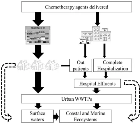
Figure 1.8: Sources and pathways for anticancer drugs into coastal waters. Dashed arrows
illustrate the additional course of wastewater in developing regions, where raw domestic and hospital effluents follow straightforward route into aquatic ecosystems. ... 17
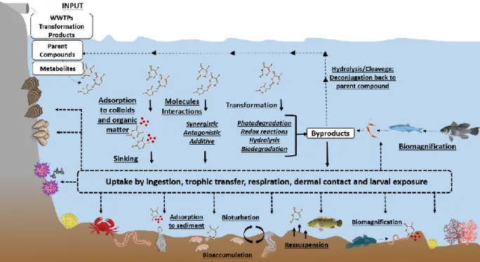
Figure 1.9: Behaviour and fate of anticancer drugs, in conjugation to abiotic and biotic
processes, in the marine environment. ... 20
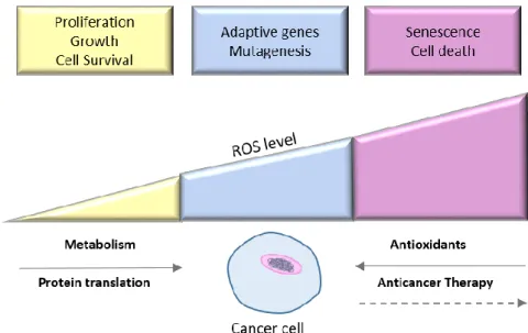
Figure 1.10: Induction and scavenging of ROS levels in cancer cells: Balance between basal
(yellow) and excessively high ROS levels (pink), into moderate and ROS-mediated mutagenic events (blue). ... 39
Figure 1.11: Number of papers published per year (Until December 2017) regarding
ecotoxicological data of anticancer drugs, considering bioassays with freshwater (FW) and seawater (SW) organisms. ... 39
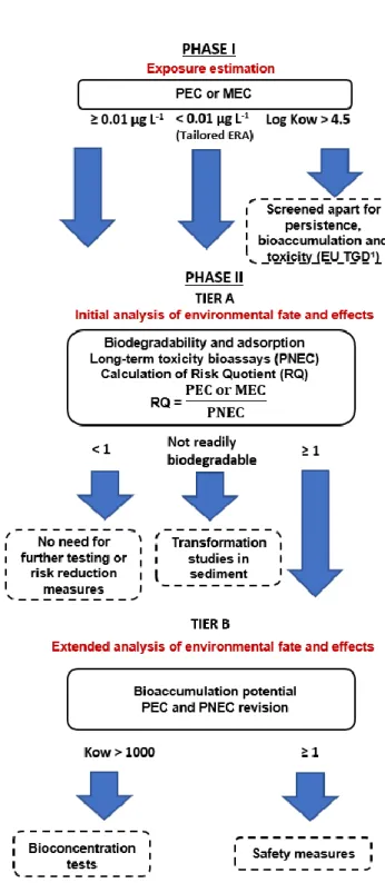
Figure 1.12: Flow chart for the ERA (Environmental Risk Assessment) based on the European
Medicines Agency Guideline (EMA, 2006). ... 43
C
HAPTER2.
Ecotoxicological assessment of the anticancer drug cisplatin in the polychaete Nereis diversicolor.Figure 2.1: Inhibition of sarcoplasmic reticulum calcium pump by CisPt. ... 68 Figure 2.2: Burrowing behaviour of N. diversicolor unexposed (CT14 – control at day 14) and
exposed to CisPt (0.1, 10 and 100 ng Pt L-1), expressed as percentage of unburied organisms over time (minutes). ... 69
Figure 2.3: AChE activity (mean ± standard deviation) (ATC.min-1.mg-1 protein) in N. diversicolor unexposed (CT0 and CT14) and exposed to CisPt (0.1, 10 and 100 ng Pt L-1) for
14 days. Different letters indicate statistically significant differences among treatments (One-way ANOVA; p < 0.05). ... 70
vi
Figure 2.4: Mean ± standard deviation of (A) SOD, (B) CAT, (C) GPx Se-dependent, (D) GPx
Se-Total, (E) GST and (F) MT in N. diversicolor unexposed and exposed to CisPt (0.1, 10 and 100 ng Pt L-1) for 14 days. Different letters indicate statistically significant differences among
treatments (One-way ANOVA (A, B, C, D, F) and Kruskal Wallis (E); p < 0.05). ... 71
Figure 2.5: LPO activity (mean ± standard deviation) (MDA+ 4-HNE nmol.mg-1 protein) in N.
diversicolor unexposed (CT0 and CT14) and exposed to CisPt (0.1, 10 and 100 ng Pt L-1) for 14 days. Different letters indicate statistically significant difference among treatments (One-way ANOVA; p < 0.05). ... 72
Figure 2.6: DNA (mean ± standard deviation) in tail, in N. diversicolor unexposed (CT0 and
CT14) and exposed to CisPt (0.1, 10 and 100 ng Pt L-1) for 14 days. Different letters indicate statistically significant differences among treatments (One-way ANOVA; p < 0.05). ... 73
Figure 2.7: Principal Component Analysis (PCA) of biochemical biomarkers and DNA
damage analysed in N. diversicolor at controls (CT0 and CT14) and CisPt-contaminated conditions, for 14 days. ... 74
C
HAPTER3.
Environmental relevant levels of the cytotoxic drug cyclophosphamide produce harmful effects in the polychaete Nereis diversicolor.Figure 3.1: CP metabolism pathway. Enzymatic activation by cytochrome P-450 enzyme
system generates 4-hydroxycyclophosphamide, coexisting with the non-cytotoxic adophosphamide metabolite. It decomposes into acrolein, that is toxic, and the DNA-binding phosphoramide mustard. ... 87
Figure 3.2: Burrowing behaviour of N. diversicolor from control conditions (day 0: CT0; day
14: CT14) and those exposed to CP-contaminated systems (10, 100, 500 and 1000 ng L-1), expressed as percentage of unburied organisms over time (lines with symbols). Continuous lines represent least-square best-fit regression lines, with respective equations. ... 98
Figure 3.3: AChE activity (mean ± S.D.) (ATC.min-1.mg-1 protein) in N. diversicolor
unexposed (CT 0 and CT 14) and exposed to CP (10, 100, 500 and 1000 ng L-1) for 14 days. Absence of letters indicates no significant differences among treatments (Kruskal-Wallis, p > 0.05). ... 99
Figure 3.4: Antioxidant enzymes activities (mean ± S.D.) of (A) SOD, (B) CAT, (C) GPx
Se-dependent, (D) T-GPx in N. diversicolor of control conditions (CT0 and CT14) and exposed to CP (10, 100, 500 and 1000 ng L-1) for 14 days. Different letters indicate significant difference among treatments (ANOVA: SOD, CAT; Kruskal-Wallis: GPx Se-dependent, T-GPx; p < 0.05). Absence of letters indicates no significant differences among treatments (p > 0.05). 100
Figure 3.5: Biotransformation enzyme activity (mean ± S.D.) in N. diversicolor of control
conditions (CT0 and CT14) and exposed to CP (10, 100, 500 and 1000 ng L-1) for 14 days.
Different letters indicate significant differences among treatments (Kruskal-Wallis, p < 0.05). ... 100
Figure 3.6: Principal Component Analysis (PCA) integrating responses of antioxidant enzymes
(SOD, CAT, Se-GPx, T-GPx, GST), AChE, DNA Tail, oxidative damage (LPO) and burrowing behaviour of N. diversicolor, exposed to control conditions (CT0: day 0; CT14: day 14) and to different CP concentrations (10, 100, 500 and 1000 ng L-1). ... 103
vii
C
HAPTER4.
Impacts of tamoxifen exposure to Nereis diversicolor.Figure 4.1: Metabolic pathways of tamoxifen in humans, following activation by cytochrome
P450. ... 113
Figure 4.2: Percentage of unburied N. diversicolor over time (minutes) in controls (CT0, CT14)
and exposed to DMSO and to TAM concentrations (0.5, 10, 25, 100 ng L-1). Lines with symbols indicate the behaviour during the 30-min experiment. ... 120
Figure 4.3: N. diversicolor AChE activity (mean ± S.D.) (ATC.min-1 mg-1 protein) in unexposed (CT0, CT14) and exposed to DMSO and to TAM concentrations (0.5, 10, 25 and 100 ng L-1). Different letters indicate significant differences among treatments (One-way ANOVA; p < 0.05). ... 121
Figure 4.4: N. diversicolor antioxidant enzymes activities (mean ± S.D.) expressed in
unexposed polychaetes (CT0, CT14) and exposed to DMSO and to a range of TAM concentrations (0.5, 10, 25 and 100 ng L-1): (A) SOD, (B) CAT, (C) GPx Se-dependent, (D) T-GPx. Different letters indicate significant differences among treatments (One-way ANOVA; p < 0.05). ... 122
Figure 4.5: N. diversicolor GST activity (mean ± S.D.) in unexposed (CT0, CT14) and exposed
to DMSO and to TAM concentrations (0.5, 10, 25 and 100 ng L-1) Absence of letters indicate no differences among treatments (One-way ANOVA, Kruskal-Wallis; p > 0.05). ... 123
Figure 4.6: N. diversicolor LPO levels (mean ± STD) (MDA+ 4-HNE nmol.mg-1 protein) in unexposed (CT0, CT14) and exposed to DMSO and to different TAM concentrations (0.5, 10, 25 and 100 ng L-1). Different letters indicate significant differences among treatments (One-way ANOVA; p < 0.05). ... 123
Figure 4.7: DNA damage (mean ± standard deviation) in coelomocytes of polychaetes N. diversicolor unexposed (CT0, CT14) and exposed to DMSO and to different TAM
concentrations (0.5, 10, 25 and 100 ng L-1). Different letters indicate significant differences
among treatments (One-way ANOVA; p < 0.05). ... 124
C
HAPTER5.
Effects of mixtures of anticancer drugs in the benthic polychaete Nereis diversicolorFigure 5.1: Burrowing behaviour of N. diversicolor from control conditions (CT0; CT14;
0.01% DMSO) and exposed to a combination of anticancer drugs, at increasing concentrations (Mix A, B, C and D). Results were expressed as percentage of unburied organisms over time (lines with symbols). ... 141
Figure 5.2: AChE activity (mean ± S.D.) (ATC.min-1.mg-1 protein) (mean ± S.D.) in N. diversicolor unexposed (CT0; CT14; 0.01% DMSO) and exposed to a combination of
anticancer drugs, at increasing concentrations (Mix A, B, C and D), over 14 days. Different letters indicate significant differences among treatments (One-way ANOVA; p < 0.05). .... 142
Figure 5.3: Antioxidant enzymes activities (mean ± S.D.) of (A) SOD, (B) CAT, (C) Se-GPx
and (D) T-GPx (mean ± S.D.) in N. diversicolor unexposed (CT0; CT14; 0.01% DMSO) and exposed to increasing concentrations of mixtures of anticancer drugs (Mix A- D), over 14 days. Different letters indicate significant differences among treatments (One-way ANOVA; p < 0.05). ... 143
viii
Figure 5.4: Lipid Peroxidation (LPO) levels (mean ± STD) (MDA+ 4-HNE nmol.mg-1 protein) in N. diversicolor unexposed (CT0; CT14; 0.01% DMSO) and exposed to increasing concentrations of mixtures of anticancer drugs (Mix A-D), over 14 days. Different letters indicate significant differences among treatments (One-way ANOVA; p < 0.05). ... 144
Figure 5.5: PCA biplot of PC1 vs. PC2 integrating biomarkers responses ( AChE, SOD, CAT, Se-GPx, T-GPx, GST, LPO, DNA damage) from polychaetes N. diversicolor unexposed and exposed to different mixtures concentrations of anticancer drugs ( Mix). ... 148
Figure 5.6: PCA biplot from (A) PC1 vs. PC2 and (B) PC1 vs. PC3 integrating biomarkers
responses ( AChE, SOD, CAT, Se-GPx, T-GPx, GST, LPO, DNA damage) in polychaetes
N. diversicolor from bioassays conducted with singular exposure of anticancer drugs in (
CisPt; CP; TAM) and in combination ( Mix), at different concentrations jointly with control conditions. ... 149
Figure 5.7: Biomarkers profile in N. diversicolor exposed to tertiary mixtures (A, B, C and D),
according to the single-drug responses. Arrows ↓ indicate inhibition; arrows ↑ induction; absence of arrows indicate effects similar to controls, in each individual anticancer bioassay. Green and red boxes indicate, respectively, similar and distinct effects in relation to the corresponding mixture. ... 153
C
HAPTER6.
Toxicity of anticancer drugs: a first insight to tropical species.Figure 6.1: Percentage of normal pluteus of E. lucunter after exposure of fertilized eggs to
single anticancer drugs. Asterisks indicate significant differences between drug treatments and control (T-test, p < 0.05). ... 174
Figure 6.2: Survival rates of the amphipod T. viscana (mean and S.D) exposed over 10 days to
control conditions (CT and DMSO), and tertiary mixtures of the anticancer drugs. Different letters indicate significant differences (p < 0.05). ... 175
Figure 6.3: Survival rates of the polychaete S. squamata (mean and S.D) exposed over 10 days
to control conditions (CT and DMSO), and tertiary mixtures of the anticancer drugs. Different letters indicate significant differences (p < 0.05). ... 175
ix
T
ABLE
I
NDEX
C
HAPTER1.
General IntroductionTable 1.1: Classification of anticancer drugs ... 9
Table 1.2: Ecotoxicity effects of anticancer drugs in marine species. ... 24
Table 1.3: Ecotoxicity effects of anticancer drugs in freshwater species. ... 27
Table 1.4: Physico-chemical characteristics of the drugs investigated. ... 52
C
HAPTER2.
Ecotoxicological assessment of the anticancer drug cisplatin in the polychaete Nereis diversicolor. Table 2.1: Concentrations of total Pt from Pt-based anticancer drugs (e.g. cisplatin; carboplatin; oxaliplatin) and Pt detected from cisplatin drug (ng L-1), in aquatic compartments. ... 59C
HAPTER3.
Environmental relevant levels of the cytotoxic drug cyclophosphamide produce harmful effects in the polychaete Nereis diversicolor. Table 3.1: Concentrations of CP in water (ng L-1) and sediments (ng g-1)... 90Table 3.2: Genotoxic effects in polychaetes N. diversicolor unexposed and after 14 days of exposure to CP. DNA damage (average ± SEM) is expressed as tail DNA %. Frequency of coelomocytes distributed by grade of DNA damage (%). Different letters represent statistical differences between treatments, during the exposure period. Percentage of coelomocytes distributed through damage criteria (i.e. Minimal to Extreme). ... 102
C
HAPTER4.
Impacts of tamoxifen exposure to Nereis diversicolor. Table 4.1: TAM Concentrations in water (ng L−1), suspended solids and sediments (ng g−1) from ground, riverine and marine waters. ... 115Table 4.2: Ecotoxicological effects of TAM reported in coastal and marine species. ... 128
C
HAPTER5.
Effects of mixtures of anticancer drugs in the benthic polychaete Nereis diversicolor Table 5.1: Genotoxicity in polychaetes N. diversicolor unexposed (CT0; CT14; 0.01% DMSO) and after 14 days of exposure to mixtures (Mix A-D), expressed as percentage of DNA in tail (mean ± SEM). Frequency of coelomocytes distributed by grade of DNA damage (%) (i.e. minimal to mid). ... 145C
HAPTER6.
Toxicity of anticancer drugs: a first insight to tropical species. Table 6.1: Physico-chemical characteristics, structure and metabolism of the selected anticancer drugs. ... 165x
A
BBREVIATIONS
4-HNE 4-hydroxyalkenals ACh Acetylcholine AChE Acetylcholinesterase ANOVA Analysis of variance
API Active pharmaceutical ingredient ATC Acetylcholine
BCF Bioconcentration factor BHT Butylated hydroxy toluene BSA Bovine albumin serum
CAGR Compound annual growth rate CAT Catalase
CDNB 1-chloro-2,4-dinitrobenzene
CETESB Companhia Ambiental do Estado de São Paulo CHMP Committee for Medicinal Products for Human Use CisPt Cisplatin
CNPq National council for scientific and technological development
CP Cyclophosphamide
CT Control
CTR 1 Copper transporter 1
CYP P450 Cytochrome P450 superfamily DAPI 4,6-diamidino-2-phenylindole DNA Deoxyribonucleic acid
DTNB 5,5-dithiobis-2-nitrobenzoic acid DTT 1,4-dithiothreitol
EC Emerging contaminant
EC50 Half maximal effective concentration
EDC Endocrine disrupting compounds EDTA Ethylenediaminetetraacetic acid EMA European Medicines Agency ER Estrogen receptor
xi ERA Environmental risk assessment
FDA Food and Drug Administration FMO Flavin-containing monooxygenases
FW Freshwater GPx Glutathione peroxidase GSH Glutathione GSH-T Total glutathione GSSG Glutathione disulphide GSSH Oxidized glutathione GST Glutatione-S-transferase H2O2 Hydrogen peroxide
IARC International agency for research on cancer IC50 Half maximal inhibitory concentration ICL Inter-strand crosslink
KH Henry´s coefficient
Koc Organic carbon partition coefficient LC50 Median lethal concentration
LECZ Low-elevation coastal zones LMA Low melting point agarose LPO Lipid peroxidation
LOAEC Lowest observable adverse effect concentration LOD Limit of detection limit
LOEC Lowest effect concentration LOQ Limit of quantification MBR Membrane bioreactors MDA Malondialdehyde
MEC Measured environmental concentration MET Methotrexate
MoA Modes of action MT Metallothionein
MTLP Metallothionein-like protein NAT N-acetyltransferase
xii NCD Non-communicable diseases
NMA Normal melting point agarose
NOAEC No observed adverse effect concentration NOEC No observed effect concentration
O2•- Superoxide anion radical
PAM Phosphoramide mustard PCA Principal component analysis
PEC Predicted environmental concentration PNEC Predicted no effect concentration
Pt Platinum
REACH Registration, Evaluation, Authorization and Restriction of Chemicals ROS Reactive oxygen species
ROW Rest of the world
SDG Sustainable development goals
Se-GPx Glutathione peroxidase selenium-dependent SERM Selective estrogen receptor modulators SOD Superoxide dismutase
SPE Solid phase extraction SR Sarcoplasmic reticulum
SRV Sarcoplasmic reticulum vesicles SULT Sulfotransferase
SW Seawater
TAM Tamoxifen
TBARS Tiobarbituric acid reactive substances T-GPx Total GPx
TP Transformation products
UGT Uridine 5´- diphospho (UDP)-glucoronosyltransferase USEPA United States Environmental Protection Agency WHO World Health Organization
WWTP Waste water treatment plant
1
Chapter
1
General Introduction
Published in:
Bebianno, M. J. and Fonseca, T. G.,
Fate and effects of cytostatic pharmaceuticals in the marine environment. In: Fate and effects of cytostatic pharmaceuticals in the environment. Eds: Heath E. & Isidori M.
Chapter 14 Prepared for submission to:
Fonseca, T. G., Rocha T.L., Maranho, L.A., Bebianno, M. J.
The polychaete Nereis diversicolor as a biomonitor species of emergent contaminants in the marine environment: a critical review of pharmaceuticals and nanomaterials.
1
1.1 World trends of pharmaceuticals production and usage
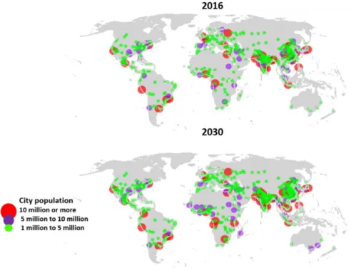
The global demographic outlook demonstrates the fast pace to which population have increased 1.1% per year), reaching a record of 7.7 billion people in 2017, with projections to a further mark of 8.6 billion in 2030 (United Nations, 2017). Moreover, the current scenario of an increasingly interconnected world indicates that over half of this population lives in urban areas. By 2030, 43 cities around the world will have 10 million or more inhabitants (i.e. megacities), which correspond to an increase of around 30% compared to 2016 (United Nations, 2016) (Figure 1.1). Another feature of the exceptional urbanization transition addressed in the 21st century is that two thirds of these megacities are established in zones near the coast, where 41% of humankind is settled (Burke et al., 2001; Martínez et
al., 2007; Von Glasow et al., 2013).
Figure 1.1: Cities with one million or more inhabitants, in 2016 and projections for 2030.
Adapted from United Nations (2016).
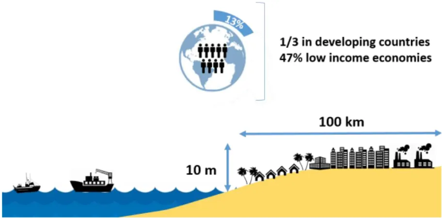
Low-elevation coastal zones (LECZ) (those with 10 m above sea level) represent about 2% of total land area where 13% of global population inhabit (Barbier, 2015). LECZ are expanding faster than any other area through immigration and demographic changes
2 (Figure 1.2). The milestone shift of global demographic profiles associated to population aging, projected to continue and even accelerate, mainly driven by declining fertility rates and remarkable increases in life expectancy (WHO, 2014; Ferlay et al., 2015).
Figure 1.2: Representation of urban settlement in low-elevation coastal zones (LECZ),
where 13% of the world population lives.
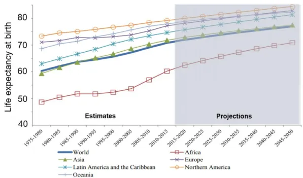
In 2010, it was estimated that 524 million people worldwide were aged 65 or older, depicting 8% of global population. By 2050, this number is expected to nearly triple to about 1.5 billion, thus represents 16% of the total. Although there are variations in patterns of population growth and levels of urbanization across regions, gains in longevity have been especially dramatic in cities of developing countries (WHO, 2016) (Figure 1.3). These trends have profound implications in society, once a transition to older populations on the demographic structure will occur, followed by health problems associated to chronic non-infectious and non-communicable diseases (NCD). NCDs mainly comprise cardiovascular and respiratory chronic illnesses, as well as cancer diseases, accounting for 67.9% of total deaths that occurred worldwide, in 2012, and currently represent the greatest burden on health of low, middle- and higher- income countries. However, the present rise of NCDs has shown to derive not only from older health status but also from the improvement of living standards and broaden socioeconomic, cultural and environmental underlying determinants, fitted by risk factors individually managed (e.g. unappropriated diet, tobacco, physical inactivity, harmful use of alcohol, infectious agents) (Jemal et al., 2011; American Cancer Society, 2014). A rising rate of diseases burden will drive up production and consumption
3 of pharmaceuticals required for different therapeutic applications in a way that global medicines spending will reach 1.4 trillion dollars by 2020, an increase of 29-32% compared to 2015, in which 63% of the spending will be led by developed nations (Aitken and Kleinrock, 2015) (Figure 1.4).
Figure 1.3: Life expectancy (years) by region: estimates 1975-2015 and projections
2015-2050. Adapted from World Population Prospects: The 2017 Revision (WHO, 2017).
Figure 1.4: Projections of global spending (US$) in pharmaceuticals in 2020, by therapeutic
4
1.2 The global scenario of cancer diseases
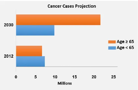
Neoplastic or cancer diseases depict a leading concern in global health condition responsible for an estimated 9.6 million deaths in 2018, with worrying projections of cancer incidence and mortality, by 2030 (Figure 1.5) (WHO, 2014; Ferlay et al., 2015; WHO, 2017). In 2015, 8.8 million people died from cancer, which represents one in six deaths globally reaching countries of all income levels. However, actions aiming to bring cancer rates under control are better managed in developed nations through decreasing the prevalence of risk factors, early detection measures and prevention action, whereas economically less developed countries continue to bear disproportionate burden of infection-related cancers cases and a rise in exposure to hazard external determinants associated to lifestyle (Torre et al., 2015). Therefore, approximately two thirds of global cancer deaths are in less developed countries, especially due to late-stage detection and less accessible treatment (WHO, 2017).
Figure 1.5: Projection of cancer cases between 2012 and 2030 (millions of people). All types
5 As a result, the United Nations has recognized cancer as a priority not only for health but also as a threat to economies and societies, thus placing this disease high on the set of commitments of a global agenda of health coverage (i.e. Global Action Plan for the Prevention and Control of NCDs) (WHO, 2014). In this scope, nations aim to monitor the trends of NCDs and evaluate their progress in an attempt to design preventive and control measures. Therefore, substantial increase in availability of affordable basic technologies and essential pharmaceutical compounds are required to treat cancer in both public and private facilities (WHO, 2014). Since cancer consists of a group of heterogeneous diseases that can affect almost any part of the body and has several anatomic and molecular subtypes, specific management of prevention, early diagnosis, treatment, palliative and survivorship care are enforced, regardless the zone of incidence (Torre et al., 2015). Cancer therapy usually involves the alternative of surgery resection, and body´s exposure to agents that kill cancer cells, via radiotherapy and the administration of anticancer medicines through systemic therapies, in a way that these may be applied individually or in combination, depending of the type and stage of the tumour (Helleday et al., 2008; Barczak et al., 2018). Anticancer drugs may correspond to different pharmaceutical modalities, regarding cytotoxic chemotherapy, in addition to hormonal, targeted and immunotherapies (Palumbo et al., 2013).
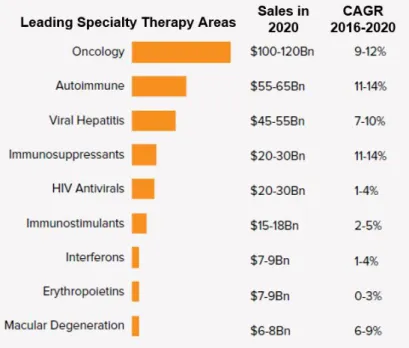
It is possible to link the increase of cancer incidences in recent years with the rise of drugs production and application, also reflected in global spending on oncology medicine as a leading key therapy area driving pharmaceutical expenditure over the next five years, including treatment and supportive care (Figure 1.6) (Aitken and Kleinrock, 2015). The pipeline of oncology drugs expanded by 45% over the past 10 years (Pineau ad Rink, 2017). By 2021, spending on oncology medicine is expected to grow in a range of 9-12% annually, compared to the 11% seen over 2016, as the demand of new and targeted therapy options raised (Aitken and Kleinrock, 2015) (Figure 1.7). Since 2011, 68 new drugs have been approved for 22 indications, including immune-oncology agents that have considerably changed the paradigm of cancer treatment. Advances in drug discovery have supported the movement towards a more personalized care that relies on the use of validated predictive biomarkers. These novel tools are able to identify genetic attributes of a population presenting a specific cancer case, thus far more likely to respond to therapy and benefits
6 from a particular drug (Pineau and Rink, 2013). However, conventional treatments, which are less effective for specific drivers of cancer, are still widely applied worldwide. They are represented by cytotoxic drugs which have been prescribed, since the 1960´s, for various solid and hematological malignancies in chemotherapies administered to adults and children (Helleday et al., 2008).
Figure 1.6: Sales in 2020 for leading therapy areas of specialty medicines and compound
annual growth rate (CAGR).
Figure 1.7: Global forecast of spending of oncology pharmaceuticals: EU5 (Germany, Italy,
France, Spain, UK); United States; Japan; Pharmerging countries (Brazil, Mexico, Venezuela, Argentina, China, India, Indonesia, Vietnam, etc.); ROW (Rest of the World).
7 Despite addressed as efficient agents against tumour cells by means of producing high levels of DNA damage, those pharmaceuticals are not selective enough to interact only with highly proliferative cells by providing cell-cycle arrest and cell death in normal and healthy tissues. Such mechanisms often cause undesirable side effects to patients during the chemotherapy and may be potent harmful compounds to all eukaryote organisms (Novak et
al., 2017).
1.3 Anticancer medicines and the rationale for chemotherapy
Chemotherapy may be administered to cancer patients through usual routes of intravenous, intramuscular or subcutaneous injection. Classification of anticancer drugs is established by the Anatomical Therapeutic Classification (ATC), where they are ranked as Antineoplastic Agents; Endocrine Therapy; Immunostimulants; and Immunosuppressants, as can be seen in Table 1.1 (Booker et al., 2014; Xie et al., 2015). Antineoplastic agents are designed to interact directly or indirectly with DNA, by damaging its structure, inhibiting, altering and disrupting mechanisms of its transcription, replication and synthesis. Derivatives of nitrogen mustards were developed, including the DNA alkylating agents cyclophosphamide, chlorambucil and melphalan, widely used in clinical therapeutics (Cheung-Ong et al., 2013. Xie et al., 2015). Other examples of DNA-alkylating agents used in cancer treatment include nitrosoureas (e.g., carmustine, lomustine, and semustine) and triazenes (e.g., dacarbazine and temozolomide) (Cheung-Ong et al., 2013).
Since most of these pharmaceuticals do not strictly act over tumour drivers, but also in mechanisms regarding all growing cells (Kosjek and Heath, 2011; Caley and Jones, 2012; Toolaram et al., 2014; Heath et al., 2016), usually leading to detrimental effects in patients owing from adverse toxicity to non-targeted tissues. In contrast, there are compounds accountable for the disruption of biological processes through mechanisms of blocking cells replication factors, or indirectly by recruiting macrophages and monocytes cells (Besse et al., 2012; Caley and Jones 2012), thus, not directly involved with DNA. Endocrine therapy manipulates hormone-dependent receptors with tissue-selective interactions that lead to agonist or antagonist activities, represented by the group of selective estrogen receptor modulators (SERMs), like tamoxifen (Paterni et al., 2014; An, 2016). Attachment of these endocrine disrupting compounds (EDC) to respective binding sites acts competitively and subsequently
8 inhibiting the transcription of estrogen responsive genes responsible for cell replication of estrogen receptor positive cells, such as seen in breast and prostate cancer (Pinto et al., 2014).
Currently, the aforementioned groups of drugs are some of the main used in cancer treatments. Most of chemotherapy regimens in clinical practice consist of a combination of several agents from different pharmaceutical groups, in an attempt to provide additive or synergistic effects to achieve maximal tumour cell death and avoid resistance (Caley and Jones, 2012). In spite to their overall application in chemotherapy, there are strong differences in the chemical structure of the several anticancer groups that also have distinct modes of action (MoA) (Booker et al., 2014).
9
Table 1.1: Classification of anticancer drugs
Classification Subcategory Compounds MoA
Antineoplastic agents
Alkylating agents
Nitrogen mustards Cyclophosphamide, chlorambucil, ifosfamide
Alkylating agents react with nitrogen (N) and extracyclic oxygen (O) atoms of DNA bases to generate a variety of covalent adducts.
Induce a range of cytotoxic and mutagenic adducts onto DNA
Nitrosoureas Carmustine, lomustine
Alkyl sulfonates Busulfan
Other alkylating agents Temozolomide
Antimetabolites
Folic acid analogues Methotrexate
Structurally similar to endogenous nucleic acids, can be incorporated into the metabolic pathways instead of the endogenous purine and pyrimidines, thereby affecting the enzyme
dependent synthesis of DNA and cell reproduction Purine analogues Mercaptopurine, tioguanine, cladribine
Pyrimidine analogues 5-fluorouracil, gemcitabine, capecitabine
Plant Alkaloids
Vinca alkaloids and
analogues Vinblastine, vincristine Interacts with microtubules or tubulins leading to inhibition of synthesis of proteins and nucleic acids, disruption of mitotic
spindle and eventually cell death Podophyllotoxin derivatives Etoposide
Taxanes Paclitaxel
Cytotoxic Antibiotics and related substances
Actinomycines Dactinomycin
Involve direct toxic action on cellular DNA, interfering with DNA replication and protein synthesis
Anthracyclines and related
substances Daunorubicin, doxorubicin, epirubicin
Other antineoplastic agents
Platinum compounds Cisplatin, carboplatin
E.g. Cisplatin products highly electrophilic. Act towards
nucleophilic sites in genomic and mitochondrial DNA, producing DNA inter- and intra-strand adducts that result in DNA distortion, inhibition of DNA replication and interruption of cell division
10
Endocrine Therapy
Hormones and related agents
Estrogens Ethinylestradiol, diethylstilbestrol
Antitumor activity involves suppression of luteinizing hormone by inhibition of pituitary function
Progestogens Megestrol, medroxyprogesterone Gonadotropin releasing
hormone analogues Leuprorelin, buserelin
Hormone antagonists and related agents
Anti-estrogens Tamoxifen, toremifene Diverse group of compounds bind to oestrogen α (ERα) and β (ERβ) receptors and produce oestrogen agonist effects in some tissues, but oestrogen antagonist activity in others. Activity determined in part by formation of oestrogen receptor–molecule complexes that vary in their ability to activate genes when bound
to ERα or ERβ Anti-androgens Flutamide, nilutamide
Aromatase inhibitors Letrozole, anastrozole
Immunostimulants Immunostimulants
Colony stimulating factors Filgrastim, lenograstim
Immune modulation is based on the stimulation of T-cell function with antibodies that block or activate regulatory receptors is
enough to cause the regression of some tumours. Interferons Interferon-α natural, interferon-β natural
Interleukins Aldesleukin
Other immunostimulants Lentinan, roquinimex
Immunosuppressants Immunosuppressants
Selective
immunosuppressants Mycophenolic acid, sirolimus Interleukin inhibitors Daclizumab, basiliximab Calcineurin inhibitors Ciclosporin, tacrolimus Other immunosuppressants Methotrexate, azathioprine
11
1.4 Drug metabolism and excretion
The major function of drug metabolism in the body is to provide the breakdown of the compound into chemically simpler metabolites for elimination, thereby preventing unwanted accumulation, as well as potentially toxic levels of endogenous molecules (Jančová and Šiller, 2012). In general, the fate of chemical biotransformation includes the generation of metabolites, which may be desired for the therapeutic purpose or not, due to toxicity. Briefly, drug pharmacokinetics occurs in two phases of biotransformation, comprised by the functionalization (Phase I) and conjugation (Phase II) of the compound load, in order to increase its polarity and facilitate its elimination through excretion (King, 2009). Phase I relies on an enzymatic system of defense against most of the xenobiotic compounds, composed by members of the cytochrome 450 gene family (CYP 450), flavin-containing monooxygenases (FMO), monoamine oxidases (MAOs), and xanthine oxidase/aldehyde oxidase (Penner et al., 2012). Those enzymes are differently expressed in tissues, but broadly distributed in liver, kidney and intestine, acting by means of introducing, modifying or unmasking reactive functional groups at the parent drug.
Phase II reactions occur with the introduction of acetyl, sulfate, glucoronid acid, glutathione and amino acids functional groups, either in the parent molecule or in a phase I metabolite structurally changed enabling binding sites for conjugation (Liska, 1998; Jančová and Šiller, 2012). These reactions are mostly catalyzed by the enzyme uridine 5´- diphospho (UDP)-glucoronosyltransferase (UGTs), sulfotransferases (SULTs), glutathione S-transferase (GSTs) and N-acetylS-transferases (NATs). Biotransformation mechanisms present low specificity to pharmaceuticals and, in general, enhance polarity of compounds, thus favoring the excretion (King, 2009). Nevertheless, this change is often incomplete, with parent compounds excreted together with the metabolites in a variable proportion (Kümmerer, 2010).
12
1.5 Anticancer compounds as a threat to the aquatic environment
1.5.1 Pharmaceuticals as Emerging Contaminants (ECs)
Pharmaceuticals are substances intended for the use in diagnosis, cure, mitigation, treatment or prevention of diseases, in humans and animals, also designed to provide therapeutic effects in biological systems by their key molecular structures, the active pharmaceutical ingredients (APIs) (Daughton and Ternes, 1999; Khetan and Collins, 2007). The pharmaceutical compounds also include excipients, additives, inorganic salts and other organic chemicals, such as sugars, scents, pigments and dyes (Kummerer, 2010). A wide range of drugs is displayed by the pharmaceutical industry and are often classified according to their APIs and purpose (e.g. antibiotics, analgesic, lipid regulators, antidepressants, antineoplastics) (Williams, 2005). Classification by similarity in chemical structure and behaviour are also usually applied (e.g. antibiotics: penicillins, cephalosporin, quinolones), which may or not coincide with biological activity. In addition, sorting can be based on the drugs MoA, referring to physiological system to be targeted (i.e. digestive system), or to specific binding receptor to which active ingredient interacts (Kummerer, 2008).
Pharmaceuticals are designed to affect targeted biomolecules and biological critical mechanisms via their specific MoA, triggered even at low doses (Fent et al., 2006b; Gunnarsson et al., 2008). However, since the end of the last century, new findings regarding pharmaceuticals´ molecular particularities and consumption have emerged questions whether these chemicals are relevant only to human benefits and healthcare (Williams, 2005; Roberts and Bersuder, 2006). According to the societal and health status outlooks, jointly with economic forecasts, it is possible to portray not only the hallmark of the increment usage of anticancer drugs in coastal urban areas, but also outline their continuous loads into aquatic ecosystems worldwide. Considering that, pharmaceuticals have been addressed as ubiquitous compounds detected at trace levels (ng – µg L-1) in aquatic compartments, and their persistent input has brought biota to a long-term exposure (Smital, 2008). These compounds, which profile of distribution, behaviour and biological interactions in the aquatic environment are poorly known (Murnyak et al., 2011; Pal et al., 2010; Robles-Molina et al., 2014) are considered emerging contaminants (ECs), and regulation by legal framework are currently under construction or still absent (Rivera-Utrilla et al., 2013). The
13 term “emerging” does not necessarily concern a new phenomenon, but a recognition of compounds that have been recently identified in the aquatic environment by the improvement of analytical methods, which allowed lowering the detection limit in environmental matrices (Glassmeyer et al., 2008; Nikolaou, 2013).
1.5.2 Environmental Background of anticancer drugs
The first publication regarding the issue of anticancer drugs in the environment dates from the 1980´s and aimed to depict the distribution profile of several classes of pharmaceuticals in different aquatic compartments (e.g. sewage influent and effluent, surface water and drinking (Aherne et al., 1985; Richardson and Bowron, 1985). These authors outlined that anticancer drugs are likely to yield environmental risks, particularly to surface waters that receive discharges from sewage treatment plant, since the confirmation of hazards verified to nurses after occupational exposure to pharmaceuticals during chemotherapy manipulation. Following the improvement of analytical technologies, a set of cytotoxic pharmaceuticals were identified in environmental screenings, at a range of ng L-1 or below (Kummerer et al., 1997; Buerge et al., 2006; Zuccato et al., 2006; Kovalova et al., 2009;Toolaram et al., 2014). Relevant reviews outlined physico-chemical parameters that determine the fate and behaviour of cytotoxics and predict their pathways in aquatic compartments according to their chemical structure namely, pKa, bioconcentration factor (BCF), octanol/water partition coefficient (Kow), organic carbon partition coefficient (Koc), solubility, Henry´s coefficient (KH) and vapor pressure (Kosjek and Heath, 2011; Xie, 2012; Toolaram et al., 2014).
Transformation products (TPs) are by-products of parent compounds and metabolites formed through physico-chemical (e.g. photodegradation, hydrolysis, redox reactions) and biological factors (e.g. biodegradation) undergone in WWTPs or over the water bodies (Mompelat et al., 2009). Those chemicals can also be as toxic or even more toxic than their parent compounds (Loffler et al., 2005; Li et al., 2014). Despite their potential importance, the formation and fate of TPs from pharmaceuticals has only been scarcely investigated (Mompelat et al., 2009). Although a significant number of drugs are excreted and released as inactive conjugates, such as glucoronates and sulfates, deconjugation provided by bacterial metabolism in WWTPs and in aquatic environment turn metabolites back to the
14 biologically active parent compounds (Noppe et al., 2007; Behera et al., 2011). Therefore, mixtures of parental forms, metabolites and TPs started to be also evaluated (Fatta-Kassinos
et al., 2011; Toolaram et al., 2014; Haddad et al., 2015; Zhang et al., 2017). The studies of
Kovalova et al. (2009), Negreira et al. (2013, 2014a, 2014b, 2015) and Zhang et al. (2013) brought advances in analytical chemistry and processes that underline technologies in WWTPs able to remove anticancer drugs, conform to molecules physico-chemical properties, stability and metabolism.
1.5.3 Sources and routes of anticancer drugs to the marine environment
Cancer treatment is shifting from hospital treatment and medical facilities to home treatment (i.e. outpatients) (Johnson et al., 2008; Kosjek and Heath, 2011; Besse et al., 2012). The generalization of this modality and the availability of molecules in pharmacies are growing in order to improve the patient treatment comfort. Consequently, a more diffuse discard of parental and metabolized anticancer agents is prone to succeed during home treatment, compared to discharges from hospital wastewater effluents, which is directly introduced into WWTPs (Kümmerer, 2010; Mater et al., 2014; Zhang et al., 2013) (Figure 1.8). In general, private households are indicated as the most important sources for pharmaceuticals emission into aquatic environment, rather than hospitals (Schuster et al., 2008; Toolaram et al., 2014). In the case of chemotherapy drugs, although the substantial contribution, there is still no clear evidence if household origination is stepping up to or even overtake hospital emissions as their distribution patterns depend on various parameters, such as the size of hospitals, size of catchment area, population density, purchase of pharmaceuticals in town pharmacies, among others (Zhang et al., 2013). WWTPs are primarily designed to serve the purpose of removing pathogens, suspended solids, gross organic and inorganic matter, rather than removing the vast array of modern chemicals like pharmaceuticals, present at trace concentrations of ng to g L-1 in influents (Lenz et al., 2005; Rowney et al., 2009; Besse et al., 2012).
In general, European countries count with conventional sewage treatment processes equipped with screening, degritting, sedimentation, biodegradation and adsorption to biomass as the main elimination mechanisms for various organic contaminants during the passage of conventional activated sludge and confined units (Carballa et al., 2004; Conn et
15
al., 2006; Zhang et al., 2013), although those have shown to be ineffective to remove
pharmaceuticals (Yang et al., 2017). Combination of advanced and non-conventional technologies in WWTPs and water reclamation are considered alternatives to improve the elimination performance of elimination of chemotherapy drugs, including electrolysis, UV radiation with H2O2, ozonation and membrane bioreactors (MBR) (Česen et al., 2015). The
cytostatic drug 5-FU was eliminated from hospital wastewaters to levels below the limits of detection by means of biodegradation processes occurring in the MBR system, whereas the major pathway by which anthracyclines (doxorubicin, epirubicin and daunorubicin) were mainly removed was through adsorption to sewage sludge, able to yield an elimination of >90% from the liquid phase (Mahnik et al. 2007). The percentage of removal achieved at the end of a batch bioreactor WWTP for tamoxifen, ciprofloxacin and etoposide reached 91, 84 and 100%, respectively (Ferrando-Climent et al., 2015).
In contrast, ifosfamide, cyclophosphamide, vincristine, docetaxel and paclitaxel showed to be recalcitrant due to the inefficiency of conventional biological treatments of WWTPs. Likewise, the alkylating agent cyclophosphamide was also considered resistant to mechanical and conventional biological treatment with suspended biomass, after its feasible detection in WWTP effluents at 17 ng L-1 (Česen et al., 2015). Alternatively, the use of ozonation technology combined to UV and H2O2 (5 g L-1) treatments showed to remove 99%
of cyclophosphamide and 94% of ifosfamide from a non-treated wastewater (Česen et al., 2015). Other studies demonstrated that cyclophosphamide can withstand degradation processes over the aquatic pathway, until concentrations detected in surface waters reach a range of 0.15 to 17 ng L-1 (Buerge et al. 2006; Moldovan 2006; Busetti et al., 2009).
In fact, literature review demonstrated inconsistencies in the removal performed through biological treatments for the alkylating agents ifosfamide and cyclophosphamide, with reports indicating a range from 0 to 72% (Buerge et al., 2006; Kovalova et al., 2012). This suggests that the extent to which WWTPs are successful in their removal depends not only on how developed are the selected treatment technologies, but also on the experimental design and on the physico-chemical properties of the effluent (Česen et al., 2015). Moreover, Martín et al. (2012) identified a removal rate of cytotoxic drugs in WWTPs, ranging from 0% (e.g. doxorubicin, gemcitabine, paclitaxel, cisplatin and vinorelbine) to 100% (e.g. etoposide, fluorouracil, methotrexate).