RESUMO.- [Strain e strain rate por meio de ecocardio-graia speckle traking bidimensional em um lobo-gua-rá.] A obtenção de parâmetros cardiovasculares em ani-mais selvagens são importantes de serem avaliados, assim como em animais de companhia, para a obtenção da função miocárdica e determinação precoce de alterações cardíacas que poderiam evoluir para insuiciência cardíaca . A ecocar-diograia speckle tracking (2D STE) é uma ferramenta nova que tem sido utilizada em medicina veterinária, a qual tem demonstrado várias vantagens quanto ao seu uso, como a
1 Received on March 13, 2012.
Accepted for publication on September 1, 2012.
2 Setor de Clínica Médica de Pequenos Animais, Serviço de Cardiologia Veterinária, Departamento de Medicina Veterinária, Universidade Federal de Lavras, Campus Universitário s/n, Cx. Postal 3037, Lavras, MG 37200-000, Brazil. *Corresponding author: [email protected]
Strain and strain rate by two-dimensional speckle tracking
echocardiography in a maned wolf
1Matheus M. Mantovani2, Adriana C. Silva2*, Ruthnéa A.L. Muzzi2, Guilherme
Oberlender2, Rosane M. Resende2, Leonardo A.L. Muzzi2, Antonio C.C. Lacreta Junior2
and Rodrigo B. Nogueira2
ABSTRACT.- Mantovani M.M., Silva A.C., Muzzi R.A.L., Oberlender G., Resende R.M., Mu-zzi L.A.L., Lacreta Junior A.C.C. & Nogueira R.B. 2012. Strain and strain rate by two-dimensional speckle tracking echocardiography in a maned wolf. Pesquisa Veteriná-ria Brasileira 32(12):1336-1340. Setor de Clínica Médica de Pequenos Animais, Serviço de Cardiologia Veterinária, Departamento de Medicina Veterinária, Universidade Federal de Lavras, Campus Universitário s/n, Caixa Postal 3037, Lavras, MG 37200-000, Brazil. E-mail: [email protected]
The measurement of cardiovascular features of wild animals is important, as is the me-asurement in pets, for the assessment of myocardial function and the early detection of cardiac abnormalities, which could progress to heart failure. Speckle tracking echocardio-graphy (2D STE) is a new tool that has been used in veterinary medicine, which demons-trates several advantages, such as angle independence and the possibility to provide the early diagnosis of myocardial alterations. The aim of this study was to evaluate the left myocardial function in a maned wolf by 2D STE. Thus, the longitudinal, circumferential and radial strain and strain rate were obtained, as well as, the radial and longitudinal velo-city and displacement values, from the right parasternal long axis four-chamber view, the left parasternal apical four chamber view and the parasternal short axis at the level of the papillary muscles. The results of the longitudinal variables were -13.52±7.88, -1.60±1.05, 4.34±2.52 and 3.86±3.04 for strain (%), strain rate (1/s), displacement (mm) and velocity (cm/s), respectively. In addition, the radial and circumferential Strain and Strain rate were 24.39±14.23, 1.86±0.95 and -13.69±6.53, -1.01±0.48, respectively. Thus, the present study provides the irst data regarding the use of this tool in maned wolves, allowing a more com-plete quantification of myocardial function in this species.
INDEX TERMS: Cardiac function, Chrysocyon brachyurus, maned wolf, systolic function, wild canid.
primei-ros dados a respeito do uso desta ferramenta em lobos gua-rás, permitindo uma quantiicação da função miocárdica de forma mais completa nesta espécie.
TERMOS DE INDEXAÇÃO: Canídeo selvagem, lobo-guará, Chry-socyon brachyurus, função cardíaca, função sistólica.
INTRODUCTION
The maned wolf (Chrysocyon brachyurus) is the largest wild canid of South America and although it is native to the Brazilian cerrado region, it can also be found in Peru, Argentina and Paraguay (Dietz 1985, Estrada et al. 2009, Vasconcellos et al. 2011). Due to dificulties in examining wild animals, there is little information on cardiovascular features in maned wolves. A study by Estrada et al. (2009) demonstrated the radiographic, electrocardiographic and echocardiographic data in captive maned wolves; however there is no data regarding left myocardial contractile func-tion obtained by two-dimensional speckle tracking echo-cardiography (2D-STE) in this species.
2D-STE is a new echocardiographic modality that allo-ws the assessment of global and regional myocardial func-tion, and has several advantages over tissue Doppler, such as the independence of the insonation angle (Chetboul 2010). This tool provides several parameters that can help to quantify left ventricular myocardial function, including strain (St), which is the magnitude of the deformation (%) and strain rate (StR), which is the rate of myocardial defor-mation (1/s) (Pavlopoulos & Nihoyannopoulos 2008).
In veterinary medicine there have been studies that re-ported the use of 2D-STE in domestic animals (Chetboul et al. 2007, Schefer et al. 2010, Decloedt et al. 2011, Grifiths et al. 2011, Takano et al. 2011, Wess et al. 2011). In dogs it was demonstrated that 2D-STE is a feasible and useful tool to assess myocardial function (Chetboul et al. 2007, Wess et al. 2011), as it is able to detect early alterations (Takano et al. 2011). However, in wild animals, such as maned wolves, there is no data reporting the use of this echocardiographic modality to evaluate left ventricular myocardial function.
Thus, the aim of this study was to evaluate the myocar-dial function of a free ranging maned wolf by STE, obtai-ning its longitudinal, circumferential and radial St and StR, as well as the radial and longitudinal velocity and displace-ment, given that, to our knowledge, similar data have not been reported previously.
MATERIALS AND METHODS
A male maned wolf, weighting 29 kg and about 2 years old, was referred to the cardiology service of the veterinary teaching hos-pital of the Institution for clinical valuation. Ketamine (10mg/kg/ IM) associated with midazolam (0.5mg/kg/IM) was used for se-dation and further clinical evaluation and echocardiographic as-sessment, due to de fact that there is no information at literature about STE in this species.
Echocardiographic studies were performed using an Esaote Mylab 40® ultrasound unit equipped with a 4-10MHz
phased-ar-ray transducer and simultaneous electrocardiogram.
Standard echocardiographic views were obtained in right and left lateral recumbence (Thomas et al. 1993, Boon 2011). Conventional echocardiographic variables included
ventricu-lar measurements taken from the right parasternal short-axis view using the two-dimensional (2D) guided M-mode, such as the left ventricular diastolic diameter (LVDd), left ventricular systolic diameter (LVSd), posterior wall at diastole (PWd) and systole (PWs), inter-ventricular septum at diastole (IVSd) and at systole (IVSs) and fractional shortening (FS %). Ejection frac-tion (EF%) was obtained using the Simpson´s modiied equa-tion from the right parasternal long axis view. Aortic (Ao) and left atrial (LA) diameter were measured by the 2D mode, and the LA/Ao ratio was calculated. All valves were examined using color Doppler and the velocities over the valves were measured using pulsed wave Doppler examinations. At least ive measure-ments were performed for all echocardiographic variables and the mean value of the measurements was calculated for each parameter.
The 2D echocardiographic loops used for STE analysis were acquired and recorded using the same ultrasound unit as for the standard examinations, as described previously for dogs (Chet-boul et al. 2007, Wess et al. 2011). Three to ive consecutive heart cycles using a continuous monitoring ECG were stored for off--line analysis. Cine loops were acquired from the left apical four chamber view (LAFCV) and the right parasternal long axis view (RPLAV) to obtain longitudinal function, as well as the right pa-rasternal short axis view (RPSAV) at the papillary muscles level to obtain circumferential and radial function. The frame rate was 50 to 110 frames/s and six measurements were obtained in two different Cine loops (three for each Cine loop) at end-systole for all of the studied variables, following which an average was ob-tained. Semi-automatic tissue tracking and analysis for STE was performed using Esaote® Xstrain software version 10.1. For
myo-cardial tracking, the endomyo-cardial border was marked manually and the epicardial border was then automatically traced by the software, which performed myocardial wall tracking, and velo-city vectors were displayed for the region of interest (Fig.1). Ma-nual adjustment of the myocardial wall borders was performed when necessary. Graphics and curves of studied variables were automatically displayed and the peak systolic values of longitudi-nal St (LSt), StR (LStR), velocity (Lvel), displacement (Ldisp), for radial St (RSt), StR (RStR), velocity (Rvel), displacement (Rdisp), as well as for circumferential St (CSt) and StR (CStR), were sho-wn for the endocardial and epicardial regions of the myocardium (Fig.2).
All data (conventional echocardiography and speckle tra-cking) were expressed as mean ± standard deviation (SD). For
the analysis of data of the variables LSt and LStR in two different echocardiographic views (LAFCV and RPLAV) and at two myocar-dial regions (epicardium and endocardium), the variables were tested for normality (Kolmogorov-Smirnov), and the values ob-tained in each view were compared using the Mann-Whitney test (P<0.05). All analyses were performed using the statistical packa-ge SPSS for Windows version 17.0.3
RESULTS
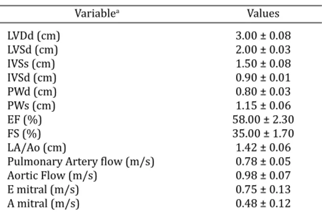
Conventional echocardiography data are displayed as mean and standard deviation (Table 1).
The values of LSt, LStR, Lvel and Ldisp obtained from LAFCV and RPLAV are shown as mean and SD for endocar-dial and epicarendocar-dial regions, as well as the global mean. En-docardial values were numerically higher than epicardial ones for all variables in both echocardiographic views, ex-cept for LSt at LAFCV. In addition, the global mean for all va-riables were numerically lower at RPLAV when compared with LAFCV (Table 2).
The values of LSt and LStR were compared between the echocardiographic RPLAV and LAFCV views, and it was observed that LSt was similar at the endocardial region (P>0.05) and different at the epicardial region (P<0.05). LStR values were signiicantly different (P<0.05) between both views (Table 3).
Table 4 gives the mean and SD values of RSt, RStR, Rvel, Rdisp, CSt and CStR obtained from RPSAV. It was shown that RSt and RStR were numerically higher than those of CSt and CStR.
Fig.2. A two-dimensional image from right parasternal short axis view of a maned wolf. Notice the graphics and curves of the radial strain and strain rate automatically displayed after myocardial tracking.
3 SPSS Statistics 17.0, Rel. 17.0.1. 2008, SPSS Inc., Chicago, IL.
Table 1. Variables of standard echocardiography parameters (mean ± SD) obtained in a maned wolf
(Chrysocyon brachyurus)
Variablea Values
LVDd (cm) 3.00 ± 0.08
LVSd (cm) 2.00 ± 0.03
IVSs (cm) 1.50 ± 0.08
IVSd (cm) 0.90 ± 0.01
PWd (cm) 0.80 ± 0.03
PWs (cm) 1.15 ± 0.06
EF (%) 58.00 ± 2.30
FS (%) 35.00 ± 1.70
LA/Ao (cm) 1.42 ± 0.06
Pulmonary Artery low (m/s) 0.78 ± 0.05 Aortic Flow (m/s) 0.98 ± 0.07
E mitral (m/s) 0.75 ± 0.13
A mitral (m/s) 0.48 ± 0.12
DISCUSSION
The values of the diameters and thicknesses of the heart chambers obtained by M-mode were within the normal range, as described by Estrada et al. (2009), indicating an absence of myocardial remodeling.
An EF of 58%, acquired by Simpson’s method, indica-ted the preservation of systolic function. This method was chosen to determine EF, because it is more accurate for de-tecting echocardiographic changes when compared to the conventional M-mode (Wess et al. 2010).
Although no data regarding mitral peak velocity was found in the literature, the velocities of transmitral peak early (E) and transmitral peak late (A) diastole are identical to those found by Pereira et al. (2009) and similar to those described in German Shepherd dogs by Muzzi et al. (2006) and Schober & Fuentes (2001). An E/A ratio greater than 1 and less than 2 indicates a normal relaxation pattern, as seen in this case. In addition, valve insuficiency by Dop-pler examinations was not observed in this case, however, mitral regurgitation is probably common in older wolves, similarly to dogs (Estrada et al. 2009).
Regarding 2D STE, this tool was found to be adequate and useful for obtaining the myocardial systolic function in the maned wolf. The images acquired were compatible with 2D STE and, due to the fact that the animal was under anesthesia, it facilitated the capture of high quality images. In addition, this technique demonstrates to be easy to per-form, since its software automatically provides the measu-rements of the myocardial variables.
The longitudinal variables St, StR, velocity and displace-ment obtained from LAFCV were similar to those obtained in dogs by Wess et al. (2011), being the global means of our study -13.52±7.88, -1.60±1.05, 3.86±3.04, 4.34±2.52, res-pectively. For all of the variables obtained in this plane, the endocardial values were higher than the epicardial, except for LSt, which was similar in both regions, a fact previously reported in humans by Leitman et al. (2010). In a study by Decloedt et al. (2011), the values observed for horses were much higher than those observed for the wolf in our study. This can be justiied by anatomical differences, such as the size of the heart.
In our study, the longitudinal variables were acquired from both the RPLAV and LAFCV, due to the fact that there are no studies comparing data obtained from these views, given that the RPLAV is not commonly used to obtain longi-tudinal function. Data demonstrated that there is no diffe-rence for endocardial LSt in both planes; however, for the other variables, signiicantly lower values were observed for RPLAV compared to LAFCV, which is the standard plane to obtain left ventricular myocardial longitudinal function. These data show that the RPLAV is not adequate to obtain LStR, Ldisp and Lvel, but is a good choice to measure LSt. Nonetheless, a study with a large population of maned wol-ves is necessary to disclose the reliability of acquiring lon-gitudinal function using the RPLAV.
Radial and circumferential variables were obtained using RPSAV. The values of RSt were lower than previous studies in dogs (Chetboul et al. 2007, Takano et al. 2011) and in horses (Schwarzwald et al. 2009), which can be at-tributed to technical differences between the ultrasound units, since the software used in those studies, was based on a different algorithm (block-matching), while in our stu-dy was used an optical low method. Species particularities and anatomical differences, as body weight and heart size, also can inluence the results. On the other hand, the values found were similar to those observed in normal human Table 2. Values of LSt, LStR, Lvel and Ldisp obtained from LAFCV and
RPLAV in a maned wolf (Chrysocyon brachyurus) and displayed as mean and SD for endocardial and epicardial regions, as well as, the global mean
View Region Longitudinal Longitudinal Longitudinal Longitudinal Strain Strain rate Displacement Velocity
(%) (1/s) (mm) (cm/s)
Endocardium -11.09 ± 8.71 -1.25 ± 0.73 1.11 ± 1.24 2.20 ± 1.50 RPLAVa Epicardium -5.00 ± 4.26 -0.73 ± 0.45 0.79 ± 0.94 2.04 ± 1.71 Mean -8.94 ± 7.33 -0.99 ± 0.66 1.21 ± 1.11 2.12 ± 1.59 Endocardium -13.14 ± 9.36 -2.05 ± 1.30 4.81 ± 2.88 4.58 ± 3.69 LAFCVb Epicardium -13.89 ± 6.22 -1.14 ± 0.39 3.87 ± 2.04 3.15 ± 2.03 Mean -13.52 ± 7.88 -1.60 ± 1.05 4.34 ± 2.52 3.86 ± 3.04 a RPLAV = Right parasternal long axis view, b LAFCV = Longitudinal apical four chamber
view.
Table 3. Values (mean ± SD) of LSt e LStR at the myocardial regions (endocardium and epicardium) compared regarding the
RPLAV and LAFCV in a maned wolf (Chrysocyon brachyurus)
Region RPLAV LAFCV P Value
Strain (%)
Endocardium -11.09 ± 8.71 -13.14 ± 9.36 0.4690 Epicardium -5.00 ± 4.26 b -13.89 ± 6.22 a <0.01*
Strain Rate (1/s)
Endocardium -1.25 ± 0.73 b -2.05 ± 1.30 a 0.0070* Epicardium -0.73 ± 0.45 b -1.14 ± 0.39 a <0.01* a Means followed by different letters in the line differ by the
Mann-Whi-tney test (P<0.05). RPLAV = Right parasternal long axis view, LAFCV = Longitudinal apical four chamber view.
Table 4. Variables of 2D STE obtained from right parasternal short axis view in a maned wolf
(Chrysocyon brachyurus)
Variable Values
subjects (Hurlburt et al. 2007). Also, the values obtained for CSt were found to be lower than previous data in dogs (Takano et al. 2011) and in humans (Hurlburt et al. 2007), yet similar to data reported in horses (Schwarzwald et al. 2009). The variables RStR and CStR were similar to those obtained by Schwarzwald et al. (2009) in a study of horses. In that same study Rdisp was assessed, and was shown to be much higher than the values found in our study.
The limitations of this investigation must be clariied. Firstly, the study was performed on a single wolf, meaning that a larger study population needs to be investigated in order to establish more accurate reference values and to identify the inluences of sex and age on 2D STE parame-ters in maned wolves. Nonetheless, this study provides preliminary reference values for longitudinal and radial St, StR, velocity and displacement, as well as circumferential St and StR for a maned wolf, and also demonstrated the feasibility of using the technique in this species. Another consideration is the fact that the sedation protocol might inluence the myocardial function and ketamine is likely to reduce the contractile function, as reported previously in other species, such as domestic cats and oncilla (Leopardus tigrinus) (Dümel et al. 1996, Carvalho et al. 2007).
CONCLUSION
2D STE can be considered a reliable technique for measu-ring longitudinal, radial and circumferential myocardial function in maned wolves. These measurements provide new insights into ventricular deformation and motion, allowing a more complete quantification of myocardial function in this species. However, further studies should be performed in a larger population of healthy and diseased wolves to evaluate the role and reliability of this technique in this species.
Acknowledgments.- To Fundação de Amparo a Pesquisa do Estado de Minas Gerais (FAPEMIG), Coordenação de Aperfeiçoamento de Pessoal de Nível Superior (CAPES) and to Conselho Nacional de Desenvolvimento Cientíico e Tecnológico (CNPq) for the inancial support.
REFERENCES
Boon J.A. 2011. The two-dimensional echocardiographyc exam, p.37-100. In: Boon J.A. (Ed.), Veterinary Echocardiography. Wiley-Blackwell, Iowa. Carvalho P.S.L., Pereira G.G., Petrus L.C., Soares E.C., Michina L.E. & Larsson
M.H.M.A. 2007. Evaluation of some echocardiographic parameters of Oncilla (L. tigrinus), kept in captivity and submitted to anesthesia with xilazine and ketamine. Arq. Bras. Med. Vet. Zootec. 59:695-699. Chetboul V., Serres F., Gouni V., Tissier F. & Pouchelon J.L. 2007. Radial
strain and strain rate by two-dimensional speckle tracking echocardio-graphy and tissue velocity based technique in the dog. J. Vet. Cardiol. 9:69-81.
Chetboul V. 2010. Advanced techniques in echocardiography in small ani-mals. Vet. Clin. North Am., Small Anim. Pract. 40:529-543.
Decloedt A., Verheyen T., Sys S., De Clercq D. & Van Loon G. 2011. Quantification of left ventricular longitudinal strain, strain rate, velocity, and displacement in healthy horses by 2-dimensional speckle tracking. J. Vet. Intern. Med. 25:330-338.
Dietz J.M. 1985. Chrysocyon brachyurus. Mammalian Species 234:1-4. Dümmel C., Neu H., Hüttig A. & Failing K. 1996. Echocardiographic
referen-ce ranges of sedated cats. Tierärztl. Praxis 24:190-196.
Estrada A.H., Gerlach T.J., Schmidt M.K., Siegal-Willott J.L., Atkins A.L., Gil-der J.V., Cintino S.B. & Padilla L.R. 2009. Cardiac evaluation of clinically healthy captive maned wolves (Chrysocyon brachyurus). J. Zoo Wildl. Med. 40:478-486.
Grifiths L.J., Fransioli J.R. & Chigerwe M. 2011. Echocardiographic asses-sment of interventricular and intraventricular mechanical synchrony in normal dogs. J. Vet. Cardiol. 13:115-126.
Hurlburt H.M., Aurigemma G.P., Hill J.C., Narayanan A., Gaasch W.H., Vinch C.S., Meyer T.E. & Tighe D.A. 2007. Direct ultrasound measurement of longitudinal, circumferential, and radial strain using 2-dimensional strain imaging in normal adults. Echocardiography 24:723-731. Leitman M., Lysiansky M., Lysyansky P., Friedman Z., Tyomkin V., Fuchs
T., Adam D., Krakover R. & Vered Z. 2010. Circumferential and longitu-dinal strain in 3 myocardial layers in normal subjects and in patients with regional left ventricular dysfunction. J. Am. Soc. Echocardiogr. 23:64-70.
Muzzi R.A.L., Muzzi L.A.L., Araujo R.B. & Cherem M. 2006. Echocardiogra-phic indices in normal German Shepherd dogs. J. Vet. Sci. 7:193-198. Pavlopoulos H. & Nihoyannopoulos P. 2008. Strain and strain rate
defor-mation parameters: from tissue Doppler to 2D speckle tracking. Int. J. Cardiovasc. Imaging 24:479-491.
Pereira G.G., Petrus L.C., Santos A.L.F., Yamaki F.L & Larsson M.H.M.A. 2009. Evaluation of left ventricular diastolic echocardiographic parameters in healthy dogs by pulsed-wave Doppler. Pesq. Vet. Bras. 29:291-294. Schefer K.D., Bitschnau C., Weishaupt M.A. & Schwarzwald CC. 2010.
Quan-titative analysis of stress echocardiograms in healthy horses with 2-di-mensional (2D) echocardiography, anatomical M-mode, tissue Doppler imaging, and 2D speckle tracking. J. Vet. Intern. Med. 24:918-931. Schober K.E. & Fuentes V.L. 2001. Effects of age, body weight, and heart
hate on transmitral and pulmonary venous low in clinically nor-mal dogs. Am. J. Vet. Res. 62:1447-1454.
Takano H., Fujii Y., Yugeta N., Takeda S. & Wakao Y. 2011. Assessment of left ventricular regional function in affected and carrier dogs with Duchene muscular dystrophy using speckle tracking echocardiography. BMC Car-diovasc. Disord. 11:2-8.
Thomas W.P., Gaber C.E., Jacobs G.J., Kaplan P.M., Lombard C.W., Moise N.S. & Mases B.L. 1993. Recommendations for standards in transthoracic two-dimensional echocardiography in the dog and cat. Echocardiogra-phy Committee of the Specialty of Cardiology, American College of Vete-rinary Internal Medicine. J. Vet. Intern. Med. 7:247-252.
Vasconcellos A.S., Chelini M.O.M., Palme R., Guimarães M.A.B.V., Oliveira C.A. & Ades C. 2011. Comparison of two methods for glucocorticoid eva-luation in maned wolves. Pesq. Vet. Bras. 31:79-83.
Wess G., Mãuer J., Simak J. & Hartmann K. 2010. Use of Simpson’s method of disc to detect early echocardiographic changes in Doberman Pins-chers with dilated cardiomyopathy. J. Vet. Intern. Med. 24:1069-1076. Wess G., Keller L.J., Klausnitzer M., Killich M. & Hartmann K. 2011.